ABSTRACT
Photooxidative sunburn, a third type that is distinctly different from sunburn necrosis and sunburn browning, is described for apple (Malus domestica Borkh.). Shaded (nonacclimated) apples suddenly exposed to sunlight became photobleached and eventually necrotic. Electron micrographs and electrolyte leakage studies indicated that cell death occurred with photobleaching. Photobleaching occurred at fruit surface temperatures (FST) below 31°C. Sunburn browning and sunburn necrosis require FST above 45°C. Ultraviolet-B radiation is involved in induction of sunburn browning, but blocking UV-A and UV-B radiation did not protect nonacclimated apples from photobleaching and necrosis. Infrared radiation (>700 nm) was ruled out as an induction factor. We suggest that visible radiation and the production of reactive oxygen species are the induction factors for photooxidative sunburn.
INTRODUCTION
Sunburn affects many fruits that are grown in areas with high solar radiation and high temperatures. CitationMacMillan (1918) reported sunburn in beans (Phaseolus vulgaris L.) and determined it was the result of exposure to the sun. Later, CitationMacMillan (1923) investigated heat and light as possible causes and concluded that the disorder was caused by light of short wavelength but was unable to give any specific values.
Since then, sunburn has been noted and studied in many fruits including banana (Musa spp.), cucumber (Cucumis sativus L.), pepper (Capsicum annum L.), tomato (Lycopersicon esculentum Mill.), and apple. CitationRabinowitch et al. (1986, Citation1974) found evidence that both heat and light were needed for sunburn to occur in tomatoes, cucumber, and pepper. CitationRetig and Kedar (1967) determined that tomatoes at the mature green stage were most susceptible and red tomatoes were completely resistant, which corresponds with others' observations that full-sized green tomatoes are more susceptible than ripe tomatoes (CitationCook, 1921; CitationHarvey, 1924; CitationRamsey and Link, 1932). The resistance of red fruit to sunburn was also noted in peppers (CitationRamsey and Link, 1932) and has been attributed to the thermal stability of carotenoids once they have been synthesized (CitationRetig and Kedar, 1967).
This evidence along with studies conducted with cucumber (CitationRabinowitch and Sklan, 1981) suggests that at least one type of sunburn is a photooxidative process that is associated with photosynthetic tissue and chlorophyll pigments. In leaves it is known that excess light that cannot be used in photochemistry has potential to cause damage (CitationNiyogi, 1999). This indicates that excess photosynthetic photon flux density in green fruit may cause over reduction of the electron transport chain leading to the formation of reactive oxygen species (ROS), which then cause oxidative damage.
Many investigators have characterized and discussed different types of sunburn, but some used fruits detached from the tree or plant. Some separated them by their induction factors, whereas others grouped them according to their appearance or their severity. CitationRetig and Kedar (1967) discussed two types of sunburn damage in tomatoes (referred to by them as sunscald): discoloration and sunscald. Discoloration referred to mild damage characterized by yellowing that remained during ripening. Sunscald referred to a more severe damage that was characterized by a white and sunken area. CitationBarber and Sharpe (1971) classified different types of sunburn of Capsicum annuum and Curcurbita pepo based on different induction factors. They described three types of sunburn (referred to by them as sunscald): heat injury sunscald, ultraviolet sunscald, and photodynamic sunscald of heated tissue. Heat injury sunscald was due to the heating of the tissue resulting in a cooked appearance. Ultraviolet radiation sunscald was seen in fruits grown at high altitudes, presumably resulting from higher levels of ultraviolet radiation. Photodynamic sunscald of heated tissue resulted from the absorption of visible light by heated photosynthetic cells.
Studies on sunburn of apples attached to trees have led to the identification and characterization of two types of sunburn: sunburn necrosis and sunburn browning (CitationSchrader et al., 2001). Sunburn necrosis occurred when fruit surface temperatures (FST) reached 52 ± 1 °C for only 10 min, and was the result of heat-induced cell death (L.E. CitationSchrader et al., 2001; L. CitationSchrader et al., 2003). Sunburn necrosis can occur naturally in sunlight but also was induced experimentally on attached fruit at an FST of 52 ± 1°C imposed in the dark (L.E. CitationSchrader et al., 2001). Sunburn necrosis appeared as a dark brown or black spot on the surface of the apple. This spot was often several millimeters thick.
The second type of sunburn, sunburn browning, did not result in cell death and was characterized by a yellowing or browning of the skin. The discoloration associated with sunburn browning has been correlated to decreases in chlorophyll and anthocyanin concentrations and increases in carotenoid and quercetin glycoside concentrations (CitationFelicetti and Schrader, 2008). The discoloration was superficial and did not penetrate into the flesh of the apple. Sunburn browning occurred at FST between 46 and 49°C, depending on the cultivar, and did not occur when apples were heated in the dark (L.E. CitationSchrader et al., 2001). The minimum FST needed to induce sunburn browning when fruits were exposed to sunlight for 1 h was referred to as the threshold temperature.
Because sunburn browning did not occur in the dark, different components of sunlight were suggested as factors in its formation. Ultraviolet-B radiation (UV-B, 280–320 nm) was implicated because the incidence of sunburn browning was reduced by application of UV-B filtering compounds such as RAYNOX® (L. CitationSchrader et al., 2008) or PABA (p-aminobenzoic acid; unpublished observations). Visible radiation was suspected because of its ability to form ROS in higher plants when absorbed in excess.
Our prior attempts to induce sunburn browning on shaded apples by exposing them to sunburn browning conditions had been unsuccessful throughout the summer and fall. Instead of developing sunburn browning, apples became photobeached and eventually necrotic. We observed this photobleaching and subsequent necrosis on apples that were suddenly exposed to sunlight at various stages of development as a result of different horticultural practices (i.e., thinning, summer pruning, and turning of apples in the fall to improve color). The conditions under which the photobleaching and necrosis occurred and the type of damage incurred were not consistent with the characteristics of sunburn necrosis or sunburn browning. We conducted this study to determine whether this disorder was a different type of sunburn. The specific objectives were to 1) determine if a threshold FST existed for the observed photobleaching and subsequent necrosis; 2) determine if either ultraviolet-A (UV-A, 320–400 nm) or UV-B were involved in causing the disorder; 3) determine the extent and type of damage that occurred; and 4) compare and contrast this disorder to sunburn necrosis and sunburn browning.
MATERIALS AND METHODS
Site and Plant Material
The following experiments were performed on ‘Fuji’ apples located at the Washington State University Tree Fruit Research and Extension Center (WSU-TFREC) Columbia View Orchards, located 19 km north of Wenatchee, Wash. The block contained both ‘Royal Gala’ and ‘Red Fuji’, alternating every tree. All trees were planted on Malling 9 (M.9) rootstock, were irrigated with micro-sprinklers, and were maintained using commercial production practices.
Temperature Measurements
FST was measured with fine wire copper-constantan (0.13 mm dia., type-T) thermocouples. Thermocouples were placed on the fruit surface and held in place by a tan-colored fabric adhesive bandage no larger than 2.5 cm2. This technique was shown to provide FSTs similar to FSTs obtained by inserting a thermocouple under the skin and avoided the problems (e.g., invasive pathogens and loss of water) associated with puncturing the skin and leaving the thermocouple for several days. Thermocouples were connected to a Campbell Scientific data logger (CR10X; Logan, Utah) that scanned the FST every 5 s and recorded the 5-min average. The FSTs of 23 ‘Fuji’ apples were monitored for 7 d starting on 16 Oct. 2002 and the fruit were photographed on days 1, 3, and 5.
Solar Radiation Measurements
Total solar radiation (300 to 3000 nm) was obtained from the Washington State University Public Agricultural Weather System (WSU-PAWS) monitoring station located at the WSU-TFREC in Wenatchee, Wash. This site uses LI-COR silicon pyranometers that have been calibrated against an Eppley Precision Spectral Pyranometer under natural daylight conditions. Total photosynthetically active radiation (PAR) was calculated from total solar radiation by first dividing total solar radiation by 1.71 × 10⁵ J mol−1 and then multiplying by 0.45. The average energy per mole of photons of terrestrial radiation is 1.71 × 10⁵ J mol−1 and was used only to convert the units of total solar radiation from J m−2 to mol photons m−2. CitationCampbell and Norman (1998) indicated that 45% of total solar radiation is PAR.
Assessment of Sunburn Damage
The damage incurred was rated on a scale of zero to four on day 7. A damage rating of 0 indicated no damage, 1 indicated mild photobleaching, 2 indicated moderate photobleaching with no brown discoloration, 3 indicated moderate photobleaching with a slight brown discoloration, and 4 indicated photobleaching with a severe brown discoloration.
Reduction of UV-B
The amount of UV-B radiation was reduced by securing 20 × 20 cm sheets of Mylar® (Northwest Laminating, Seattle, Wash.) 6 to 10 cm in front of 12 of the apples on 16 Oct. 2002. The tops and the bottoms of the Mylar® sheets were secured to the tree with string to prevent movement. After securing the Mylar®, shaded apples or the shaded sides of apples were exposed to sunlight. This was achieved by either pruning overhanging limbs or by turning apples to expose the shaded side. The Mylar® was left in place for 7 d, after which the apples were evaluated. The UV-A and UV-B transmittance of Mylar® was determined using calibrated sensors with a Solar Light Company radiometer (PMA 2100; Glenside, Pa.; ).
TABLE 1. The amount of UV-B and UV-A radiation passing through Mylar® and UV-blocking plexiglass filters. Measurements were taken with a Solar Light Company PMA 2100 radiometer on 20 June 2002 at 1300 hr. UV-B was measured with a PMA 2101 detector and UV-A was measured with a PMA 2110 detector
Exclusion of UV-A and UV-B
The exclusion of both UV-A and UV-B was achieved by using 19 × 28 cm sheets of UV-blocking Plexiglas™ acrylic glass (hereafter referred to as plexiglass; Cyro Industries, Rockaway, N.J.; ). The plexiglass sheets were attached to tripods and placed 3 to 5 cm above 11 of the apples on 16 Oct. 2002. After placement of the plexiglass, apples were exposed to sunlight in the same manner as described above and left in place for 7 d, after which the apples were evaluated. The UV-A and UV-B transmittance of the UV-blocking plexiglass was determined using a Solar Light Company radiometer (PMA 2100; Glenside, Pa.; ).
Electrolyte Leakage
Determination of electrolyte leakage was adapted from L.E. CitationSchrader et al. (2001). Apples of two classes were used: those that received a damage rating of zero and those that received a damage rating of four. Three replicates were used for the green undamaged tissue and five replicates were used for brown photooxidized tissue. The peel was removed from the apple and the cortex scraped off with a razor blade. After removal of the cortex, 0.8-cm-diameter disks were made with a cork borer. The three replicates of green undamaged tissue contained between 0.33 and 0.35 g of tissue per replicate. The five replicates of the browned photooxidized tissue contained between 0.44 and 0.67 g of tissue per replicate.
Electron Microscopy
Apples were gently rinsed in deionized water at 1°C to remove any loose surface material. Other than cold water, the area selected for examination was untouched. The fruit was left to dry at 20°C for 30 min, after which a thin section of peel tissue about 0.3 mm thick and 5 mm in diameter was excised by hand with a razor blade and flash frozen on an aluminum block held at −190°C with liquid nitrogen. Together, the tissue and cold aluminum block were transferred to a small vacuum dessicator in which the sample was freeze-dried for 36 h. Freeze-dried peel sections were fixed to a 12-mm aluminum stub with carbon tape and coated with a thin film of platinum using a cold sputter coater (Desk II; Moorestown, N.J.). Samples were kept under low vacuum until viewed with a scanning electron microscope (S-530, Hitachi Instruments, Inc., San Jose, Calif.). All samples were examined at 20 Kev.
Statistical Analyses
Linear regression analyses were performed using statistical analysis system software (Proc REG; SAS Institute Inc., Cary, N.C.).
RESULTS
Temperature and Solar Radiation Measurements
During the 7-day period of measurements, the maximum FST of all 13 photobleached apples ranged from 30.5 to 37.6°C. The maximum FST of apples that did not photobleach ranged from 31.2 to 35.9°C. When the maximum FST was plotted against the level of sunburn damage a linear R2 of 0.11 (P = 0.13) was obtained. When the difference between maximum FST and the concurrent air temperature was plotted against the level of damage, a linear R2 of 0.03 (P = 0.43) was obtained. Total daily PAR during the 7-day induction period ranged from 21.8 to 31.8 mol m−2 d−1 and PAR did not exceed 1350 μmol m−2 s−1
Reduction of UV-B
Of the 12 apples covered with Mylar®, the peels of 5 were photobleached after 3 d (). After an additional 2 d of exposure a total of 6 apples were photobleached. No additional apples were photobleached by day 7. Six apples were rated as zero (damage rating), 2 apples rated one, 1 apple rated two, 2 apples rated three, and 1 apple rated four. All necrotic areas were within the photobleached area. The area underneath the adhesive bandage was undamaged on all 12 apples. After an additional 6 d of exposure without Mylar®, 4 of the damaged apples had an increase in the size of the necrotic area and none had a decrease in the amount of peel that was photobleached or brown.
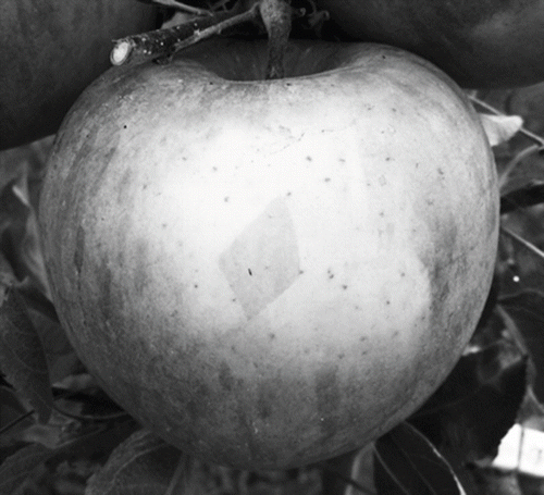
FIGURE 1. Photobleaching on ‘Fuji’ 3 d after the initial exposure of the shaded side to sunlight. The diamond shape in the center of the photobleached area is undamaged peel that was covered by adhesive bandage used to attach thermocouple to the apple.
Exclusion of UV-A and UV-B
Of the 11 apples covered with UV-blocking plexiglass, the peels of 6 were photobleached after 3 d. After 5 d of exposure a total of 7 apples were photobleached. By day 7 no additional apples were photobleached. Four apples were rated as zero, 2 apples were rated one, 3 apples were rated two, 1 apple was rated three, and 1 apple was rated four. All necrotic areas were within the photobleached area. The area underneath the adhesive bandage was undamaged on all 11 apples.
Electrolyte Leakage
The average relative electrical conductivity for the control and the damaged peel was 36.3% ± 0.66% and 73.1% ± 1.6%, respectively.
Electron Microscopy
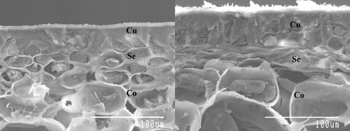
The micrographs of transverse sections show that the subepidermal cells in the photobleached peel have collapsed or flattened while the cortical cells appear to be undamaged ().
FIGURE 2. Electron micrographs of transverse section of green undamaged peel (left) and photobleached peel (right). Cu, cuticle; Se, sub-epidermal tissue; Co, cortex.
DISCUSSION
The monitoring of FST demonstrated that photobleaching and later necrosis can occur at an FST below 31.0°C. Observations that the area under the adhesive bandage remained undamaged in all apples despite having an FST above ambient temperature suggested that solar radiation, not temperature, was the main causal factor. Additionally, the degree of damage was not correlated to the maximum FST or the difference between maximum FST and the ambient temperature. These results indicate that maximum FST is not a factor in the formation of the photobleached area.
Cellular breakdown is suggested by both the high relative EC and electron micrographs. The relative EC for the damaged region is twice that of green tissue. Low relative EC of tissue is an indicator of cell membrane integrity. The relative EC of the damaged peel was within 2% of values published for sunburned necrotic peel while the relative EC of the green tissue was within 2% of the values published for sunburn browning (L.E. CitationSchrader et al., 2001). Sunburn necrosis is thought to result from thermally induced cell death. The relative EC of the so-called necrotic areas within the photobleached peel in this experiment suggests that cell death had indeed occurred. Unlike sunburn necrosis, where necrosis is induced by thermal death, the cell death observed in these experiments is likely not due to thermal death because the maximum FSTs achieved by eight of the damaged apples during the induction period did not exceed 34°C. Ambient temperatures during the summer in this region regularly exceeded 37°C, thus exposing even shaded peels to much higher temperatures than achieved during October on sun-exposed peels during the induction period. Additionally, if the necrosis observed in this experiment were caused by thermal death it would be expected that apples whose peel achieved higher FSTs would have more severe damage. This was not the case and is shown by the extremely low R2 and P values when correlating maximum FST with the damage rating.
As mentioned earlier, the area under the adhesive bandage remained undamaged suggesting solar radiation is the main cause of photooxidative sunburn. The formation of this type of sunburn on apples exposed to sunlight lacking UV-A and UV-B wavelengths as well as relatively low levels of terrestrial UV radiation during the month of October indicate that UV-A and UV-B are not necessary for induction of the observed disorder. This leaves visible and infrared radiation as possible causes of damage.
Infrared radiation has less energy per photon than visible radiation and, aside from far-red radiation (710–740 nm), causes nothing more than warming of the object that it irradiates. Although an unlikely cause, far-red radiation does interact with phytochrome, which is a pigment that can be interconverted between a red-light absorbing form and a far-red absorbing form. The phytochrome response in plants is typically associated with helping plants adjust to changing light conditions and is related to the changing ratio of red to far-red light (which increases from shade to sun). Additionally, there is no evidence to suggest a mechanism that would harm the plant or cause cell death through the phytochrome system.
Solar radiation in the visible range is a more likely candidate because it causes formation of reactive oxygen species (ROS) when in excess. Lipids, proteins, and DNA can be damaged by ROS. The formation of ROS in this instance may occur via a process similar to chronic photoinhibition (CitationNiyogi, 1999; CitationOsmond, 1994), which occurs when shaded leaves are exposed to sunlight. Shade leaves have a higher capacity for light harvesting by chlorophyll than capacity for electron transport and energy use in carbon assimilation. The sudden exposure to sunlight provides an excessive amount of light energy, causing damage to the photosynthetic mechanism, which in turn can lead to the production of singlet oxygen. Singlet oxygen is very reactive and oxidizes amino acids and lipids found in the cell. The formation of singlet oxygen and other ROS (e.g., superoxide) as a result of excess visible light provides a very plausible explanation. Data obtained from the WSU-PAWS indicate that during the induction period the total daily PAR did not exceed 32 mol m−2 d−1 and PAR did not exceed 1350 μmol m−1 s−1. These light levels are approximately 50% and 65%, respectively, of the light levels observed in Wenatchee during July and indicate that even moderate irradiance levels can cause photooxidative damage if the tissue is not properly acclimated.
The evidence gathered in these experiments strongly suggests that photobleaching of nonacclimated apples is due to visible solar radiation and that UV-A, UV-B, and high temperatures are not primary factors. These characteristics distinguish it from sunburn browning, which requires UV-B radiation and an FST between 46 and 49°C. It differs from sunburn necrosis in that sunburn necrosis does not require light directly but requires an FST of 52 ± 1°C. Although this disorder may result in necrosis, it is not sunburn necrosis. Sunburn necrosis refers specifically to thermally induced necrotic sunburn. However, at the time of its naming there was only one known type of sunburn in apples that resulted in necrosis. Therefore, there was no need to differentiate it from other types of sunburn that may become necrotic. Additionally, photooxidative sunburn does not inevitably result in necrosis.
Based on evidence presented above, we have named the observed disorder photooxidative sunburn. Because photooxidative sunburn is a distinct type of sunburn with its own characteristics and induction requirements, preventative measures currently being used to control sunburn browning and sunburn necrosis may not be effective in controlling photooxidative sunburn. Based on the induction factors of photooxidative sunburn elucidated in this research and the modes of action of current sunburn prevention practices, it is not unreasonable to theorize whether these practices would also prevent photooxidative sunburn formation. Evaporative cooling is unlikely to be effective at controlling photooxidative sunburn because it works by reducing FST, which does not appear to be a factor in photooxidative sunburn. Sunburn protectants such as Surround® and RAYNOX® reflect light and therefore may provide some protection. However, it is not known whether they reflect enough light to be effective. If they do reflect enough light, the ability to apply appropriate amounts to susceptible peel—that is, the back side of apples and shaded apples—may not be possible with current application methods.
Other practices that are used to prevent sunburn of apple are bagging and shade cloth. These two methods of sunburn protection are aimed at reducing the amount of sunlight reaching the apples and may seem to be good preventative measures for photooxidative sunburn. However, based on the observations in this study it is likely that such practices may increase the amount of photooxidative sunburn because they reduce acclimation of the entire apple, making it vulnerable to high light conditions when protection is removed (i.e., removal of bags or the rolling back of shade cloth near maturity to induce color development). Furthermore, these practices are not likely to be used for control of photooxidative sunburn in the United States due to their cost, which has already sharply diminished their use by growers for protection from sunburn browning.
Growers should be cautious late in the season when turning fruit to improve color or when leaving bins of apples uncovered in the sun after harvest, as many of the fruit have been shaded while hanging on the tree. Additionally, growers should be careful not to expose shaded apples when hand-thinning clusters and when summer pruning and should take care that limbs bearing a heavy crop do not suddenly move, exposing shaded apples.
The authors gratefully acknowledge financial support from the Washington Tree Fruit Research Commission. We thank Eric Curry, USDA-ARS-Tree Fruit Research Laboratory, for the electron micrographs.
LITERATURE CITED
- Barber , N.H. and Sharpe , P.J.H. 1971 . Genetics and physiology of sunscald of fruits . Agr. Meteorol. , 8 : 175 – 191 .
- Campbell , G. and Norman , J. 1998 . An introduction to environmental biophysics , 2nd , New York, N.Y : Springer .
- Cook , M.T. 1921 . Sunburn and tomato fruit rots . Phytopathology , 11 : 379 – 380 .
- Felicetti , D.A. and Schrader , L.E. 2008 . Changes in pigment concentrations associated with the degree of sunburn browning of “Fuji” apple . J. Am. Soc. Hort. Sci. , 133 : 27 – 34 .
- Harvey , R.B. 1924 . Sunscald of tomatoes . Minn. Studies Plant Sci. , 4 : 229 – 234 .
- Macmillan , H.G. 1918 . Sunscald of beans . J. Agr. Res. , 13 : 647 – 650 .
- MacMillan , H.G. 1923 . Causes of sunscald of beans . Phytopathology , 13 : 376 – 380 .
- Niyogi , K.K. 1999 . Photoprotection revisited: Genetic and molecular approaches . Annu. Rev. Plant Physiol. Plant Mol. Biol. , 50 : 333 – 359 .
- Osmond , C.B. 1994 . “ What is photoinhibition? Some insights from comparisons of shade and sun plants ” . In Photoinhibition of Photosynthesis , Edited by: Baker , N.R. and Bowyer , J.R. 255 – 271 . Oxford : BIOS Scientific .
- Rabinowitch , H.D. , Ben-David , B. and Friedmann , M. 1986 . Light is essential for sunscald induction in cucumber and pepper fruits, whereas heat conditioning provides protection . Scientia Hort. , 29 : 21 – 29 .
- Rabinowitch , H.D. , Kedar , N. and Budowski , P. 1974 . Induction of sunscald damage in tomatoes under natural and controlled conditions . Scientia Hort. , 2 : 265 – 272 .
- Rabinowitch , H.D. and Sklan , D. 1981 . Superoxide dismutase activity in ripening cucumber Cucumis sativus and pepper fruit Capsicum annuum . Physiol. Plant , 52 : 380 – 384 .
- Ramsey , R.B. and Link , G.K.K. 1932 . Market diseases of fruit and vegetables: Tomatoes, peppers, and eggplants , Washington, D.C : U.S. Dept of Agr .
- Retig , N. and Kedar , N. 1967 . The effect of stage of maturity on heat absorption and sunscald of detached tomato fruits . Israel J. Agri. Res. , 17 : 77 – 83 .
- Schrader , L. , Sun , J. , Zhang , J. , Felicetti , D. and Tian , J. 2008 . Heat and light-induced apple skin disorders: Causes and prevention . Acta Hort. , 772 : 51 – 58 .
- Schrader , L. , Zhang , J. and Sun , J. 2003 . Environmental stresses that cause sunburn of apple . Acta Hort. , 618 : 497 – 503 .
- Schrader, L.E., J. Zhang, and W.K. Duplaga. 2001. Two types of sunburn in apple caused by high fruit surface (peel) temperature. Plant Health Prog http://hort.tfrec.wsu.edu/les/Temperature.pdf