
Following the publication of this paper on 04 Jun 2018, it has come to the authors’ attention that the images () acquired in transmission electron microscopy (TEM) might not have convincingly displayed extracellular vesicles (EVs). During consultations with editors of the Journal of Extracellular Vesicles as well as experts in the field, it became clear that the initial protocol for TEM sample preparation lacked a methyl cellulose embedding step that is essential to maintain the structural integrity of EVs [1].
Figure 7. Morphology of serum EVs by transmission electron microscopy. Images are representative for at least two biological replicates for both volunteers (top panel) and sepsis patients (bottom panel). Scale bars within each panel are 250 nm (top row) and 100 nm (bottom row).
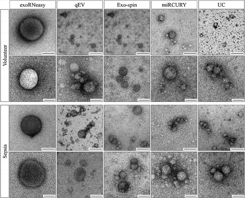
The authors have added this step for the improved protocol detailed below and herein present a revised , which now convincingly depicts EVs. The morphology of EVs from each isolation method can be observed more clearly, and differences in their size, shape and levels of background contamination are more apparent. The structures observed in exoRNeasy isolates look distinctly different from those isolated by all other methods and reflect the increased particle diameters detected in nanoparticle tracking analysis (Figure 6).
The published online version has been corrected to display a revised .
A correction has also been made to the Materials and methods section, paragraph Transmission electron microscopy:
EVs were adsorbed onto formvar/carbon-coated 200-mesh nickel grids (Electron Microscopy Sciences) for 20 min at RT. Next, grids were fixed with 2% paraformaldehyde for 20 min, washed with PBS three times and fixed with 1 % glutaraldehyde for 5 min. After six washing steps with distilled water, grids were negative stained with 4 % uranyl acetate for 5 min. Finally, grids were embedded in 0.4 % uranyl acetate/0.2 % methyl cellulose for 10 min on ice in the dark and air-dried overnight. Images were acquired on a Zeiss EM900 (Carl Zeiss Microscopy GmbH) with a wide-angle dual-speed 2K-CCD camera at 80 kV.
The authors apologize for any inconvenience caused by this discrepancy.
Related Research Data
Reference
- Thery C, Amigorena S, Raposo G, Clayton A. Isolation and characterization of exosomes from cell culture supernatants and biological fluids. Curr Protoc Cell Biol. 2006;Chapter 3: Unit 3 22. Epub 2008/ 01/30.