
abstract
Galleria mellonella (greater wax moth or honeycomb moth) has been introduced as an alternative model to study microbial infections. G. mellonella larvae can be easily and inexpensively obtained in large numbers and are simple to use as they don't require special lab equipment. There are no ethical constraints and their short life cycle makes them ideal for large-scale studies. Although insects lack an adaptive immune response, their innate immune response shows remarkable similarities with the immune response in vertebrates.
This review gives a current update of what is known about the immune system of G. mellonella and provides an extensive overview of how G. mellonella is used to study the virulence of Gram-positive and Gram-negative bacteria. In addition, the use of G. mellonella to evaluate the efficacy of antimicrobial agents and experimental phage therapy are also discussed. The review concludes with a critical assessment of the current limitatons of G. mellonella infection models.
Introduction
One of the most commonly used models for studying microbial infections is the murine model. However, there are ethical, budgetary and logistical hurdles associated with the use of rodents as infection models. Firstly, maintaining a sufficient number of animals required to obtain statistically relevant data is expensive and often regarded as ethically objectionable. Secondly, mammals have lengthy reproduction times, which slow the progress of experimentation. More recently, Galleria mellonella (greater wax moth or honeycomb moth) has been introduced as an alternative model to study microbial infections. More than 1000 articles have been published on PubMed about the G. mellonella infection model, of which >200 were published in 2014–2015 alone demonstrating the increasing popularity of this infection model.
G. mellonella is an insect from the order Lepidoptera and the family Pyralidae (snout moths).Citation1 It is in fact the caterpillar larvae, or wax worm, and not the adult moth that is used as an animal model. When compared with traditional mammalian model hosts, G. mellonella larvae are cheaper to establish and easier to maintain, as they don't require special lab equipment.Citation2 Additionally, the use of G. mellonella does not require ethical approval and their short life span makes them ideal for high-throughput studies.
With the completion of many microbial genome projects over the last decade, there is now a large number of so-called “hypothetical proteins” in the databases. These annotations are based on identified open reading frames, sometimes with predicted functions after bioinformatics analyses, but often without an experimentally confirmed function. Many of these novel proteins are believed to be virulence factors and deciphering their functions will increase our understanding in disease mechanisms and ultimately provide a base for the development of novel therapeutic agents. Unlike other invertebrate models such as Caenorhabditis elegans and Drosophila melanogaster, G. mellonella larvae can survive at 37°C and therefore allow the investigation of temperature-dependent microbial virulence factors.Citation3,4
Insects have diverged from vertebrates approximately 500 million years ago. Although vertebrates have developed an adaptive immune response, their innate immune response still retains remarkable similarities with the immune response in insects.Citation5 Over recent years, G. mellonella has been widely used as an infection model to study bacterial and fungal infections and for assessing the efficacy of novel antimicrobial drugs.
The Galleria mellonella immune system
The innate immune response of insects consists of 2 major parts, the cellular and the humoral immune response. The cellular response is mediated by phagocytic cells, termed hemocytes. These are found within the hemolymph, which functions analogously to mammalian blood. These cells are not only involved in phagocytosis, but also in encapsulation and clotting. The humoral response is orchestrated by soluble effector molecules that immobilize or kill the pathogen and includes complement-like proteins, melanin, and anti-microbial peptides (summarized in ).
Table 1. Components of the G. mellonella innate immune response.
Cellular immune response
At least 8 types of hemocytes are found in insects, of which 6 different types have been identified in G. mellonella.Citation6 These include prohemocytes, plasmatocytes, granular cells, coagulocytes, spherulocytes and oenocytoids. In G. mellonella, plasmatocytes and granular cells play a key role in cellular defense and are involved in phagocytosis, nodule formation and encapsulation.Citation7 Plasmatocytes are the most common hemocyte and are characterized by a leaf-like shape and lysosomal enzymes within their cytoplasm. Granular cells are smaller and contain many granules in the cytoplasm. Encapsulation begins with the attachment of granular cells to the foreign target triggering the release of material (e.g. plasmatocyte spreading peptides, PSP) that promotes the attachment of multiple layers of plasmatocytes around the foreign target resulting in a smooth capsule.Citation8,9 Encapsulation is mainly associated with immune responses against larger microbes, such as protozoa and nematodes (including eggs and larvae).Citation6
Phagocytosis in insects and mammals is believed to be very similar and involves both plasmatocytes and granular cells.Citation7 G. mellonella expresses on hemocytes a protein with high homology to human calreticulin found on neutrophils, which is believed to be involved in non-self recognition in cellular defense reactions.Citation10 Once phagocytosed, pathogens are killed by several mechanisms including reactive oxygen species (e.g., superoxide) generated by the oxidative burst, which is initiated by the NADPH oxidase complex. In neutrophils, the functional NADPH complex is formed after translocation of proteins p47phox and p67phox from the cytosol to the plasma membrane. Homologous proteins p47 and p67 were identified in G. mellonella hemocytes and it was shown that superoxide production in both hemocytes and human neutrophils could be triggered by 12-myristate 13 acetate (PMA) and inhibited by diphenyleneiodonium chloride.Citation11,12
Humoral immune response
Opsonins
G. mellonella produces several plasma proteins that serve as opsonins that recognize and bind to conserved microbial components similar to pattern recognition receptors in mammals. Apolipophorin-III (apoLp-III), a major exchangeable lipid transport molecule plays a crucial role in the innate immune response as a pattern recognition molecule. ApoLp-III shows high affinity for hydrophobic ligands such as bacterial lipopolysaccharide (LPS) and lipoteichoic acid (LTA).Citation13,14 Furthermore, binding to β-1, 3 glucan and to fungal conidia resulting in increased cellular encapsulation was reported.Citation15 ApoLp-III shows high homology with mammalian apolipoprotein E (apoE), which is involved in LPS detoxification, the stimulation of phagocytosis and nitric oxide (NO) release from platelets.Citation16,17 A multifunctional role has also been demonstrated for apoLp-III. For example, apoLp-III stimulates increases in hemolymph antibacterial activity and superoxide production by hemocytesCitation18 and enhances the activity of the antimicrobial peptide cecropin.Citation19 More recently, it was shown that apoLp-III acts synergistically with G. mellonella lysozyme increasing the permeabilizing activity of lysozyme against Gram-negative bacteria.Citation20
Peptidoglycan recognition proteins (PGRPs) bind to peptidoglycan via a conserved domain homologous to T4 bacteriophage lysozyme.Citation21 G. mellonella PGRPs were identified during the analysis of LPS induced genes in hemocytes using subtractive hybridization.Citation22 PGRPs in some other insect species are also able to hydrolyze peptidoglycan, but this has not been demonstrated for PGRPs from G. mellonella.
A novel opsonin with homology to the cationic protein 8 (CP8) of Manduca sexta (tobacco hornworm) has been identified from the hemolymph of the G. mellonella larvae and termed GmCP8 (G. mellonella CP8). GmCP8, which is produced in the fat body (a biosynthetic organ analogous to the mammalian liver), midgut, and integument and is secreted into the hemolymph showed marked binding activity to LPS, LTA, and β-1,3-glucan.Citation23
Hemolin, a member of the immunoglobulin protein superfamily binds to LPS and LTA and associates with hemocytes.Citation24 Hemolin is found in several lepidopteran species, including G. mellonella, where it is expressed in several organs, including the silk gland of the larvae and is up-regulated during bacterial infectionCitation25 or after exposure to low doses of β-glucan (3.75μg/larva).Citation26
Antimicrobial peptides (AMPs)
Antimicrobial peptides (AMPs), or host defense peptides, are found among all classes of life and play a major part in innate immunity showing broad-spectrum microbicidal activity. An analysis of the AMP repertoire of G. mellonella hemolymph identified 18 known or putative AMPs: lysozyme, 5 moricin-like peptides, 2 cecropins, gloverin, Gm proline-rich peptides 1 and 2, Gm anionic peptide 1 and 2, galiomycin, gallerimycin, inducible serine protease inhibitor 2, x-tox and heliocin-like peptide.Citation27 Another AMP, an insect defensin, named Galleria defensin, was purified from the larval hemolymph of G. mellonella immunized against E. coli.Citation28 Insect AMPs are mainly produced in the fat body, hemocytes, the digestive tract, salivary glands and the reproductive tract. In mammals, AMPs are secreted from epithelial surfaces and from phagocytic cells, where they are stored within intracellular granules.
Lysozyme degrades cell wall peptidoglycan by hydrolyzing the β-1, 4 linkage between N-acetylglucosamine and N-acetylmuramic acid. In addition, G. mellonella lysozyme also shows non-enzymatic activity against fungi resembling the mode of action of cationic defense peptides. The exact mechanism is unclear, but a synergistic effect with anionic peptide 2 (AP2) has been suggested.Citation29
Cecropins and moricins both belong to the family of amphipathic α-helical AMPs, which penetrate bacterial cell walls and form cytoplasmic membrane pores resulting in ion leakage. They are active against both Gram-negative and Gram-positive bacteria. G. mellonella cecropin is expressed as a prepropeptide, with a putative 22-residue signal peptide, a 4-residue propeptide and a 39-residue mature peptide.Citation30 Moricin-like peptides in G. mellonella were first identified by Brown et al, who showed that these AMPs have a particular strong activity against filamentous fungi.Citation31 Defensins are cysteine-rich cationic peptides that act by forming voltage-dependent ion channels in the cytoplasmic membrane resulting in ion leakage and cell lysis. Insect defensins act against Gram-positive and certain Gram-negative bacteria.Citation32
Proline-rich peptides are small peptides between 2–4 KDa and appear to increase membrane permeability of bacteria. Gm proline-rich peptide 1 was also shown to inhibit the growth of yeast.Citation33 Gloverin belongs to the family of glycin-rich AMPs that binds to LPS on Gram-negative bacteria and inhibits the synthesis of vital outer membrane proteins resulting in a permeable outer membrane.Citation34 Gallerimycin is a defensin-like antifungal peptide, which was first cloned in G. mellonella from hemocytes isolated from LPS-pretreated larvae. It has no measurable effects on Gram-positive and Gram-negative bacteria or yeast, but shows activity against filamentous fungi.Citation35 X-tox is an atypical inducible defensin-like peptide that lacks detectable antimicrobial activity suggesting a yet unknown immune function.Citation36
A comparative study with 8 G. mellonella AMPs (Gm proline-rich peptides 1 and 2, Galleria defensin, Gm defensin-like peptide, Gm anionic peptides 1 and 2, Gm cecropin and Gm apolipophoricin) revealed varying activity against Gram-positive bacteria, fungi and yeast. The most effective was Gm defensin-like peptide, which inhibited yeast, fungi and sensitive bacteria at concentrations of <3 μM. In contrast, Gm apolipophoricin and Gm proline-rich peptide 2 showed the lowest antimicrobial activity.Citation33
Phenoloxidase pathway and melanization
The melanization response can be described as the synthesis and deposition of melanin to encapsulate pathogens at the wound site followed by hemolymph coagulation and opsonization and is analogous to abscess formation in mammalian infections.Citation37 Melanin formation is catalyzed by phenoloxidase (PO), which is produced as the inactive zymogen pro-phenoloxidase (ProPO) in hemocytes.Citation38 Insect ProPO is an important innate immunity protein due to its involvement in cellular and humoral defense (reviewed inCitation39). Melanization is initiated after the engagement of soluble PRRs with target surfaces, such as LTACitation13 or thermolysin,Citation40 triggering the serine protease cascade that results in the cleavage of ProPO to PO. The activated PO converts monophenols and phenols to quinines, which polymerize non-enzymatically to form melanin around invading pathogens and wounds. PO can also produce cell damaging reactive oxygen species and therefore PO activation is highly controlled by protease inhibitors.Citation38 It has recently been reported that lysozyme, Galleria defensin, proline-rich peptide 1, and anionic peptide 2 decreased the hemolymph PO activity considerably suggesting a role of these AMPs in immune modulation.Citation41
Extracellular nucleic acid traps
Upon stimulation with LPS, PMA or interleukin-8 (IL-8), neutrophils release chromosomal DNA spiked with bactericidal proteins to form an extracellular fibril matrix known as neutrophil extracellular traps (NETs) due to their ability to trap and kill bacteria.Citation42 Microscopic ex-vivo analyses of G. mellonella hemolymph clotting reactions has shown that oenocytoids represent a source of endogenously derived extracellular nucleic acids. It has been suggested that actively released nucleic acid from neutrophils and hemocytes have comparable roles in entrapping pathogens and enhancing innate immune responses.Citation43
Experimental aspects of G. mellonella infection models
The last instar larvae, which develop from the egg in about 5 weeks are used for experimental studies. The larvae are 2 to 2.5 cm long and have a creamy color (). The larvae can be stored at 15°C before use and it is recommended to starve the larvae for 24h before infection.Citation2 The most common infection route is by intrahemocoelic injection through the last left pro-legCitation44 or through the skin.Citation2 Oral infection has also been described,Citation45 but has the draw back that exact infection doses are difficult to obtain. This problem can be overcome by using a technically more challenging force-feeding method.Citation2 Microbial inoculums should be washed prior to infection to minimize the introduction of virulence factors secreted during in-vitro growth of the microorganism. It is also recommended to apply a placebo inoculum as a control for potential physical trauma due to the injection.Citation46 At least 10–20 larvae for each experimental condition should be used. After infection, the larvae may be maintained at temperatures up to 37°C.Citation3,4
Figure 1. Photographic images of G. mellonella larvae. A: Image of a healthy G. mellonella last instar larvae with a typical creamy color and a size of 2 to 2.5 cm. B: Images of infected larvae showing different stages of disease. Melanization, which comprises the synthesis and deposition of melanin to encapsulate pathogens at the wound site followed by hemolymph coagulation and opsonization typically starts with distinctive black spots on the cream colored larvae (third image from left). Complete melanization (black larvae, left image) correlates with death of the larvae soon after. A decrease in cocoon formation can also be used as a marker for disease in G. mellonella larvae (right image) The numbers on the bottom of Fig. 1B are the health index scores (see also ).
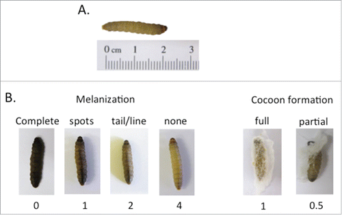
Table 2. The G. mellonella Health Index Scoring System.Citation49
Microbial virulence in the G. mellonella infection model is typically assessed within 5 d and the most commonly used end point is the survival rate at different time points. When larvae are inoculated with a variety of doses, a half-maximum lethal dose (LD50) can be calculated. Other end points include the expression of anti-microbial proteins in response to the infectionCitation47 and production of lactate dehydrogenase as a marker of cell damage.Citation48 More recently, a health index scoring system () was introduced which assesses the larvae health status by assigning scores according to 4 major observations: larvae mobility, cocoon formation, melanization and survival.Citation49 Melanization typically starts with distinctive black spots on the cream colored larvae. Complete melanization (black larvae) correlates with death of the larvae soon after ().
Microbial virulence can also be assessed by measuring the proliferation of the microorganism inside the larvae during infection. This is typically done by plating larval extracts on agar plates for enumerationCitation48,49 or, more recently, by using bioluminescent microorganisms to detect the pathogen load by biophotonic imaging.Citation50,51
G. mellonella larvae as a model to study virulence of gram-positive bacteria
The G. mellonella infection model has been used to study a variety of Gram-positive bacteria, including Streptococcus pyogenes (group A streptococcus),Citation49,52 Streptococcus pneumoniae (pneumococcus),Citation53 Enterococus faecalis,Citation50,51,54-62 Enterococcus faecium,Citation63-65 Staphylococcus aureusCitation46,66,67 and Listeria monocytogenesCitation68-72 (summarized in Table S1).
Infection of larvae with the S. pyogenes reference strain SF370 (a serotype M1 strain) killed the larvae in a dose-dependent manner with a LD50 of 6 × 10⁶ CFU.Citation49 Infection with different M-type strains (serotypes M1, M2, M3, M4, M6, M18, M28, M49) gave a wide range of responses in the G. mellonella model with serotype M18 being the most virulent and serotype M2 the least virulent strains.Citation49 There were also significant differences between a M3 carrier strain (MGAS12501) and a M3 invasive strain (MGAS315), which showed higher killing at doses of 106 CFU and a more rapidly progressing melanin accumulation and hemolymph coagulation. Approximately 45% of larvae survived after 24h and ˜25% after 96h when infected with the invasive strain, compared to survival rates of ˜75% after 24h and ˜55% after 96h for larvae infected with the carrier strain. Interestingly, survival in G. mellonella strongly correlated with survival in mice. Furthermore, a new GAS strain lineage (subclone 8 strains), which is associated with necrotizing fasciitis had significantly higher mortality compared with subclone 5 strains, which are epidemiologically associated with decreased necrotizing fasciitis cases in humans and have a significantly decreased capacity to cause necrotizing fasciitis in mice.Citation52 The G. mellonella/S. pyogenes model was further improved by the introduction of a health index that enabled measurement of more subtle differences, including cocoon formation and melanizationCitation49 (see also under Experimental aspects of G. mellonella infection models, and ).
Differences in virulence between S. pneumoniae serotypes (2, 4, 14, 19A, 19F) could also be observed in the G. mellonella model and correlated with the presence or absence of known virulence factors. LD50 values ranged from 1.56×10⁶ CFU (England14-9 strain) to 1.36×10⁵ CFU (strain Portugal19F-21), when determined 48h after inoculation with ˜106 CFU, and were generally higher than those that have been observed in murine models of infection.Citation53
The first article describing a G. mellonella infection model with E. faecalis was published in 2007. In this study, larvae were infected via injection of 5×10⁵ CFU E. faecalis into the hemocoel resulting in intense melanization within 5 min and death 30 min later. Extracellular gelatinase (GelE) was identified as a major virulence factor in G. mellonella infections. Purified GelE injected into the hemolymph degraded the inducible antimicrobial peptide Gm cecropin analogous to its ability to degrade human complement proteins.Citation61
Recently, the E. faecalis/G. mellonella infection model was improved by the introduction of a bioluminescent marker allowing for visualization of the infecting bacteria by biophotonic imaging. The luxABCDE operon was introduced into E. facalis by a plasmid that carries a toxin-antitoxin cassette providing segregational stability in the absence of antibiotic selection.Citation51 No difference in killing was detected between larvae infected with wildtype strain and the bioluminescent strain. A decrease of bioluminescence was observed in the first 2 hours, before the signal increased and reached a peak after 4 hours consistent with disease progression. The signal then remained stable until the death of the larvae after 24h. By expressing the lux-cassette under the control of either the cytolysin or the gelatinase promotor, this model was later used to demonstrate temporal regulation of gelatinase and cytolysin in response to the host environment.Citation50
In contrast to E. faecalis, Enterococcus faecium causes only weak lethality as demonstrated by monitoring the bacterial load after infection of G. mellonella. However, virulence was significantly increased when the antibiotic and stress response regulator (AsrR) was deleted.Citation65 The parenteral load markedly decreased from 1×10⁶ to 3×10⁴ CFU after 72h, whereas the asrR- mutant load only slightly decreases to 2.8×10⁵ CFU.
Desbois and Coote have shown that G. mellonella larvae infected with S. aureus were killed in a dose-dependent manner. Infection with 1×10⁷ CFU resulted in complete killing after 24h, whereas infection with 1×10⁵ CFU resulted in ∼20% killing after 120h. Virulence was temperature-dependent with more efficient killing at increasing temperatures when tested at 25°C, 30°C, and 37°C.Citation46 Over recent years, the G. mellonella model was mainly used for screening potential antistaphylococcal drugs (see section 6), but analysis of selected virulence factors has also been reported. Deletion of the Sec pathway (SecDF−) significantly reduced virulence in the G. mellonella model and this was consistent with reduced cytotoxicity and invasion of human umbilical vein endothelial cells.Citation67
Mukherjee et al. investigated the suitability of the G. mellonella model to study L. monocytogenes infections and found that at infection doses of 106 CFU, the model could distinguish between pathogenic L. monocytogenes and nonpathogenic Listeria species, such as L. innocua. The model also proved to be a valuable tool to study attenuated L. monocytogenes strains. Deletion mutants with deficiencies in phospholipase B (PLB), listeriolysin (LLO, Hly), metalloproteinase Mpl (an important spreading factor) and Act (a protein that directs polymerization of actin in the host cell), as well as isogenic mutants lacking the virulence gene cluster (vgc) and the virulence gene regulator PrfA all showed increased survival rates of G. mellonella larvae. Virulence of the mutant strains correlated well with previous results obtained from mouse models.Citation70 Infection of larvae with L. monocytogenes results in increased production of lysozyme, galiomycin, gallerimycin and insect metalloproteinase inhibitor (IMPI),Citation70 activation of the phenoloxidase system and hemocyte destruction.Citation68 Increased expression of cecropin D has also been reported and these AMPs had a strong inhibitory effect on L. monocytogenes.Citation69 L. monocytogenes also elicits a cellular immune response with formation of nodules (melanized cellular aggregates) that contained entrapped bacteria on the surface of the brains. These nodules are similar to structures on the brain of humans infected with L. monocytogenes.
G. mellonella larvae as a model to study virulence of gram-negative bacteria
Gram-negative bacteria investigated by using the G. mellonella infection model include Pseudomonas aeroginosa,Citation73-86 Escherichia coli,Citation87-91 Klebsiella pneumonia,Citation48,92-94 Legionella pneumophila,Citation95-97 Francisella tularensis,Citation98,99 Acinetobacter baumaniiCitation100,101 and various species of BurkholderiaCitation102-106 (summarized in Table S1).
One of the first studies of P. aeruginosa in a G. mellonella infection model was published in 1975 and demonstrated that rough mutants (mutants with deficiencies in LPS expression) of 2 P. aeruginosa strains were 8-62-fold less pathogenic than the smooth wt strains.Citation79 Since then, this model was used in several studies to analyze P. aeruginosa virulence and G. mellonella immune defense mechanisms. P. aeruginosa is highly virulent in G. mellonella and it was shown that injection of as little as 25 CFU of strain NCTC13437 resulted in 100% killing of the larvae after 24h.Citation77 Elastase B injected into the larvae at sublethal doses resulted in an increase in antibacterial activity and up-regulation of lysozyme and AMPs, in particular apoLp-III.Citation74 Similarly, apoLp-III levels are increased after infection of G. mellonella with P. aeruginosa followed by proteolytic degradation. In vitro and in-vivo studies suggest that both elastase B and serine protease IV play a role in apoLp-III degradation.Citation73,75 A more recent study using an entomopathogenic and 2 clinical strains found differences in the humoral immune responses, in particular the levels of lysozyme, phenoloxidase and AMPs. Notably, high levels of elastase A activity was detected in the entomopathogenic strain, but not in the 2 clinical strains.Citation76 Infection of G. mellonella with an entomopathogenic P. aeruginosa strain also showed significant changes in morphology and spreading ability and eventually apoptotic death of granylocytes and plasmatocytes.Citation82
The first study reporting the use of the G. mellonella infection model to study pathogenic E. coli was published in 2012.Citation89 Leuko and Raivio demonstrated that G. mellonella larvae could be killed by enteropathogenic E. coli (EPEC) in a dose-dependent manner with a LD50 value of 2.57×10³ CFU at 48h post-infection. The bacteria were injected into the hemocoel, but disappeared shortly thereafter and became localized to melanized capsules. Infections resulted in an increase in the AMPs gloverin and cecropin.
A recent study showed a remarkable correlation between virulence gene repertoire and virulence potential of extraintestinal pathogenic E. coli (ExPEC) in the G. mellonella model. ExPEC isolates with higher number of virulence genes resulted in significantly faster killing of the larvae.Citation91 In a similar study, 40 well-characterized ExPEC strains were analyzed in a G. mellonella infection model. The study which measured larvae survival, melanization, and cell damage found increased virulence in isolates from community-associated infections, complicated urinary tract infections (UTIs) and urinary-sourced bacteremia in particular in isolates belonging to the ST131 lineage.Citation88
The suitability of G. mellonella as an infection model for K. pneumoniae has only recently been demonstrated. Survival of infected larvae was dose and strain dependent. For example, infection with 106 CFU of the O1:K2 serotype strain 52145 caused 75% death after 24h and 100% death after 72h, whereas infection with the same dose of the O1:K2 serotype strain 43816 caused 95% death after 24h. Infections resulted in host responses similar to innate immune responses in mouse pneumonia models including cell death associated with bacterial replication and inhibition of phagocytosis and AMP production.Citation93 Similar results were reported from another study that compared a selection of 50 clinical isolates and reference strains at challenge doses of 105 CFU. Survival rates after 24h ranged from 0% (3 isolates) to almost 100%, and 68% of the strains caused greater than 50% mortality. In addition, lactate dehydrogenase as a marker of cell damage, melanisation, and bacterial proliferation was analyzed and broadly correlated with survival rates.Citation48
Comparison of 15 clinical K. pneumoniae producing carbapenemase (KPC(+) strains with 60 KPC(−) strains revealed decreased virulence of KPC(+) strains in the G. mellonella model, which was opposite from disease severity found in patients. This suggests some limitation of the G. mellonella model for K. pneumoniae infections.Citation94 However, a more recent study has shown a strong variability in virulence among carbapenem-resistant K. pneumoniae (CR-Kp). The differences were associated with the type of KPC gene and the capsular polysaccharide (CPS) type, and differences in serum resistance correlated with virulence in G. mellonella.Citation92
The G. mellonella infection model has only recently been used to study virulence and pathogenesis of L. pneumophila. Harding et al. have demonstrated that 3 commonly used serogroup 1 strains caused death of at least 70% of the larvae that was strain, infectious dose and growth phase dependent. After infection, L. pneumophila was found within hemocytes inside a vacuole that showed resemblance with the Legionella-containing vacuole (LCD) observed in macrophages. Severe organ damage accompanied by melanization, nodule formation and increased AMP production was also observed.Citation96
A G. mellonella infection model for F. tularensis was established by Aperis et al. using a live vaccine strain (LVS).Citation99 Infection of larvae with 3×10⁵ CFU resulted in 100% mortality after 3 days, whereas infection with 3×10⁴ CFU led to ˜70% mortality after 10 d. The authors also observed that >90% of hemocytes were associated with bacteria 48 hours post-infection, which correlates well with the infection of macrophages in mammals. Virulence was found to be higher when larvae were incubated at 37°C compared to 30°C. Until today, this model has mainly been used for screening antimicrobial agents effective against F. tularensis (see section 6).
The utility of the G. mellonella infection model to study A. baumanii virulence was investigated by Peleg et al. Using the reference strain ATCC 17978, the authors showed that 75% of larvae infected with 3.5×10⁵ CFU died within 48h.Citation101 A study comparing 5 different A. baumanii isolates found significant differences in virulence. Six days post-infection with 1×10⁵ CFU, larvae survival rates were between 16% (isolate AB5075) and 85% (AB5711). Notably, AB5075 was also the most virulent strain in a mouse pulmonary infection model.Citation100
B. mallei and B. pseudomallei were found to be highly virulent in the G. mellonella model. Only 10 CFU of B. mallei killed >90% of larvae after 4 days, whereas 10 CFU of B. pseudomallei killed >80% larvae after 2 d Both species caused severe paralysis starting 12 hours before death, similar to what can be observed in infected hamsters. At the time of death, ˜106 CFU/ml of hemolymph were recovered indicating a very fast growth rate of the bacteria. In contrast, infection with 105 CFU of a cystic fibrosis epidemic B. cenocepacia strain only resulted in 15% mortality after 6 days.Citation103 Strong virulence in the G. mellonella model was also found in other Burkholderia species. Two strains of B. cepacia showed LD50 values of 1 CFU and 30 CFU, respectively, 48h postinfection, whereas 2 B. cenocepacia strains had LD50 values of 900 CFU and 4000 CFU, respectively.Citation104 Varying virulence within the same species was also reported for B. pseudomallei and B. thailandensis. Two B. pseudomallei strains (576 and K96243) with low median dose values in mice had 100% mortality rates 24h after the challenge of G. mellonella larvae with 100 CFU, whereas a third strain (708a), which is attenuated in mice, was avirulent (100% survival). Similar, 2 B. thailandensis strains (CDC272 and CDC301) were highly virulent (100% mortality), whereas strains Phuket and E264 were significantly less virulent (80% and 50% mortality, respectively) when infected with 100 CFU and the differences reflect the observed virulence in mouse infection models.Citation106 B. thailandensis is rapidly phagocytosed by hemocytes; bacteria were shown to be associated with hemocytes at 0.5 hours post-infection, and intracellular bacteria could be seen 1 hour after infection.Citation105
Use of G. mellonella to evaluate efficacy of antimicrobial agents
The rate of antibiotic resistance among important human pathogens, such as Pseudomonas, Klebsiella and Acinetobacter species, has accelerated dramatically in recent years. The discovery and development of novel antimicrobial agents is therefore of the utmost importance. Novel compounds are generally screened in vitro first to assess their effectiveness and potential toxicity. Eventually, successful candidates will have to be evaluated in an animal model, usually in murine and other rodent models, before their potential application in humans. In vivo tests are important to identify potential loss of activity due to host factors, e.g. degradation by host enzymes and the effect of physiological conditions such as pH.Citation107 However, experiments using mammalian hosts are time-consuming, expensive and often regarded as ethically objectionable. In contrast, the G. mellonella model is a simple and inexpensive alternative for the rapid evaluation of antimicrobial drug effectiveness in vivo and reduces the likelihood of an antimicrobial agent that performed well in in vitro studies from progressing to an unsuccessful performance in a mammalian model. The G. mellonella model can therefore serve as an additional pre-screening experiment to lower the number of antimicrobial drugs proceeding to tests in mammalian models (summarized in Table S2).
G. mellonella larvae can be accurately injected with defined doses of bacteria resulting in consistent survival/mortality rates. It is therefore easy to determine a dose that will not kill the larvae immediately, but leads to increased mortality over an appropriate time course, e.g., one to 3 d. Antimicrobial agents can be administered in different treatment regimes, including the total dose given, the number of doses and the treatment schedule. Most studies have used single treatment doses, usually given between 30min and 2 hours after infection of the larvae with the test pathogen. In some cases, the agent was given immediately after infectionCitation108,109 or even before the infection.Citation105 The antimicrobial agent can be delivered systemically by injecting it directly into the hemocoel, which closely mimics the conventional administration route used in mammalian models.
Several studies have used combinations of antibiotics and shown synergism when administered to infected G. mellonella larvae. For example, gentamycin and daptomycin injected 1h after infection with vancomycin-sensitive E. faecalis or vancomycin-resistant E. faecium were significantly more effective than either antibiotic given alone at the same doses.Citation110 Krezdorn et al. have tested a variety of single, dual and triple antibiotic combinations against the multidrug-resistant P. aeruginosa strain NCTC13437 and showed synergistic effects for several combinations, like cefotaxime and piperacillin, amikacin and meropenem, or the triple combination of piperacillin, amikacin and meropenem, which was particularly effective.Citation111 Interestingly, there was little correlation between antibiotic combinations that showed synergy in in-vitro screens and those that showed enhanced effects in the G. mellonella model. This further emphasizes the role of the G. mellonella model as a useful tool for large-scale screen of antibiotic efficacy in an in-vivo model. The authors have also shown that antibiotic efficacy can be increased with administration of multiple doses. For example, larvae infected with 2.5×10³ CFU and treated with a single dose of amikacin plus meropenem 2 hours post-infection showed increased survival after 24h (100%) and 48h (˜20%) compared to no survival after 24h in untreated larvae. However, treatment of larvae infected with 2.5×10⁶ CFU and treated with a triple dose of amikacin plus meropenem 2 hours, 5 hours and 8 hours post-infection had survival rates of >50 % after 72h.Citation111 The importance of the correct timing of drug administration was shown for G. mellonella larvae infected with F. tularensis. Although, azithromycin, ciprofloxacin, levofloxacin and streptomycin all increased survival of larvae when injected as a single dose 2 hours after infection, only ciprofloxacin and streptomycin were effective when administered as 2 doses 24h and 48h after infection.Citation99
Some more unconventional compounds tested in the G. mellonella model include the antibiofilm compound hamamelitannin, the estrogen receptor antagonist tamoxifen, the antihistamine terfenadine, the antimicrobial peptide LL-37 (and the enantiomer D-LL-37), the quorum sensing inhibitor and antibiofilm compound baicalin hydrate, cinnamaldehyde (isolated from cinnamon oil), a mutant form of P. aeruginosa acyl-homoserine lactone acylase PvdQ, a carbene silver(I) acetate derivative (SBC3), and metal ions (silver and zinc). See Table S2.
PvdQ is an effective quorum-quenching enzyme from P. aeruginosa and it has recently been shown that a variant with 2 point mutations, PvdQ (Lα146W, Fβ24Y), showed strong activity toward C8-HSL, the major communication molecule expressed by Burkholderia species. Injection of PvdQ (Lα146W, Fβ24Y) into G. mellonella larvae 1h before infection with B. cenocepacia significantly increased the survival of the larvae compared to untreated control animals.Citation102
Of particular interest are the results obtained with hamamelitannin and baicalin hydrate. These are drugs that reduce the activity of bacterial virulence factors, in this case the ability to form biofilms, without possessing direct antimicrobial activity. The survival rates of G. mellonella larvae were further increased when these antivirulence agents were combined with antibiotics (hamamelitannin with vancomycin in larvae infected with S. aureus and baicalin hydrate with tobramycin in larvae infected with B. cenocepacia).Citation112 In many cases the expression of virulence factors by the pathogens is controlled by temperature.Citation3,4 G. mellonella is therefore perfectly suited for the screening of antivirulence drug, as the larvae can be incubated at a variety of temperatures that are necessary for expression of virulence factors.
The G. mellonella model has also been used to evaluate the combination of antibiotic therapy and antimicrobial photodynamic therapy (aPDT). PDT is based on photoactive dye molecules (photosensitizers) that produce reactive oxygen species when irradiated with visible light. Injection of methylene blue into larvae infected with E. faecium followed by whole body illumination increased the survival rates of the larvae compared to untreated controls. In addition, treatment of larvae infected with vancomycin-resistant E. faecium with aPDT followed by administration of vancomycin significantly reduced mortality rates when compared to aPDT or antibiotic treatment alone.Citation63 The photosensitizer can be injected into the haemocoel of the larvae and their relatively translucent body facilitates light delivery activating the photosensitizer, which makes G. mellonella an excellent model to evaluate these forms of therapy.
Use of G. mellonella to evaluate experimental phage therapy
Phage therapy describes the therapeutic application of bacteriophages to treat pathogenic bacterial infections. Multiple studies have demonstrated the effectiveness of phage therapy in animal models for the treatment of various bacterial pathogens. However, apart from Russia and Georgia, this treatment has not been approved to treat infections in humans. The G. mellonella model was used to assess the efficacy of phage therapy in Burkholderia cenocepacia and in Pseudomonas aeruginosa. Injection of phages KS4-M and KS12 immediately after infection with a lethal dose of B. cenocepacia strain K56-2 resulted in significantly improved survival of the larvae. A similar result was obtained with phage KS14 after infection with strain C6433.Citation113 More recently, it was observed that the efficacy of experimental phage therapy was improved in the presence of sublethal concentrations of certain antibiotics and this effect has been termed ‘phage-antibiotic synergy' (PAS).Citation114 Supporting evidence was provided by Kamal & Dennis, who demonstrated that the use of low-dose meropenem increased the survival rates of G. mellonella larvae infected with B. cenocepacia and treated with phage K12 over controls treated with antibiotic or phage alone.Citation115 Phage therapy was also analyzed with 29 phages and 121 P. aeruginosa isolates from cystic fibrosis patients and the protective efficacy of 2 selected phages (KT28 and PA5oct) against P. aeruginosa was confirmed using the G. mellonella model.Citation116
Limitations in the use of G. mellonella as an infectious disease model
As discussed in the previous sections, G. mellonella is an excellent model for assessing the virulence for a range of microrganisms. Although, it will not replace mammalian models, G. mellonella provides a rapid and cost-effective alternative to collect initial data. However, one has to consider that the G. mellonella infection model is still in its infancy and not as well established as some other invertebrate models, such as the nematode (Caenorhabditis elegans) or the fruit fly (Drosophila melanogaster).Citation117 The G. mellonella genome has not been fully sequenced and there is no established method for generating mutant strains and no access to microarrays or RNA interference libraries. Most importantly, there are no stock centers for G. mellonella, like the Drosophila stock centers where researchers can purchase specific genotypes that were raised under standard conditions. G. mellonella larvae are usually purchased from a wide range of independent breeders who sell the larvae as pet food. Differences in genotypes, breeding conditions or maintenance of the animals might well influence their susceptibility to infections. Even after the larvae are purchased, treatment conditions might vary between research labs, e.g. housing temperature, light sources and diet. It was shown that pre-exposure of larvae to heat induces their immune response,Citation118 whereas starvation results in reduction of immune responses and increased susceptibility to infection.Citation119 Furthermore, the size of the inoculum was shown to have an effect on cellular and humoral immune responses.Citation120
Variations in supplier, breeding conditions, maintenance and handling of G. mellonella larvae might easily result in differences in mortality rates after infection with pathogens. This might explain conflicting results with some reference strains that induced variable mortality in larvae in different research labs. For example, in a study conducted in the US larvae infected with 106 CFU of the S. pyogenes serotype M3 strain MGAS315 resulted in ∼45% survival after 24h and 25% survival after 96h.Citation52 In contrast, we have used the same strain and observed lower virulence (˜90% survival after 24h and 70% survival after 96h) when infected with a higher dose of 8×10⁶ CFU.Citation49 The larvae for these studies were purchased from different suppliers (Best Bet Inc., Blackduck, MN, USA and Biosuppliers, Auckland, New Zealand) and there were also differences in maintenance conditions. The larvae from Best Bet Inc. were stored at 10-12°C without food for up to 10 d and, after infection, were incubated in the presence of 0.5% CO2. The larvae from Biosuppliers were stored at room temperature with food and infected larvae were incubated under normal atmospheric conditions. These problems might be solved with the establishment of stock centers that supply reference populations of well-defined G. mellonella genotypes.
Conclusion
Over recent years, G. mellonella has become increasingly popular as a surrogate host to study infectious diseases, as well as a screening platform for antibiotics. However, this model is still in its infancy. Major hurdles are the lack of stock centers that supply reference strains raised under standard conditions to enable comparable experiments carried out by different research groups and the limited availability of genomic information on G. mellonella. Furthermore, experimental conditions often differ between individual research labs and need to be standardized to minimize ambiguity. It is probably only a question of time until these issues are addressed, which will help to advance G. mellonella to a powerful and reliable infection model.
Disclosure of potential conflicts of interest
No potential conflicts of interest were disclosed.
KVIR_A_Supp_1135289.docx
Download MS Word (127 KB)Funding
TP and JMSL are supported by the Health Research Council New Zealand. CT is a recipient of a Dean's International PhD student scholarship from the Faculty of Medical and Health Sciences, The University of Auckland.
References
- Scoble M. Classification of the Lepidoptera. Oxford University Press, 1995
- Ramarao N, Nielsen-Leroux C, Lereclus D. The insect Galleria mellonella as a powerful infection model to investigate bacterial pathogenesis. J Vis Exp 2012 Dec 11;(70):e4392; PMID:23271509; http://dx.doi.org/10.3791/4392.
- Konkel ME, Tilly K. Temperature-regulated expression of bacterial virulence genes. Microbes Infect 2000; 2:157-66; PMID:10742688; http://dx.doi.org/10.1016/S1286-4579(00)00272-0
- Smoot LM, Smoot JC, Graham MR, Somerville GA, Sturdevant DE, Migliaccio CA, Sylva GL, Musser JM. Global differential gene expression in response to growth temperature alteration in group A Streptococcus. Proc Natl Acad Sci U S A 2001; 98:10416-21; PMID:11517341; http://dx.doi.org/10.1073/pnas.191267598
- Browne N, Heelan M, Kavanagh K. An analysis of the structural and functional similarities of insect hemocytes and mammalian phagocytes. Virulence 2013; 4:597-603; PMID:23921374; http://dx.doi.org/10.4161/viru.25906
- Boman HG, Hultmark D. Cell-free immunity in insects. Annu Rev Microbiol 1987; 41:103-26; PMID:3318666; http://dx.doi.org/10.1146/annurev.mi.41.100187.000535
- Tojo S, Naganuma F, Arakawa K, Yokoo S. Involvement of both granular cells and plasmatocytes in phagocytic reactions in the greater wax moth, Galleria mellonella. J Insect Physiol 2000; 46:1129-35; PMID:10817839; http://dx.doi.org/10.1016/S0022-1910(99)00223-1
- Pech LL, Strand MR. Granular cells are required for encapsulation of foreign targets by insect haemocytes. J Cell Sci 1996; 109 ( Pt 8):2053-60; PMID:8856501
- Schmit AR, Ratcliffe NA. The encapsulation of foreign tissue implants in Galleria mellonella larvae. J Insect Physiol 1977; 23:175-84; PMID:323370; http://dx.doi.org/10.1016/0022-1910(77)90027-0
- Choi JY, Whitten MM, Cho MY, Lee KY, Kim MS, Ratcliffe NA, Lee BL. Calreticulin enriched as an early-stage encapsulation protein in wax moth Galleria mellonella larvae. Dev Comp Immunol 2002; 26:335-43; PMID:11888648; http://dx.doi.org/10.1016/S0145-305X(01)00081-7
- Bergin D, Reeves EP, Renwick J, Wientjes FB, Kavanagh K. Superoxide production in Galleria mellonella hemocytes: identification of proteins homologous to the NADPH oxidase complex of human neutrophils. Infect Immun 2005; 73:4161-70; PMID:15972506; http://dx.doi.org/10.1128/IAI.73.7.4161-4170.2005
- Renwick J, Reeves EP, Wientjes FB, Kavanagh K. Translocation of proteins homologous to human neutrophil p47phox and p67phox to the cell membrane in activated hemocytes of Galleria mellonella. Dev Comp Immunol 2007; 31:347-59; PMID:16920193; http://dx.doi.org/10.1016/j.dci.2006.06.007
- Halwani AE, Niven DF, Dunphy GB. Apolipophorin-III and the interactions of lipoteichoic acids with the immediate immune responses of Galleria mellonella. J Invertebr Pathol 2000; 76:233-41; PMID:11112367; http://dx.doi.org/10.1006/jipa.2000.4978
- Pratt CC, Weers PM. Lipopolysaccharide binding of an exchangeable apolipoprotein, apolipophorin III, from Galleria mellonella. Biol Chem 2004; 385:1113-9; PMID:15576334; http://dx.doi.org/10.1515/BC.2004.145
- Whitten MM, Tew IF, Lee BL, Ratcliffe NA. A novel role for an insect apolipoprotein (apolipophorin III) in β-1,3-glucan pattern recognition and cellular encapsulation reactions. J Immunol 2004; 172:2177-85; PMID:14764684; http://dx.doi.org/10.4049/jimmunol.172.4.2177
- Carvalho MD, Tobias VE, Vendrame CM, Shimabukuro AF, Gidlund M, Quintao EC. Lipoproteins modify the macrophage uptake of triacylglycerol emulsion and of zymosan particles by similar mechanisms. Lipids 2000; 35:55-9; PMID:10695924; http://dx.doi.org/10.1007/s11745-000-0494-1
- Riddell DR, Graham A, Owen JS. Apolipoprotein E inhibits platelet aggregation through the L-arginine:nitric oxide pathway. Implications for vascular disease. J Biol Chem 1997; 272:89-95; PMID:8995232; http://dx.doi.org/10.1074/jbc.272.1.89
- Niere M, Meisslitzer C, Dettloff M, Weise C, Ziegler M, Wiesner A. Insect immune activation by recombinant Galleria mellonella apolipophorin III(1). Biochim Biophys Acta 1999; 1433:16-26; PMID:10446356; http://dx.doi.org/10.1016/S0167-4838(99)00148-X
- Park SY, Kim CH, Jeong WH, Lee JH, Seo SJ, Han YS, Lee IH. Effects of two hemolymph proteins on humoral defense reactions in the wax moth, Galleria mellonella. Dev Comp Immunol 2005; 29:43-51; PMID:15325522; http://dx.doi.org/10.1016/j.dci.2004.06.001
- Zdybicka-Barabas A, Staczek S, Mak P, Skrzypiec K, Mendyk E, Cytrynska M. Synergistic action of Galleria mellonella apolipophorin III and lysozyme against Gram-negative bacteria. Biochim Biophys Acta 2013; 1828:1449-56; PMID:23419829; http://dx.doi.org/10.1016/j.bbamem.2013.02.004
- Dziarski R, Gupta D. The peptidoglycan recognition proteins (PGRPs). Genome Biol 2006; 7:232; PMID:16930467; http://dx.doi.org/10.1186/gb-2006-7-8-232
- Seitz V, Clermont A, Wedde M, Hummel M, Vilcinskas A, Schlatterer K, Podsiadlowski L. Identification of immunorelevant genes from greater wax moth (Galleria mellonella) by a subtractive hybridization approach. Dev Comp Immunol 2003; 27:207-15; PMID:12590972; http://dx.doi.org/10.1016/S0145-305X(02)00097-6
- Kim CH, Shin YP, Noh MY, Jo YH, Han YS, Seong YS, Lee IH. An insect multiligand recognition protein functions as an opsonin for the phagocytosis of microorganisms. J Biol Chem 2010; 285:25243-50; PMID:20519517; http://dx.doi.org/10.1074/jbc.M110.134940
- Yu XQ, Kanost MR. Binding of hemolin to bacterial lipopolysaccharide and lipoteichoic acid. An immunoglobulin superfamily member from insects as a pattern-recognition receptor. Eur J Biochem 2002; 269:1827-34; PMID:11952784; http://dx.doi.org/10.1046/j.1432-1033.2002.02830.x
- Shaik HA, Sehnal F. Hemolin expression in the silk glands of Galleria mellonella in response to bacterial challenge and prior to cell disintegration. J Insect Physiol 2009; 55:781-7; PMID:19414015; http://dx.doi.org/10.1016/j.jinsphys.2009.04.010
- Mowlds P, Coates C, Renwick J, Kavanagh K. Dose-dependent cellular and humoral responses in Galleria mellonella larvae following β-glucan inoculation. Microbes Infect 2010; 12:146-53; PMID:19925881; http://dx.doi.org/10.1016/j.micinf.2009.11.004
- Brown SE, Howard A, Kasprzak AB, Gordon KH, East PD. A peptidomics study reveals the impressive antimicrobial peptide arsenal of the wax moth Galleria mellonella. Insect Biochem Mol Biol 2009; 39:792-800; PMID:19786100; http://dx.doi.org/10.1016/j.ibmb.2009.09.004
- Lee YS, Yun EK, Jang WS, Kim I, Lee JH, Park SY, Ryu KS, Seo SJ, Kim CH, Lee IH. Purification, cDNA cloning and expression of an insect defensin from the great wax moth, Galleria mellonella. Insect Mol Biol 2004; 13:65-72; PMID:14728668; http://dx.doi.org/10.1111/j.1365-2583.2004.00462.x
- Sowa-Jasilek A, Zdybicka-Barabas A, Staczek S, Wydrych J, Mak P, Jakubowicz T, Cytryńska M. Studies on the role of insect hemolymph polypeptides: Galleria mellonella anionic peptide 2 and lysozyme. Peptides 2014; 53:194-201; PMID:24472857; http://dx.doi.org/10.1016/j.peptides.2014.01.012
- Kim CH, Lee JH, Kim I, Seo SJ, Son SM, Lee KY, Lee IH. Purification and cDNA cloning of a cecropin-like peptide from the great wax moth, Galleria mellonella. Mol Cells 2004; 17:262-6; PMID:15179040
- Brown SE, Howard A, Kasprzak AB, Gordon KH, East PD. The discovery and analysis of a diverged family of novel antifungal moricin-like peptides in the wax moth Galleria mellonella. Insect Biochem Mol Biol 2008; 38:201-12; PMID:18207081; http://dx.doi.org/10.1016/j.ibmb.2007.10.009
- Hoffmann JA, Reichhart JM, Hetru C. Innate immunity in higher insects. Curr Opin Immunol 1996; 8:8-13; PMID:8729440; http://dx.doi.org/10.1016/S0952-7915(96)80098-7
- Cytrynska M, Mak P, Zdybicka-Barabas A, Suder P, Jakubowicz T. Purification and characterization of eight peptides from Galleria mellonella immune hemolymph. Peptides 2007; 28:533-46; PMID:17194500; http://dx.doi.org/10.1016/j.peptides.2006.11.010
- Kawaoka S, Katsuma S, Daimon T, Isono R, Omuro N, Mita K, Shimada T. Functional analysis of four Gloverin-like genes in the silkworm, Bombyx mori. Arch Insect Biochem Physiol 2008; 67:87-96; PMID:18076111; http://dx.doi.org/10.1002/arch.20223
- Langen G, Imani J, Altincicek B, Kieseritzky G, Kogel KH, Vilcinskas A. Transgenic expression of gallerimycin, a novel antifungal insect defensin from the greater wax moth Galleria mellonella, confers resistance to pathogenic fungi in tobacco. Biol Chem 2006; 387:549-57; PMID:16740126; http://dx.doi.org/10.1515/BC.2006.071
- Girard PA, Boublik Y, Wheat CW, Volkoff AN, Cousserans F, Brehelin M, Escoubas JM. X-tox: an atypical defensin derived family of immune-related proteins specific to Lepidoptera. Dev Comp Immunol 2008; 32:575-84; PMID:17988734; http://dx.doi.org/10.1016/j.dci.2007.09.004
- Tang H. Regulation and function of the melanization reaction in Drosophila. Fly (Austin) 2009; 3:105-11; PMID:19164947; http://dx.doi.org/10.4161/fly.3.1.7747
- Soderhall K, Cerenius L. Role of the prophenoloxidase-activating system in invertebrate immunity. Curr Opin Immunol 1998; 10:23-8; PMID:9523106; http://dx.doi.org/10.1016/S0952-7915(98)80026-5
- Lu A, Zhang Q, Zhang J, Yang B, Wu K, Xie W, Luan YX, Ling E. Insect prophenoloxidase: the view beyond immunity. Front Physiol 2014; 5:252; PMID:25071597
- Altincicek B, Linder M, Linder D, Preissner KT, Vilcinskas A. Microbial metalloproteinases mediate sensing of invading pathogens and activate innate immune responses in the lepidopteran model host Galleria mellonella. Infect Immun 2007; 75:175-83; PMID:17074843; http://dx.doi.org/10.1128/IAI.01385-06
- Zdybicka-Barabas A, Mak P, Jakubowicz T, Cytrynska M. Lysozyme and defense peptides as suppressors of phenoloxidase activity in Galleria mellonella. Arch Insect Biochem Physiol 2014; 87:1-12; PMID:25044335; http://dx.doi.org/10.1002/arch.21175
- Brinkmann V, Reichard U, Goosmann C, Fauler B, Uhlemann Y, Weiss DS, Weinrauch Y, Zychlinsky A. Neutrophil extracellular traps kill bacteria. Science 2004; 303:1532-5; PMID:15001782; http://dx.doi.org/10.1126/science.1092385
- Altincicek B, Stotzel S, Wygrecka M, Preissner KT, Vilcinskas A. Host-derived extracellular nucleic acids enhance innate immune responses, induce coagulation, and prolong survival upon infection in insects. J Immunol 2008; 181:2705-12; PMID:18684961; http://dx.doi.org/10.4049/jimmunol.181.4.2705
- Cotter G, Doyle S, Kavanagh K. Development of an insect model for the in vivo pathogenicity testing of yeasts. FEMS Immunol Med Microbiol 2000; 27:163-9; PMID:10640612; http://dx.doi.org/10.1111/j.1574-695X.2000.tb01427.x
- Fedhila S, Buisson C, Dussurget O, Serror P, Glomski IJ, Liehl P, Lereclus D, Nielsen-LeRoux C. Comparative analysis of the virulence of invertebrate and mammalian pathogenic bacteria in the oral insect infection model Galleria mellonella. J Invertebr Pathol 2010; 103:24-9; PMID:19800349; http://dx.doi.org/10.1016/j.jip.2009.09.005
- Desbois AP, Coote PJ. Wax moth larva (Galleria mellonella): an in vivo model for assessing the efficacy of antistaphylococcal agents. J Antimicrob Chemother 2011; 66:1785-90; PMID:21622972; http://dx.doi.org/10.1093/jac/dkr198
- Vilmos P, Kurucz E. Insect immunity: evolutionary roots of the mammalian innate immune system. Immunol Lett 1998; 62:59-66; PMID:9698099; http://dx.doi.org/10.1016/S0165-2478(98)00023-6
- Wand ME, McCowen JW, Nugent PG, Sutton JM. Complex interactions of Klebsiella pneumoniae with the host immune system in a Galleria mellonella infection model. J Med Microbiol 2013; 62:1790-8; PMID:24000226; http://dx.doi.org/10.1099/jmm.0.063032-0
- Loh JM, Adenwalla N, Wiles S, Proft T. Galleria mellonella larvae as an infection model for group A streptococcus. Virulence 2013; 4:419-28; PMID:23652836; http://dx.doi.org/10.4161/viru.24930
- La Rosa SL, Casey PG, Hill C, Diep DB, Nes IF, Brede DA. In vivo assessment of growth and virulence gene expression during commensal and pathogenic lifestyles of luxABCDE-tagged Enterococcus faecalis strains in murine gastrointestinal and intravenous infection models. Appl Environ Microbiol 2013; 79:3986-97; PMID:23603680; http://dx.doi.org/10.1128/AEM.00831-13
- La Rosa SL, Diep DB, Nes IF, Brede DA. Construction and application of a luxABCDE reporter system for real-time monitoring of Enterococcus faecalis gene expression and growth. Appl Environ Microbiol 2012; 78:7003-11; PMID:22843522; http://dx.doi.org/10.1128/AEM.02018-12
- Olsen RJ, Watkins ME, Cantu CC, Beres SB, Musser JM. Virulence of serotype M3 Group A Streptococcus strains in wax worms (Galleria mellonella larvae). Virulence 2011; 2:111-9; PMID:21258213; http://dx.doi.org/10.4161/viru.2.2.14338
- Evans BA, Rozen DE. A Streptococcus pneumoniae infection model in larvae of the wax moth Galleria mellonella. Eur J Clin Microbiol Infect Dis 2012; 31:2653-60; PMID:22466968; http://dx.doi.org/10.1007/s10096-012-1609-7
- Benachour A, Ladjouzi R, Le Jeune A, Hebert L, Thorpe S, Courtin P, Chapot-Chartier MP, Prajsnar TK, Foster SJ, Mesnage S. The lysozyme-induced peptidoglycan N-acetylglucosamine deacetylase PgdA (EF1843) is required for Enterococcus faecalis virulence. J Bacteriol 2012; 194:6066-73; PMID:22961856; http://dx.doi.org/10.1128/JB.00981-12
- Gaca AO, Abranches J, Kajfasz JK, Lemos JA. Global transcriptional analysis of the stringent response in Enterococcus faecalis. Microbiology 2012; 158:1994-2004; PMID:22653948; http://dx.doi.org/10.1099/mic.0.060236-0
- Gaspar F, Teixeira N, Rigottier-Gois L, Marujo P, Nielsen-LeRoux C, Crespo MT, Lopes Mde F, Serror P. Virulence of Enterococcus faecalis dairy strains in an insect model: the role of fsrB and gelE. Microbiology 2009; 155:3564-71; PMID:19696101; http://dx.doi.org/10.1099/mic.0.030775-0
- Hanin A, Sava I, Bao Y, Huebner J, Hartke A, Auffray Y, Sauvageot N. Screening of in vivo activated genes in Enterococcus faecalis during insect and mouse infections and growth in urine. PLoS One 2010; 5:e11879; PMID:20686694; http://dx.doi.org/10.1371/journal.pone.0011879
- Lebreton F, Riboulet-Bisson E, Serror P, Sanguinetti M, Posteraro B, Torelli R, Hartke A, Auffray Y, Giard JC. ace, Which encodes an adhesin in Enterococcus faecalis, is regulated by Ers and is involved in virulence. Infect Immun 2009; 77:2832-9; PMID:19433548; http://dx.doi.org/10.1128/IAI.01218-08
- Martini C, Michaux C, Bugli F, Arcovito A, Iavarone F, Cacaci M, Paroni Sterbini F, Hartke A, Sauvageot N, Sanguinetti M, et al. The polyamine N-acetyltransferase-like enzyme PmvE plays a role in the virulence of Enterococcus faecalis. Infect Immun 2015; 83:364-71; PMID:25385793; http://dx.doi.org/10.1128/IAI.02585-14
- Michaux C, Sanguinetti M, Reffuveille F, Auffray Y, Posteraro B, Gilmore MS, Hartke A, Giard JC. SlyA is a transcriptional regulator involved in the virulence of Enterococcus faecalis. Infect Immun 2011; 79:2638-45; PMID:21536798; http://dx.doi.org/10.1128/IAI.01132-10
- Park SY, Kim KM, Lee JH, Seo SJ, Lee IH. Extracellular gelatinase of Enterococcus faecalis destroys a defense system in insect hemolymph and human serum. Infect Immun 2007; 75:1861-9; PMID:17261598; http://dx.doi.org/10.1128/IAI.01473-06
- Zhao C, Hartke A, La Sorda M, Posteraro B, Laplace JM, Auffray Y, Sanguinetti M. Role of methionine sulfoxide reductases A and B of Enterococcus faecalis in oxidative stress and virulence. Infect Immun 2010; 78:3889-97; PMID:20566694; http://dx.doi.org/10.1128/IAI.00165-10
- Chibebe Junior J, Fuchs BB, Sabino CP, Junqueira JC, Jorge AO, Ribeiro MS, Gilmore MS, Rice LB, Tegos GP, Hamblin MR, et al. Photodynamic and antibiotic therapy impair the pathogenesis of Enterococcus faecium in a whole animal insect model. PLoS One 2013; 8:e55926; PMID:23457486; http://dx.doi.org/10.1371/journal.pone.0055926
- Lebreton F, Le Bras F, Reffuveille F, Ladjouzi R, Giard JC, Leclercq R, Cattoir V. Galleria mellonella as a model for studying Enterococcus faecium host persistence. J Mol Microbiol Biotechnol 2011; 21:191-6; PMID:22286046; http://dx.doi.org/10.1159/000332737
- Lebreton F, van Schaik W, Sanguinetti M, Posteraro B, Torelli R, Le Bras F, Verneuil N, Zhang X, Giard JC, Dhalluin A, et al. AsrR is an oxidative stress sensing regulator modulating Enterococcus faecium opportunistic traits, antimicrobial resistance, and pathogenicity. PLoS Pathog 2012; 8:e1002834; PMID:22876178; http://dx.doi.org/10.1371/journal.ppat.1002834
- Peleg AY, Monga D, Pillai S, Mylonakis E, Moellering RC, Jr., Eliopoulos GM. Reduced susceptibility to vancomycin influences pathogenicity in Staphylococcus aureus infection. J Infect Dis 2009; 199:532-6; PMID:19125671; http://dx.doi.org/10.1086/596511
- Quiblier C, Seidl K, Roschitzki B, Zinkernagel AS, Berger-Bachi B, Senn MM. Secretome analysis defines the major role of SecDF in Staphylococcus aureus virulence. PLoS One 2013; 8:e63513; PMID:23658837; http://dx.doi.org/10.1371/journal.pone.0063513
- Joyce SA, Gahan CG. Molecular pathogenesis of Listeria monocytogenes in the alternative model host Galleria mellonella. Microbiology 2010; 156:3456-68; PMID:20688820; http://dx.doi.org/10.1099/mic.0.040782-0
- Mukherjee K, Abu Mraheil M, Silva S, Muller D, Cemic F, Hemberger J, Hain T, Vilcinskas A, Chakraborty T. Anti-Listeria activities of Galleria mellonella hemolymph proteins. Appl Environ Microbiol 2011; 77:4237-40; PMID:21531838; http://dx.doi.org/10.1128/AEM.02435-10
- Mukherjee K, Altincicek B, Hain T, Domann E, Vilcinskas A, Chakraborty T. Galleria mellonella as a model system for studying Listeria pathogenesis. Appl Environ Microbiol 2010; 76:310-7; PMID:19897755; http://dx.doi.org/10.1128/AEM.01301-09
- Mukherjee K, Raju R, Fischer R, Vilcinskas A. Galleria mellonella as a model host to study gut microbe homeostasis and brain infection by the human pathogen listeria monocytogenes. Adv Biochem Eng Biotechnol 2013; 135:27-39; PMID:23708825
- Seifart Gomes C, Izar B, Pazan F, Mohamed W, Mraheil MA, Mukherjee K, Billion A, Aharonowitz Y, Chakraborty T, Hain T. Universal stress proteins are important for oxidative and acid stress resistance and growth of Listeria monocytogenes EGD-e in vitro and in vivo. PLoS One 2011; 6:e24965; PMID:21980369; http://dx.doi.org/10.1371/journal.pone.0024965
- Andrejko M, Cytrynska M, Jakubowicz T. Apolipophorin III is a substrate for protease IV from Pseudomonas aeruginosa. FEMS Microbiol Lett 2005; 243:331-7; PMID:15686832; http://dx.doi.org/10.1016/j.femsle.2004.12.024
- Andrejko M, Mizerska-Dudka M. Elastase B of Pseudomonas aeruginosa stimulates the humoral immune response in the greater wax moth, Galleria mellonella. J Invertebr Pathol 2011; 107:16-26; PMID:21236262; http://dx.doi.org/10.1016/j.jip.2010.12.015
- Andrejko M, Mizerska-Dudka M. Effect of Pseudomonas aeruginosa elastase B on level and activity of immune proteins/peptides of Galleria mellonella hemolymph. J Insect Sci 2012; 12:88; PMID:23421724; http://dx.doi.org/10.1673/031.012.8801
- Andrejko M, Zdybicka-Barabas A, Cytrynska M. Diverse effects of Galleria mellonella infection with entomopathogenic and clinical strains of Pseudomonas aeruginosa. J Invertebr Pathol 2014; 115:14-25; PMID:24513029; http://dx.doi.org/10.1016/j.jip.2013.10.006
- Hill L, Veli N, Coote PJ. Evaluation of Galleria mellonella larvae for measuring the efficacy and pharmacokinetics of antibiotic therapies against Pseudomonas aeruginosa infection. Int J Antimicrob Agents 2014; 43:254-61; PMID:24361354; http://dx.doi.org/10.1016/j.ijantimicag.2013.11.001
- Jander G, Rahme LG, Ausubel FM. Positive correlation between virulence of Pseudomonas aeruginosa mutants in mice and insects. J Bacteriol 2000; 182:3843-5; PMID:10851003; http://dx.doi.org/10.1128/JB.182.13.3843-3845.2000
- Kropinski AM, Chadwick JS. The pathogenicity of rough strains of Pseudomonas aeruginosa for Galleria mellonella. Can J Microbiol 1975; 21:2084-8; PMID:814978; http://dx.doi.org/10.1139/m75-297
- McLaughlin HP, Caly DL, McCarthy Y, Ryan RP, Dow JM. An orphan chemotaxis sensor regulates virulence and antibiotic tolerance in the human pathogen Pseudomonas aeruginosa. PLoS One 2012; 7:e42205; PMID:22870303; http://dx.doi.org/10.1371/journal.pone.0042205
- Miyata S, Casey M, Frank DW, Ausubel FM, Drenkard E. Use of the Galleria mellonella caterpillar as a model host to study the role of the type III secretion system in Pseudomonas aeruginosa pathogenesis. Infect Immun 2003; 71:2404-13; PMID:12704110; http://dx.doi.org/10.1128/IAI.71.5.2404-2413.2003
- Mizerska-Dudka M, Andrejko M. Galleria mellonella hemocytes destruction after infection with Pseudomonas aeruginosa. J Basic Microbiol 2014; 54:232-46; PMID:23456635; http://dx.doi.org/10.1002/jobm.201200273
- Pustelny C, Brouwer S, Musken M, Bielecka A, Dotsch A, Nimtz M, Häussler S. The peptide chain release factor methyltransferase PrmC is essential for pathogenicity and environmental adaptation of Pseudomonas aeruginosa PA14. Environ Microbiol 2013; 15:597-609; PMID:23278968; http://dx.doi.org/10.1111/1462-2920.12040
- Ryan RP, Lucey J, O'Donovan K, McCarthy Y, Yang L, Tolker-Nielsen T, Dow JM. HD-GYP domain proteins regulate biofilm formation and virulence in Pseudomonas aeruginosa. Environ Microbiol 2009; 11:1126-36; PMID:19170727; http://dx.doi.org/10.1111/j.1462-2920.2008.01842.x
- Sonnleitner E, Hagens S, Rosenau F, Wilhelm S, Habel A, Jager KE, Bläsi U. Reduced virulence of a hfq mutant of Pseudomonas aeruginosa O1. Microb Pathog 2003; 35:217-28; PMID:14521880; http://dx.doi.org/10.1016/S0882-4010(03)00149-9
- Whiley RA, Sheikh NP, Mushtaq N, Hagi-Pavli E, Personne Y, Javaid D, Waite RD. Differential potentiation of the virulence of the Pseudomonas aeruginosa cystic fibrosis liverpool epidemic strain by oral commensal Streptococci. J Infect Dis 2014; 209:769-80; PMID:24158959; http://dx.doi.org/10.1093/infdis/jit568
- Alghoribi MF, Gibreel TM, Dodgson AR, Beatson SA, Upton M. Galleria mellonella infection model demonstrates high lethality of ST69 and ST127 uropathogenic E. coli. PLoS One 2014; 9:e101547; PMID:25061819; http://dx.doi.org/10.1371/journal.pone.0101547
- Ciesielczuk H, Betts J, Phee L, Doumith M, Hope R, Woodford N, Wareham DW. Comparative virulence of urinary and bloodstream isolates of extra-intestinal pathogenic Escherichia coli in a Galleria mellonella model. Virulence 2015; 6:145-51; PMID:25853733; http://dx.doi.org/10.4161/21505594.2014.988095
- Leuko S, Raivio TL. Mutations that impact the enteropathogenic Escherichia coli Cpx envelope stress response attenuate virulence in Galleria mellonella. Infect Immun 2012; 80:3077-85; PMID:22710873; http://dx.doi.org/10.1128/IAI.00081-12
- Morgan JK, Ortiz JA, Riordan JT. The role for TolA in enterohemorrhagic Escherichia coli pathogenesis and virulence gene transcription. Microb Pathog 2014; 77:42-52; PMID:25448467; http://dx.doi.org/10.1016/j.micpath.2014.10.010
- Williamson DA, Mills G, Johnson JR, Porter S, Wiles S. In vivo correlates of molecularly inferred virulence among extraintestinal pathogenic Escherichia coli (ExPEC) in the wax moth Galleria mellonella model system. Virulence 2014; 5:388-93; PMID:24518442; http://dx.doi.org/10.4161/viru.27912
- Diago-Navarro E, Chen L, Passet V, Burack S, Ulacia-Hernando A, Kodiyanplakkal RP, Levi MH, Brisse S, Kreiswirth BN, Fries BC. Carbapenem-resistant Klebsiella pneumoniae exhibit variability in capsular polysaccharide and capsule associated virulence traits. J Infect Dis 2014; 210:803-13; PMID:24634498; http://dx.doi.org/10.1093/infdis/jiu157
- Insua JL, Llobet E, Moranta D, Perez-Gutierrez C, Tomas A, Garmendia J, et al. Modeling Klebsiella pneumoniae pathogenesis by infection of the wax moth Galleria mellonella. Infect Immun 2013; 81:3552-65; PMID:23836821; http://dx.doi.org/10.1128/IAI.00391-13
- McLaughlin MM, Advincula MR, Malczynski M, Barajas G, Qi C, Scheetz MH. Quantifying the clinical virulence of Klebsiella pneumoniae producing carbapenemase Klebsiella pneumoniae with a Galleria mellonella model and a pilot study to translate to patient outcomes. BMC Infect Dis 2014; 14:31; PMID:24428847; http://dx.doi.org/10.1186/1471-2334-14-31
- Aurass P, Schlegel M, Metwally O, Harding CR, Schroeder GN, Frankel G, Flieger A. The Legionella pneumophila Dot/Icm-secreted effector PlcC/CegC1 together with PlcA and PlcB promotes virulence and belongs to a novel zinc metallophospholipase C family present in bacteria and fungi. J Biol Chem 2013; 288:11080-92; PMID:23457299; http://dx.doi.org/10.1074/jbc.M112.426049
- Harding CR, Schroeder GN, Reynolds S, Kosta A, Collins JW, Mousnier A, Frankel G. Legionella pneumophila pathogenesis in the Galleria mellonella infection model. Infect Immun 2012; 80:2780-90; PMID:22645286; http://dx.doi.org/10.1128/IAI.00510-12
- Harding CR, Stoneham CA, Schuelein R, Newton H, Oates CV, Hartland EL, Schroeder GN, Frankel G. The Dot/Icm effector SdhA is necessary for virulence of Legionella pneumophila in Galleria mellonella and A/J mice. Infect Immun 2013; 81:2598-605; PMID:23649096; http://dx.doi.org/10.1128/IAI.00296-13
- Ahmad S, Hunter L, Qin A, Mann BJ, van Hoek ML. Azithromycin effectiveness against intracellular infections of Francisella. BMC Microbiol 2010; 10:123; PMID:20416090; http://dx.doi.org/10.1186/1471-2180-10-123
- Aperis G, Fuchs BB, Anderson CA, Warner JE, Calderwood SB, Mylonakis E. Galleria mellonella as a model host to study infection by the Francisella tularensis live vaccine strain. Microbes Infect 2007; 9:729-34; PMID:17400503; http://dx.doi.org/10.1016/j.micinf.2007.02.016
- Jacobs AC, Thompson MG, Black CC, Kessler JL, Clark LP, McQueary CN, Gancz HY, Corey BW, Moon JK, Si Y, et al. AB5075, a Highly Virulent Isolate of Acinetobacter baumannii, as a Model Strain for the Evaluation of Pathogenesis and Antimicrobial Treatments. MBio 2014; 5:e01076-14; PMID:24865555; http://dx.doi.org/10.1128/mBio.01076-14
- Peleg AY, Jara S, Monga D, Eliopoulos GM, Moellering RC, Jr., Mylonakis E. Galleria mellonella as a model system to study Acinetobacter baumannii pathogenesis and therapeutics. Antimicrob Agents Chemother 2009; 53:2605-9; PMID:19332683; http://dx.doi.org/10.1128/AAC.01533-08
- Koch G, Nadal-Jimenez P, Reis CR, Muntendam R, Bokhove M, Melillo E, Dijkstra BW, Cool RH, Quax WJ. Reducing virulence of the human pathogen Burkholderia by altering the substrate specificity of the quorum-quenching acylase PvdQ. Proc Natl Acad Sci U S A 2014; 111:1568-73; PMID:24474783; http://dx.doi.org/10.1073/pnas.1311263111
- Schell MA, Lipscomb L, DeShazer D. Comparative genomics and an insect model rapidly identify novel virulence genes of Burkholderia mallei. J Bacteriol 2008; 190:2306-13; PMID:18223084; http://dx.doi.org/10.1128/JB.01735-07
- Seed KD, Dennis JJ. Development of Galleria mellonella as an alternative infection model for the Burkholderia cepacia complex. Infect Immun 2008; 76:1267-75; PMID:18195031; http://dx.doi.org/10.1128/IAI.01249-07
- Thomas RJ, Hamblin KA, Armstrong SJ, Muller CM, Bokori-Brown M, Goldman S, Atkins HS, Titball RW. Galleria mellonella as a model system to test the pharmacokinetics and efficacy of antibiotics against Burkholderia pseudomallei. Int J Antimicrob Agents 2013; 41:330-6; PMID:23402703; http://dx.doi.org/10.1016/j.ijantimicag.2012.12.009
- Wand ME, Muller CM, Titball RW, Michell SL. Macrophage and Galleria mellonella infection models reflect the virulence of naturally occurring isolates of B. pseudomallei, B. thailandensis and B. oklahomensis. BMC Microbiol 2011; 11:11; PMID:21241461; http://dx.doi.org/10.1186/1471-2180-11-11
- Zak O, O'Reilly T. Animal models in the evaluation of antimicrobial agents. Antimicrob Agents Chemother 1991; 35:1527-31; PMID:1929323; http://dx.doi.org/10.1128/AAC.35.8.1527
- Betts JW, Phee LM, Hornsey M, Woodford N, Wareham DW. In vitro and in vivo activities of tigecycline-colistin combination therapies against carbapenem-resistant Enterobacteriaceae. Antimicrob Agents Chemother 2014; 58:3541-6; PMID:24687491; http://dx.doi.org/10.1128/AAC.02449-14
- Dean SN, Bishop BM, van Hoek ML. Susceptibility of Pseudomonas aeruginosa Biofilm to Alpha-Helical Peptides: D-enantiomer of LL-37. Front Microbiol 2011; 2:128; PMID:21772832; http://dx.doi.org/10.3389/fmicb.2011.00128
- Luther MK, Arvanitis M, Mylonakis E, LaPlante KL. Activity of daptomycin or linezolid in combination with rifampin or gentamicin against biofilm-forming Enterococcus faecalis or E. faecium in an in vitro pharmacodynamic model using simulated endocardial vegetations and an in vivo survival assay using Galleria mellonella larvae. Antimicrob Agents Chemother 2014; 58:4612-20; PMID:24867993; http://dx.doi.org/10.1128/AAC.02790-13
- Krezdorn J, Adams S, Coote PJ. A Galleria mellonella infection model reveals double and triple antibiotic combination therapies with enhanced efficacy versus a multidrug-resistant strain of Pseudomonas aeruginosa. J Med Microbiol 2014; 63:945-55; PMID:24928215; http://dx.doi.org/10.1099/jmm.0.074245-0
- Brackman G, Cos P, Maes L, Nelis HJ, Coenye T. Quorum sensing inhibitors increase the susceptibility of bacterial biofilms to antibiotics in vitro and in vivo. Antimicrob Agents Chemother 2011; 55:2655-61; PMID:21422204; http://dx.doi.org/10.1128/AAC.00045-11
- Seed KD, Dennis JJ. Experimental bacteriophage therapy increases survival of Galleria mellonella larvae infected with clinically relevant strains of the Burkholderia cepacia complex. Antimicrob Agents Chemother 2009; 53:2205-8; PMID:19223640; http://dx.doi.org/10.1128/AAC.01166-08
- Comeau AM, Tetart F, Trojet SN, Prere MF, Krisch HM. Phage-Antibiotic Synergy (PAS): β-lactam and quinolone antibiotics stimulate virulent phage growth. PLoS One 2007; 2:e799; PMID:17726529; http://dx.doi.org/10.1371/journal.pone.0000799
- Kamal F, Dennis JJ. Burkholderia cepacia complex Phage-Antibiotic Synergy (PAS): antibiotics stimulate lytic phage activity. Appl Environ Microbiol 2015; 81:1132-8; PMID:25452284; http://dx.doi.org/10.1128/AEM.02850-14
- Olszak T, Zarnowiec P, Kaca W, Danis-Wlodarczyk K, Augustyniak D, Drevinek P, de Soyza A, McClean S, Drulis-Kawa Z. In vitro and in vivo antibacterial activity of environmental bacteriophages against Pseudomonas aeruginosa strains from cystic fibrosis patients. Appl Microbiol Biotechnol 2015; 99(14):6021-33; PMID:25758956
- Cook SM, McArthur JD. Developing Galleria mellonella as a model host for human pathogens. Virulence 2013; 4:350-3; PMID:23799664; http://dx.doi.org/10.4161/viru.25240
- Mowlds P, Kavanagh K. Effect of pre-incubation temperature on susceptibility of Galleria mellonella larvae to infection by Candida albicans. Mycopathologia 2008; 165:5-12; PMID:17922218; http://dx.doi.org/10.1007/s11046-007-9069-9
- Banville N, Browne N, Kavanagh K. Effect of nutrient deprivation on the susceptibility of Galleria mellonella larvae to infection. Virulence 2012; 3:497-503; PMID:23076277; http://dx.doi.org/10.4161/viru.21972
- Fallon JP, Troy N, Kavanagh K. Pre-exposure of Galleria mellonella larvae to different doses of Aspergillus fumigatus conidia causes differential activation of cellular and humoral immune responses. Virulence 2011; 2:413-21; PMID:21921688; http://dx.doi.org/10.4161/viru.2.5.17811
- Halwani AE, Dunphy GB. Apolipophorin-III in Galleria mellonella potentiates hemolymph lytic activity. Dev Comp Immunol 1999; 23:563-70; PMID:10579385; http://dx.doi.org/10.1016/S0145-305X(99)00037-3
- Kopacek P, Weise C, Gotz P. The prophenoloxidase from the wax moth Galleria mellonella: purification and characterization of the proenzyme. Insect Biochem Mol Biol 1995; 25:1081-91; PMID:8580908; http://dx.doi.org/10.1016/0965-1748(95)00040-2
- Abranches J, Miller JH, Martinez AR, Simpson-Haidaris PJ, Burne RA, Lemos JA. The collagen-binding protein Cnm is required for Streptococcus mutans adherence to and intracellular invasion of human coronary artery endothelial cells. Infect Immun 2011; 79:2277-84; PMID:21422186; http://dx.doi.org/10.1128/IAI.00767-10
- Bitoun JP, Liao S, Yao X, Ahn SJ, Isoda R, Nguyen AH, Brady LJ, Burne RA, Abranches J, Wen ZT. BrpA is involved in regulation of cell envelope stress responses in Streptococcus mutans. Appl Environ Microbiol 2012; 78:2914-22; PMID:22327589; http://dx.doi.org/10.1128/AEM.07823-11
- Buckley AA, Faustoferri RC, Quivey RG, Jr. β-Phosphoglucomutase contributes to aciduricity in Streptococcus mutans. Microbiology 2014; 160:818-27; PMID:24509501; http://dx.doi.org/10.1099/mic.0.075754-0
- Purves J, Cockayne A, Moody PC, Morrissey JA. Comparison of the regulation, metabolic functions, and roles in virulence of the glyceraldehyde-3-phosphate dehydrogenase homologues gapA and gapB in Staphylococcus aureus. Infect Immun 2010; 78:5223-32; PMID:20876289; http://dx.doi.org/10.1128/IAI.00762-10
- Michaux C, Martini C, Shioya K, Ahmed Lecheheb S, Budin-Verneuil A, Cosette P, Sanguinetti M, Hartke A, Verneuil N, Giard JC. CspR, a cold shock RNA-binding protein involved in the long-term survival and the virulence of Enterococcus faecalis. J Bacteriol 2012; 194:6900-8; PMID:23086208; http://dx.doi.org/10.1128/JB.01673-12
- Michaux C, Saavedra LF, Reffuveille F, Bernay B, Goux D, Hartke A, Verneuil N, Giard JC. Cold-shock RNA-binding protein CspR is also exposed to the surface of Enterococcus faecalis. Microbiology 2013; 159:2153-61; PMID:23955430; http://dx.doi.org/10.1099/mic.0.071076-0
- La Rosa SL, Casey PG, Hill C, Diep DB, Nes IF, Brede DA. In VivoAssessment of Growth and Virulence Gene Expression during Commensal and Pathogenic Lifestyles ofluxABCDE-Tagged Enterococcus faecalis Strains in Murine Gastrointestinal and Intravenous Infection Models. Appl Environ Microbiol 2013; 79:3986-97; PMID:23603680; http://dx.doi.org/10.1128/AEM.00831-13
- de Oliveira NE, Abranches J, Gaca AO, Laport MS, Damaso CR, Bastos Mdo C, Lemos JA, Giambiagi-deMarval M. clpB, a class III heat-shock gene regulated by CtsR, is involved in thermotolerance and virulence of Enterococcus faecalis. Microbiology 2011; 157:656-65; PMID:21148206; http://dx.doi.org/10.1099/mic.0.041897-0
- Mukherjee K, Hain T, Fischer R, Chakraborty T, Vilcinskas A. Brain infection and activation of neuronal repair mechanisms by the human pathogen Listeria monocytogenes in the lepidopteran model host Galleria mellonella. Virulence 2013; 4:324-32; PMID:23348912; http://dx.doi.org/10.4161/viru.23629
- Harding CR, Schroeder GN, Collins JW, Frankel G. Use of Galleria mellonella as a model organism to study Legionella pneumophila infection. J Vis Exp 2013:e50964; PMID:24299965
- Champion OL, Karlyshev AV, Senior NJ, Woodward M, La Ragione R, Howard SL, Wren BW, Titball RW. Insect infection model for Campylobacter jejuni reveals that O-methyl phosphoramidate has insecticidal activity. J Infect Dis 2010; 201:776-82; PMID:20113177
- Senior NJ, Bagnall MC, Champion OL, Reynolds SE, La Ragione RM, Woodward MJ, Salguero FJ, Titball RW. Galleria mellonella as an infection model for Campylobacter jejuni virulence. J Med Microbiol 2011; 60:661-9; PMID:21233296; http://dx.doi.org/10.1099/jmm.0.026658-0
- Gundogdu O, Mills DC, Elmi A, Martin MJ, Wren BW, Dorrell N. The Campylobacter jejuni transcriptional regulator Cj1556 plays a role in the oxidative and aerobic stress response and is important for bacterial survival in vivo. J Bacteriol 2011; 193:4238-49; PMID:21642451; http://dx.doi.org/10.1128/JB.05189-11
- Wand ME, Bock LJ, Turton JF, Nugent PG, Sutton JM. Acinetobacter baumannii virulence is enhanced in Galleria mellonella following biofilm adaptation. J Med Microbiol 2012; 61:470-7; PMID:22194338; http://dx.doi.org/10.1099/jmm.0.037523-0
- Iwashkiw JA, Seper A, Weber BS, Scott NE, Vinogradov E, Stratilo C, Reiz B, Cordwell SJ, Whittal R, Schild S, et al. Identification of a general O-linked protein glycosylation system in Acinetobacter baumannii and its role in virulence and biofilm formation. PLoS Pathog 2012; 8:e1002758; PMID:22685409; http://dx.doi.org/10.1371/journal.ppat.1002758
- Gaddy JA, Arivett BA, McConnell MJ, Lopez-Rojas R, Pachon J, Actis LA. Role of acinetobactin-mediated iron acquisition functions in the interaction of Acinetobacter baumannii strain ATCC 19606T with human lung epithelial cells, Galleria mellonella caterpillars, and mice. Infect Immun 2012; 80:1015-24; PMID:22232188; http://dx.doi.org/10.1128/IAI.06279-11
- Esterly JS, McLaughlin MM, Malczynski M, Qi C, Zembower TR, Scheetz MH. Pathogenicity of clinical Acinetobacter baumannii isolates in a Galleria mellonella host model according to bla(OXA-40) gene and epidemiological outbreak status. Antimicrob Agents Chemother 2014; 58:1240-2; PMID:24295983; http://dx.doi.org/10.1128/AAC.02201-13
- Muller CM, Conejero L, Spink N, Wand ME, Bancroft GJ, Titball RW. Role of RelA and SpoT in Burkholderia pseudomallei virulence and immunity. Infect Immun 2012; 80:3247-55; PMID:22778096; http://dx.doi.org/10.1128/IAI.00178-12
- Ferreira AS, Silva IN, Oliveira VH, Becker JD, Givskov M, Ryan RP, Fernandes F, Moreira LM. Comparative transcriptomic analysis of the Burkholderia cepacia tyrosine kinase bceF mutant reveals a role in tolerance to stress, biofilm formation, and virulence. Appl Environ Microbiol 2013; 79:3009-20; PMID:23435894; http://dx.doi.org/10.1128/AEM.00222-13
- Gibreel TM, Upton M. Synthetic epidermicin NI01 can protect Galleria mellonella larvae from infection with Staphylococcus aureus. J Antimicrob Chemother 2013; 68:2269-73; PMID:23711896
- Coughlan A, Scanlon K, Mahon BP, Towler MR. Zinc and silver glass polyalkenoate cements: an evaluation of their antibacterial nature. Biomed Mater Eng 2010; 20:99-106; PMID:20592447
- Jacobs AC, Didone L, Jobson J, Sofia MK, Krysan D, Dunman PM. Adenylate kinase release as a high-throughput-screening-compatible reporter of bacterial lysis for identification of antibacterial agents. Antimicrob Agents Chemother 2013; 57:26-36; PMID:23027196; http://dx.doi.org/10.1128/AAC.01640-12
- Browne N, Hackenberg F, Streciwilk W, Tacke M, Kavanagh K. Assessment of in vivo antimicrobial activity of the carbene silver(I) acetate derivative SBC3 using Galleria mellonella larvae. Biometals 2014; 27:745-52; PMID:25037059; http://dx.doi.org/10.1007/s10534-014-9766-z
- Hornsey M, Wareham DW. In vivo efficacy of glycopeptide-colistin combination therapies in a Galleria mellonella model of Acinetobacter baumannii infection. Antimicrob Agents Chemother 2011; 55:3534-7; PMID:21502628; http://dx.doi.org/10.1128/AAC.00230-11
- O'Hara JA, Ambe LA, Casella LG, Townsend BM, Pelletier MR, Ernst RK, Shanks RM, Doi Y. Activities of vancomycin-containing regimens against colistin-resistant Acinetobacter baumannii clinical strains. Antimicrob Agents Chemother 2013; 57:2103-8; PMID:23422916; http://dx.doi.org/10.1128/AAC.02501-12
- Yang H, Pan A, Hu L, Liu Y, Ye Y, Li J. Vancomycin protects against Acinetobacter baumannii infection in a Galleria mellonella model. Infect Dis (Lond) 2015; 47:433-5; PMID:25746598; http://dx.doi.org/10.3109/00365548.2014.997284
- Yang H, Chen G, Hu L, Liu Y, Cheng J, Li H, Ye Y, Li J. In vivo activity of daptomycin/colistin combination therapy in a Galleria mellonella model of Acinetobacter baumannii infection. Int J Antimicrob Agents 2015; 45:188-91; PMID:25440154; http://dx.doi.org/10.1016/j.ijantimicag.2014.10.012
- Hornsey M, Phee L, Longshaw C, Wareham DW. In vivo efficacy of telavancin/colistin combination therapy in a Galleria mellonella model of Acinetobacter baumannii infection. Int J Antimicrob Agents 2013; 41:285-7; PMID:23312607; http://dx.doi.org/10.1016/j.ijantimicag.2012.11.013
- Dean SN, van Hoek ML. Screen of FDA-approved drug library identifies maprotiline, an antibiofilm and antivirulence compound with QseC sensor-kinase dependent activity in Francisella novicida. Virulence 2015; 6:487-503; PMID:26155740; http://dx.doi.org/10.1080/21505594.2015.1046029