Article title: Sperm associated antigen 9 (SPAG9) expression and humoral response in benign and malignant salivary gland tumors.
Author: Sumit Agarwal, Deepak Parashar, Namita Gupta, Nirmala Jagadish, Alok Thakar, Vaishali Suri, Rajive Kumar, Anju Gupta, Abdul S Ansari, Nirmal Kumar Lohiya, and Anil Suri.
Journal: OncoImmunology.
Bibliometrics: Volume 3, Number 12, pages e974382
DOI: 10.4161/2162402X.2014.974382
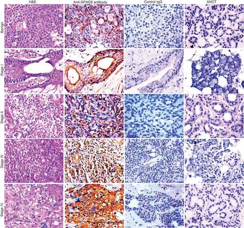
In the published version of Figure 3, Author inadvertently duplicated the ANCT panels (fourth row of the images) for benign and stage II tumors during figure assembly. This error has been corrected by replacing the ANCT panel for the benign tissue in the upper right corner of the figure. The corrected Figure 3 is enclosed. The figure legend is correct as published and is shown for reference below. This error has no impact on the interpretation of the results, nor does it influence the conclusions of the work.
Author apologize for any inconvenience that may have been caused.
Figure 3 Validation of SPAG9 protein expression in various stages of SGT by immunohistochemistry. First panel shows representative images for H&E staining in benign, malignant stage I, II, III, and IV tumor specimens. Second panel shows the representative images for the cytoplasmic localization of SPAG9 protein probed with anti-SPAG9 antibody as depicted by brown color immunoreactivity. No immunoreactivity was observed in serial tumor sections probed with control IgG, as shown in the third panel. Fourth panel depicts no immunoreactivity against SPAG9 protein in ANCT specimens when probed with anti-SPAG9 antibody. Original magnification: x400; objective: x40.