
Abstract
Cervical cancer is the third most common highest mortality in women worldwide. The use of standard chemotherapeutic drugs against cervical cancer patients received several side effects. Therefore, we focused phytoconsituents-mediated synthesis of gold nanoparticles (AuNPs) considered as greatest attention in the treatment of cervical cancer. In this present study, we reported that green synthesis of AuNPs by using with Alternanthera Sessilis aqueous extract. Synthesis of AuNPs were characterized by UV visible spectroscopy, energy dispersive X-ray (EDX), selected area diffraction pattern (SAED), Fourier transform infrared spectroscopy (FTIR), high-resolution transmission electron microscopy (HR-TEM) and atomic force microscope. Synthesized AuNPs confirmed by the UV absorption maximum at 535 and crystal structure of gold AuNPs was further confirmed by EDX and SAED. TEM and atomic force microscopy images show the size and morphological distribution of nanoparticles. FTIR analysis was confirmed the hydroxyl groups, amine and alkaline groups of biomolecules are present in the AuNPs. Moreover, AuNPs induce cytotoxicity in cervical cancer cells and also induce apoptosis through modulating intrinsic apoptotic mechanisms in cervical cancer cells. This green synthesis of AuNPs from Alternanthera sessilis approach was easy, large scaled up and eco-friendly.
Introduction
Cervical cancer is the most common type of cancer and it has occupied third highest mortality in women worldwide [Citation1]. The 99% of cervical cancers have been associated with human papiloma virus (HPV) infection. WHO report stated that cervical cancer shows approximately 12% of all type of cancers in women worldwide and most commonly it shows developing countries [Citation2]. In the annual estimation of cervical cancer for the year 2000, 470,600 new cases were identified and 233,400 deaths were observed [Citation3]. Several treatments were used to cure cervical cancer such as chemotherapy, radiotherapy, and finally surgery. When the radiotherapy to patients who have affected advanced cervical carcinoma, it resulted in a 70% progression-free survival rate [Citation4]. Moreover, cisplatin, paclitaxol, topotecan, gemicitabine, etc., have been used as chemotherapeutic drug against cervical carcinoma.
Treatment of cervical cancer by chemotherapeutic drug exhibits drug resistance-mediated side effects. However, these drugs exhibit several side effects and lower prognosis [Citation5]. However, nanotechnology- or nanomedicine-based research has been focused on the targeted delivery to cancer cells and it overcomes the drug resistance and improves the efficacy of chemotherapeutic drugs [Citation6]. Moreover, nanoparticle-mediated naturally occurring anticancer properties of drugs are safer than existing standard chemotherapeutic drugs. Furthermore, these agents are derived from various dietary supplements; therefore, these natural products prevent early stages of cancer [Citation7].
Nanotechnology refers to the physical and chemical manufacture of materials having nanosize between 1 nm and 100 nm [Citation8]. There are several nanomaterials being developed for nanomedicine applications, gold nanoparticles (AuNPs) are considered as potential tumour sensors and it effectively enhances delivery of the drug. AuNPs is the most constant metal nanoparticles and they have attractive properties such as size-related electronic, magnetic, biocompatible, and nontoxic materials [Citation9].
Alternanthera sessilis is a weed; occurring South-east Asia and it tolerates in wet lands as well as uplands based on different soil types [Citation10]. It has been found that weed of rice in tropical regions and other crops such as sugarcane, cereal and banana. Alternanthera sessilis young shoots and leaves are used as vegetables in South-east Asia. Alternanthera sessilis has found to be several active constituents such as triterpenoids, flavonols, campesterol, stigmasterol, β-sitosterol steroids, and tannins; lupeol are important constituents of A. Sessilis [Citation11,Citation12]. This species exhibits several pharmacological properties like antipyretic, antibacterial, hepatoprotective, antiulcer, hematinic, and antitumor. Previously it has been seen that A. Sessilis-assisted silver nanoparticles effectively inhibits the breast cancer cell proliferation [Citation11]. In this study, we investigated the green synthesis of AuNPs from aqueous extract of Alternanthera sessilis which was confirmed by UV, selected area diffraction pattern (SAED), transmission electron microscopy (TEM), energy dispersive X-ray (EDX), Fourier transform infrared spectroscopy (FTIR), atomic force microscopy (AFM), and their anticancer activity through apoptotic signalling which was studied in cervical cancer cell lines (HeLa cells).
Materials and methods
Chemicals
AuNPs synthesis and purification
The aqueous extract of Alternanthera sessilis used for the reduction of Au3+ ions to Au0 was made by combining systematically washed leaves of A. sessilis (50 g) in a sterile conical flask with double distilled water (100 ml). The mixture was allowed to boil for 5 min by using water bath. In brief, 5 ml of the A. sessilis extract was added to 1 mM HAuCl4 solution (45 ml). Reduction of AuCl4- was recorded by UV-vis absorption spectrum as a different function of time. After the reaction, AuNPs were centrifuged at 14,000 RPM for 30 min at ambient temperature to remove the large aggregation of particles, then the supernatants were collected and purified on PD-10 columns and eluted samples (3.5 ml) were collected and dialyzed against sodium phosphate buffer (10 mM) with a pH of 7.0 using with 20 kDa dialysis bags (Spectrum Labs, Rancho Dominguez, CA) with buffer exchange after 2 h, followed by overnight incubation for 15–18 h.
Characterization of AuNPs
After the synthesis and purification of AuNPs, they were characterized by several experiments which confirm the nanoparticles size, shape, and morphological characteristics. The synthesized AuNPs were confirmed by Ultraviolet-visible absorbance spectrum analysis at wavelength ranges of 300–700 nm (UV-1800 Shimadzu UV spectrophotometer). The synthesized AuNPs size and morphological characteristics were identified and confirmed by TEM images. Furthermore, energy dispersive spectroscopy (EDS) analysis was performed to confirm the chemical composition of synthesized AuNPs. The synthesis of nanoparticles stability and crystal nature was analyzed by EDX. SAED and dynamic light scattering (DLS) measurements were also used to determine the surface morphology and size of the nanoparticles. FTIR spectroscopy measurements were performed to analyze the active compounds present on the synthesized AuNPs. The FTIR spectra of AuNPs were recorded in the range of 4000–1000 cm−1 in KBr pellets using FTIR spectrophotometer.
Cell culture
Human HeLa cells were procured from National Centre for Cell Science, Pune, India. The cells were maintained and grown in Dulbecco’s Modified Eagle Medium (DMEM) in addition with 10% heat-inactivated foetal bovine serum (FBS), 3% l-glutamine, 100 IU penicillin, and 100 g/mL of streptomycin. The cells were incubated and grown in 5% CO2 environment at 37 °C and had 80–90% confluence before they were sub-cultured for the experiments.
Cell viability assay
AuNPs on HeLa cell line toxicity was assessed by MTT [3-[4, 5-dimethylthiazol-2-yl]-2,5-diphenyltetrazolium bromide]-based colorimetric assay [Citation13]. HeLa cells (1 × 10⁴) cells were seeded in all the well of a microtiter plate and incubated for 24 h. Then, the cells were exposed to different concentration of AuNPs (10–500 μg/ml) at 37 °C and 5% CO2 for 24 h incubation. Then, the untreated and AuNPs-treated cells were treated 100 μl MTT solution (0.5 mg/ml) at 37 °C and 5% CO2 for 4 h incubation. Finally, 100 μl of dimethyl sulfoxide (DMSO) was added to dissolve the purple formazan crystal in the reaction. The optical density (OD) was determined at 570 nm in an ELISA plate reader.
Apoptotic studies for acridine orange/propidium iodide
AuNPs-mediated apoptotic changes in HeLa cells were assessed by acridine orange and propidium iodide (AO/PI) staining. Briefly, 1 × 10⁵ cells were plated in 6-well plate then cultured cells were treated with IC50 concentrations of AuNPs at 24 h incubation. After the incubation, the treated cells were washed with ice-cold PBS then stained with 20 µL of AO/PI staining (10 µL/mg AO and 10 µL/mg PI) solution at 37 °C for 10 min. The stained apoptotic and viable cells were observed by fluorescent microscope.
Caspase 8, 9 and 3 activity assay
The caspase 8, 9 and 3 activities were measured by caspase test kit following the manufacturer’s instruction (Invitrogen). In brief, the HeLa cells were plated in 6-well plate and treated with AuNPs, and incubated for 24 h at CO2 incubator. Then the cells were treated with caspase 8, 9, and 3 assay reagents and it incubated for 2 h in dark room. Then, the absorbance for caspase 8, 9, and 3 was measured using a microplate autoreader at 400 or 405 nm.
Western blot analysis
The apoptotic markers such as Bax, Bid, and Bcl-2 were analyzed by Western blotting [Citation14]. In brief, 1 × 10⁶ cells were treated with AuNPs for 24 h incubation. Then the protein samples were prepared using with RIPA buffer containing protease cocktail. The concentration of protein was estimated by Lowry method [Citation16]. The proteins were subjected to 10% SDS-PAGE and the gel was transferred into PVDF membranes. The membrane was blocked with 5% bovine serum albumin (BSA) for 2–3 h. The membranes were treated with an appropriate primary monoclonal antibody and incubated for 24 h at 4 °C. Further, the membranes were incubated with appropriate secondary antibodies conjugated to horseradish peroxidase for 1 h. Protein bands were detected by using enhanced chemiluminescene substrates, and band intensity were analyzed using image J software.
Statistical analysis
All the experiments were carried out in three independent experiments and the results were expressed as the Mean ± Standard deviation (Mean ± SD) by using with one-way analysis of variance (ANOVA). p Values < .05 were indicative of significant differences.
Results
UV-visible spectroscopy
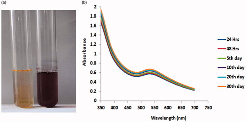
The synthesis of AuNPs from aqueous leaf extract of Alternanthera sessilis was confirmed by changes in the solution colour from light yellow to ruby red (). Furthermore, UV-visible spectroscopy results () show that typical plasmonic peak ranges from 525 to 545 nm with maximum absorbance peak was found to be approximately 535–545 nm. This result suggested the typical characteristic of spherical AuNPs with a diameter of 30–50 nm. This reaction kinetics confirmed the completion of the reaction after 30 days as evident from the stability of the plasmonic peak to no significant change beyond this time.
Figure 1. (A) The synthesis of AuNPs from aqueous leaf extract of Alternanthera sessilis was confirmed by changes in solution colour. (B). UV-visible absorption spectrum of synthesized AuNPs.
SAED pattern of AuNPs synthesized from Alternanthera sessilis
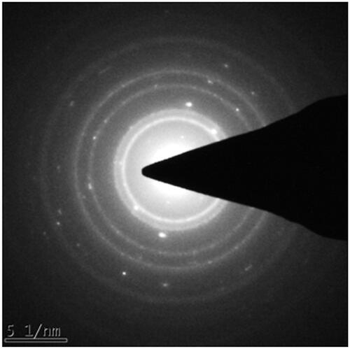
The crystalline nature of AuNPs also confirmed by SAED is shown in . In this study, we observed that bright circular pattern rings were observed. This might be the reflection from the lattice planes of crystalline Au nanoparticles.
Figure 2. SAED pattern images of gold nanoparticles synthesised from Alternanthera sessilis.
EDX and TEM analysis of AuNPs synthesized from Alternanthera sessilis
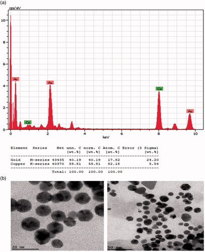
EDX analysis of the stabilized AuNPs also indicated the presence of AuNPs stabilized by plant phytochemicals. The EDX spectra of AuNPs are given in . In this study, respective relevant spectrums with strong signals of Au (0–1, 2–2.5 and 9.5–10 keV) at characteristic energy were observed. Furthermore, the shape and morphological characteristics of AuNPs were synthesized from leaf extract of A. sessilis and it was assessed by high-resolution transmission electron microscopy (HR-TEM). For HR-TEM analysis, samples were prepared as dried from colloidal AuNPs samples containing 1000 and 1500 mg/L concentration of the leaf extract of A. sessilis were recorded. illustrates HR-TEM images of 20–40 nm sized spherical-shaped AuNPs.
Figure 3. Energy dispersive X-ray (EDX) and transmission electron microscopy (TEM) analysis of gold nanoparticles synthesised from Alternanthera sessilis.
Atomic force microscopy and FTIR spectroscopy analysis of AuNPs synthesized from A. sessilis
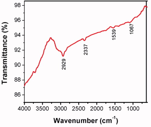
AFM was performed to analyze the three-dimensional measurement of AuNPs height (). In this study, it was observed that AuNPs are in the range of 12–18 nm. These results were closely correlated with the TEM data. Furthermore, FTIR analysis was used to identify the presence of several functional groups present in the synthesized AuNPs. FTIR spectrum showed several absorption bands at 2929, 2337, 1539 and 1067 cm−1 representing the presence of capping molecules with the nanoparticles (). The absorption bands at 2929 and 2337 cm−1 region arising CeH stretching of the aromatic compound were found. The band ranged at 1539 and 1067 cm−1 was assigned for NeH and CeN (amines) stretch vibration which was found in the amide linkages of the proteins.
Figure 4. Atomic force microscopy images for 3D structure of gold nanoparticles (AuNPs) synthesized from A. sessilis.
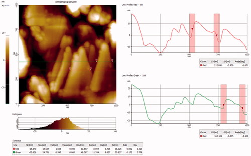
Figure 5. Fourier transforms infrared (FTIR) spectroscopy analysis of gold nanoparticles (AuNPs) synthesized from A. sessilis.
Cytotoxic potential of AuNPs from A. sessilis in HeLa cells
The cytotoxic nature of AuNPs-mediated HeLa cells was studied by MTT-based cytotoxicity assay (). In this study, we used a different concentration of AuNPs (1–15 µg/ml) for 24 h incubation. We found that increasing concentration of AuNPs significantly decreased the cell viability in HeLa cells. Moreover, AuNPs at the concentration of (10–15 µg/ml) was found to be pronounced cell death in HeLa cells.
AuNPs from A. sessilis induces apoptotic morphological changes in HeLa cells
Apoptotic morphological studies were investigated to find out the cells whether apoptotic or viable by using with acridine orange and ethidium bromide staining. In this study, we found that non-treated cells showed there is no apoptosis in HeLa cells (). Treatment with AuNPs significantly increased the apoptotic cells by observing red fluorescence staining in HeLa cells in concentration-dependent manner.
Figure 6. Cytotoxic potential of AuNPs from A. sessilis in HeLa cervical cancer cell lines.
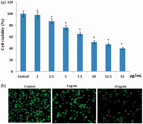
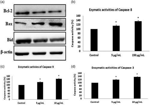
Figure 7. AuNPs from A. sessilis induces apoptotic morphological changes in HeLa cervical cancer cell lines. (A) Western blotting images for Bax, Bid and Bcl-2 protein expression in AuNPs-treated HeLa cells. (B–D) Colorimetric assay for caspase 8,9 and 3 activity expression in AuNPs treated HeLa cells.
AuNPs from A. sessilis induces apoptotic gene expressions in HeLa cells
AuNPs-mediated apoptotic marker (Bax, Bid and Bcl-2) expressions were analyzed by Western blotting. In this study, control cells showed decreased expression of Bax and increased expression of Bid and Bcl-2. In contrast, AuNPs treatment significantly increased the expression of Bax and decreased expression of Bid and Bcl-2 in HeLa cells in concentration-dependent manner. AuNPs-mediated expression of caspase 8, 9 and 3 were analyzed by the caspase kit colorimetric method. In this study, we observed that control groups showed decreased activity of caspase 8, 9 and 3 in HeLa cells. Treatment with AuNPs significantly increased the activity of caspase 8, 9 and 3 in HeLa cells in concentration-dependent manner ().
Discussion
Cervical cancer is the major common cancer in women worldwide and it is mainly caused by human papillomavirus (HPV) [Citation16]. There are several treatment methods such as chemotherapy and radiotherapy which have been utilized for cervical cancer, but they have produced significant number of undesirable side effects for the patients [Citation17]. Therefore, phytochemical-based nanoparticles are considered as successful drug delivery system [Citation18]. In this study, for the first time, we synthesized and characterized the AuNPs from aqueous leaf extract of Alternanthera sessilis which has anticancer potential against HeLa cervical cancer cells. Alternanthera sessilis was commonly known as joy weed which belongs to the family of Amarantheacea. It exhibits multipurpose herb of oriental medicine and it cures several disorders like burning sensation in stomach, dyspepsia, diarrhoea, skin diseases and haemorrhoids [Citation19]. It has several pharmacological and therapeutic effects [Citation20]. Previously, silver nanoparticles were prepared by Alternanthera Sessilis leaf extract and it showed antimicrobial activity [Citation21].
Synthesis and characterization of AuNPs gained various attentions for peculiar properties like high surface to volume ratio and biocompatibility [Citation22]. Use of AuNPs showed excellent physio-chemical properties. Green synthesis of AuNPs is an economic and environmental-friendly approach. These nanoparticles could provide an active coating of biological residues on the surface of the particle and also avoid the usage of chemicals [Citation23]. Therefore, we used green synthesis AuNPs from Alternanthera sessilis leaf extract and it was characterized by UV-visible spectroscopy, EDX spectroscopy, SAED, FTIR, HR-TEM and atomic force microscopy.
The properties of AuNPs depend on the morphological characteristics such as shape, size, crystal nature and distribution of nanoparticles [Citation24]. In this study, we synthesis AuNPs using the leaf extract of Alternanthera sessilis. This extract performs as both reducing and stabilizing agent for the nanosynthesis, established by both the visible colour change and well-known absorption peaks in the UV-visible absorption spectra of nanoparticles. The green synthesis of AuNPs from leaf extract of Alternanthera sessilis was visually observed by changes in solution colour from light-yellow to red. UV-visible spectroscopy analyses the characterization of AuNPs present in the sample. In this study, maximum absorbance was found to be approximately 525–535 nm. This reaction kinetics confirmed the completion of the reaction after 30 days as evident from the stability of the plasmonic peak to no significant change beyond this time. FTIR analysis was used to identify the presence of several functional groups present in the synthesized AuNPs. Our results show that several FTIR absorption bands were observed at 2929, 2337, 1539 and 1067 cm−1 and it indicates the active molecules present with the nanoparticles (). The previous report stated that the hydroxyl groups, amine and alkaline groups of molecules have a stronger ability to bind with Au ions [Citation25]. In our FTIR absorption spectrum, active molecules are aromatic, amine, alkaloids and amide linkage proteins present in the particles.
EDX studies were analyzed to understand the spherical nature of the particles synthesized from Alternanthera sessilis silver metal. In this study, strong signal was observed in the gold region and it confirms the synthesis of AuNPs. We also observed the strong signals of Au (0–1, 2–2.5 and 9.5–10 keV) at characteristic energy level. The typical optical absorption for metallic crystal generally found to be 3 KeV due to surface plasmon resonance [Citation26]. Furthermore, the shape and morphological characteristics of AuNPs were analyzed by HR-TEM. Different magnifications showed 20–40 nm sized spherical-shaped AuNPs. Moreover, the three-dimensional qualities of AuNPs shape and morphology were analyzed by AFM.. In this study, AuNPs are in the range of 12–18 nm. These data closely relate with the two-dimensional TEM data. The crystalline nature of AuNPs was determined by SAED and bright circular pattern rings were observed. This indicates the crystal nature of Au in the synthesized particles. This result relatively correlated with the HR-TEM analysis.
The anticancer activities of synthesized AuNPs against HeLa cells were performed by MTT cytotoxicity assay, apoptotic staining and apoptotic marker expressions. In order to find out the cytotoxic effect of synthesized AuNPs in HeLa cells. AuNPs showed the good cytotoxic activity against the cervical cancer cells in the concentration-dependent manner. MTT assay showed that 10–15 µg/ml concentrations of AuNPs effectively inhibit the cervical cancer cell growth. Previously, synthesis of AuNPs inhibits cell proliferation in A549 and Hep-2 cell lines [Citation27]. Apoptosis is a cell suicidal mechanism which can eliminate potentially unwanted harmful cells from chemically treated cells [Citation14]. There are two main kinds of pathways which include intrinsic and extrinsic pathways. The intrinsic pathways are mainly regulated by Bcl-2-mediated caspase-3 activation and it leads to apoptosis [Citation28]. AuNPs-mediated apoptotic morphological studies were analyzed by acridine orange and propidium iodide staining. In this study, we found that AuNPs increased the apoptotic stained in HeLa cells. Furthermore, we confirmed AuNPs-mediated apoptotic protein expressions were analyzed by enzymatic method and Western blotting analysis. In this study, AuNPs treatment significantly increased the expression of Bax and decreased expression of Bid and Bcl-2 in HeLa cells in concentration-dependent manner. Moreover, we observed that treatment with AuNPs significantly increased the activity of caspase 8, 9 and 3 in HeLa cells in concentration-dependent manner. Numerous documents also postulated that AuNPs from different extracts also induces apoptosis through regulating intrinsic apoptotic pathways in several types of cancer cells [Citation29,Citation30].
Conclusion
In this study, we conclude that the synthesis of AuNPs from Alternanthera sessilis was achieved and characterized. It was initially identified by the formation of the ruby red colour solution, and UV absorption spectrum also confirmed the maximum absorbance at 535 nm. Crystal structure of AuNPs was further confirmed by EDX and SAED. TEM and AFM images show the size and morphological distribution of nanoparticles. The hydroxyl groups, amine and alkaline groups of biomolecules present in the AuNPs were identified by FTIR. Moreover, AuNPs induce cytotoxicity in cervical cancer cells and also induce apoptosis through modulating intrinsic apoptotic mechanisms in HeLa. This AuNPs increases the cytotoxicity based on the concentrations in cervical cancer cells. This green synthesis of AuNPs from Alternanthera sessilis approach was easy, large scaled up and eco-friendly.
Disclosure statement
The authors declare no conflict of interest
Related Research Data
References
- Parkin DM, Bray F, Ferlay J, et al. Estimating the world cancer burden: Globocan 2000. Int J Cancer. 2001;94:153–156. TQ1
- Burd EM. Human papillomavirus and cervical cancer. Clin Microbiol Rev. 2003;16:1–17.
- Patra S, Panda D. Cervical cancer screening in developing countries. Indian J Cancer. 2010;314:47–43.
- Minig L, Patrono MG, Romero N, et al. Different strategies of treatment for uterine cervical carcinoma stage IB2-IIB. World J Clin Oncol. 2014;5:86–92.
- Ordikhani F, Erdem Arslan M, Marcelo R, et al. Drug delivery approaches for the treatment of cervical cancer. Pharmaceutics. 2016;8:23.
- Xu X, Ho W, Zhang X, et al. Cancer nanomedicine: from targeted delivery to combination therapy. Trends Mol Med. 2015;21:223–232.
- Watkins R, Wu L, Zhang C, et al. Natural product-based nanomedicine: recent advances and issues. Int J Nanomed. 2015;10:6055–6074.
- Jeevanandam J, Barhoum A, Chan YS, et al. Review on nanoparticles and nanostructured materials: history, sources, toxicity and regulations. Beilstein J Nanotechnol. 2018;9:1050–1074.
- Arvizo R, Bhattacharya R, Mukherjee P. Gold nanoparticles: opportunities and challenges in nanomedicine. Expert Opin Drug Deliv. 2010;7:753–763.
- Singh A, Thangaraj K, Bharti O. In vitro propagation of Alternanthera sessilis (sessile joyweed), a famine food plant. Afr J Biotechnol. 2009;8:21.
- Firdhouse MJ, Lalitha P. Biosynthesis of silver nanoparticles using the extract of Alternanthera sessilis – antiproliferative effect against prostate cancer cells. Cancer Nanotechnol. 2013;4:137–143.
- Sahithi B, Rajani GP, Sowjanya K, et al. Anti-inflammatory activity of ethanolic and aqueous extracts of Alternanthera sessilis lin. Pharmacologyonline. 2011;1:1039–1043.
- Balupillai A, Nagarajan RP, Ramasamy K, et al. Caffeic acid prevents UVB radiation induced photocarcinogenesis through regulation of PTEN signaling in human dermal fibroblasts and mouse skin. Toxicol Appl Pharmacol. 2018;352:87–96.
- Agilan B, Rajendra Prasad N, Kanimozhi G, et al. Caffeic acid inhibits chronic UVB-induced cellular proliferation through JAK-STAT3 signaling in mouse skin. Photochem Photobiol. 2016;92:467–474.
- Lowry OH, Rosebrough NJ, Farr AL, et al. Protein measurement with the Folin phenol reagent. J Biol Chem. 1951;193:265–275.
- Kaarthigeyan K. Cervical cancer in India and HPV vaccination. Indian J Med Paediatr Oncol. 2012;33:7–12.
- Banerjee R, Kamrava M. Brachytherapy in the treatment of cervical cancer: a review. Int J Womens Health. 2014;6:555–564.
- Niraimathi KL, Sudha V, Lavanya R, et al. Biosynthesis of silver nanoparticles using Alternanthera sessilis (Linn.) extract and their antimicrobial, antioxidant activities. Colloids Surf B Biointerfaces. 2013;102:288–291.
- Lin SC, Lin YH, Shyuu SJ, et al. Hepatoprotective effects of Taiwan folk medicine: Alternanthera sessilis on liver damage induced by various hepatotoxins. Phytother Res. 1994; 89:391–398.
- Rizvi SAA, Saleh AM. Applications of nanoparticle systems in drug delivery technology. Saudi Pharm J. 2018;26:64–70.
- Sarvamangala D, Kantipriya K, Murthy USN, et al. Green synthesis of AgNP’S using Alternanthera Sessilis leaf extract [A natural source for ocular therapy.]. Int J Innovative Res Sci Eng Technol. 2014;3:15000–15010.
- Yeh YC, Creran B, Rotello VM. Gold nanoparticles: preparation, properties, and applications in bionanotechnology. Nanoscale. 2012;4:1871–1880.
- Kunoh T, Takeda M, Matsumoto S, et al. Green synthesis of gold nanoparticles coupled with nucleic acid oxidation. ACS Sustainable Chem Eng. 2017;6:364–373.
- Khan I, Saeed K, Khan I. Nanoparticles: properties, applications and toxicities. Arabian J Chem. 2017.
- Vijayan R, Siby J, Beena M. Indigofera tinctoria leaf extract mediated green synthesis of silver and gold nanoparticles and assessment of their anticancer, antimicrobial, antioxidant and catalytic properties. Artif Cells Nanomed Biotechnol. 2018;46:861–871.
- Kaviya S, Santhanalakshmi J, Viswanathan B, et al. Biosynthesis of silver nanoparticles using citrus sinensis peel extract and its antibacterial activity. Spectrochimica Acta Part A. 2011;79:594–598.
- Rajeshkumar S. Anticancer activity of eco-friendly gold nanoparticles against lung and liver cancer cells. J Genet Eng Biotechnol. 2016;14:195–202.
- Pistritto G, Trisciuoglio D, Ceci C, et al. Apoptosis as anticancer mechanism: function and dysfunction of its modulators and targeted therapeutic strategies. Aging. 2016;8:603–619.
- Baharara J, Ramezani T, Divsalar A, et al. Induction of apoptosis by green synthesized gold nanoparticles through activation of caspase-3 and 9 in human cervical cancer cells. Avicenna J Med Biotechnol. 2016;8:75–83.
- Selim ME, Hendi AA. Gold nanoparticles induce apoptosis in MCF-7 human breast cancer cells. Asian Pac J Cancer Prev. 2012;13:1617–1620.