
Abstract
Gold nanoparticles (AuNPs) as the most excellent anticancer theranostic nanoparticles were synthesized through efficient, simple, and green synthesis method using Marsdenia tenacissima plant extracts and they are widely characterized by several techniques including ultraviolet-visible (UV) spectroscopy, atomic force microscopy (AFM), energy-dispersive X-ray spectrometers (EDS), transmission electron microscopy (TEM), and Fourier transform infrared (FT-IR) spectroscopy. From the AuNPs synthesized by M. tenacissima extracts, it was discovered that particle size around 50 nm, which is admirable nano dimension, was achieved by plant-mediated synthesis. After characterization of these nanoparticles, they performed as in vitro anticancer activity against lung cancer cell lines (A549). MTT assay revealed that AuNPs produce toxicity based on the dose-dependent A549 cells growth inhibition. AuNPs treatment activates caspase expression and down-regulates the anti-apoptotic protein expression in A549 cells. Our results point out that the AuNPs from M. tenacissima extract are apposite stabilizing agents, which serve as an effective anticancer agent against lung cancer cell lines (A549).
Introduction
Lung cancer, also called lung carcinoma, affects both male and female. It is considered as the leading cause of deaths worldwide [Citation1]. Lung cancer is distinguished into small cell and non-small cell lung cancer based on histologic studies. Generally, an advanced type of non-small cell lung cancer has been thought of as poor prognosis; therefore, new ultimate methods are urgently needed for improving the chances of patient survival [Citation2]. The development of therapies to fight against severely multiplying tumors is very difficult. Numerous treatments are available for cancer therapy but they are restricted by the lack of specificity and dose-limiting toxicity [Citation3]. Finding therapeutic drugs for the treatment of numerous types of cancer is a challenge [Citation4]. However, safest methods are of importance for the combination of controlled release, exhibiting targeted delivery, less harmful, and very effective [Citation5]. Nanomaterials are projected optimistically to develop cancer diagnosis and therapy. Nanotechnology includes the synthesis of nanoscale materials, utilization and understanding of physicochemical and optoelectronic properties. In the new millennium, nanotechnology plays a vital role in the key technologies [Citation6]. Nanoparticles, especially in metal, have obtained great attention because of its distinct physiochemical nature and have become a main active research area due to sensors, imaging, cosmetics, and cancer therapy and drug delivery [Citation7].
Gold nanoparticles, (AuNPs) as the best drug nanocarriers, have excellent characteristics with controlled size, stability, and biocompatibility, which makes them potential agent in cancer diagnosis and treatment [Citation8]. They are employed to visualize the tumors in their primary-secondary locations and can also be used as delivery vehicles for the anticancer drug [Citation9]. Gold nanoparticles are the most gorgeous nanomaterials for diverse applications like catalytic, anticancer, antimicrobial, anti-inflammation, and various biomedical research [Citation10].
Generally, chemical methods which have been applied for the synthesis of AuNPs cause environmental pollution due to the toxicity of the reagents used [Citation11]. Accordingly, researchers are looking at biological systems for nanoparticle synthesis to avoid environmental pollution and minimize the toxicity [Citation12]. So currently, AuNPs synthesized by using plant extract is more effective and useful. It also reduces environmental pollution and produces a huge amount of nanoparticles [Citation13]. Plant extracts possibly will act as reducing and stabilizing agents in the synthesis of nanoparticles. Plant extract mediated AuNPs synthesis is more advantageous than the other methods because it is very simple to use, safe, and eco-friendly in nature [Citation14].
Plants have enormous prospective for AuNPs synthesis for broad-spectrum applications with preferred morphology of the particles. The small sizes of gold nanoparticles rapidly synthesized using Acalypha indica extract have been equipped as a novel source of bio-reductants [Citation15]. Previously, AuNPs from Nepenthes Khasiana extract has the average size of the particles to be 50–100 nm [Citation16]. Moreover, 15–25 nm size of the gold nanomaterials was produced from Cassia auriculata extract [Citation17]. In this current study, we employed the synthesis and characterization of Au nanoparticles from Marsdenia tenacissima extract. Marsdenia tenacissima (Roxb.) is mainly grown in China and the medicinal usage of this plant was first recorded in “Dian Nan Ben Cao”, a medical literature written by Mao Lan in Ming Dynasty. Marsdenia tenacissima is used in Chinese traditional medicine to treat pneumonia and tracheitis. In addition, this plant has records of significant anti-proliferative activity [Citation18].
Numerous studies have recognized that different extracts from Marsdenia tenacissima have exhibited anti-tumor properties on in vitro and in vivo experimental models [Citation19,Citation20]. The anti-tumor activities of M. tenacissima extract were linked with the cell cycle arrest, cell apoptosis, and induction of anti-angiogenesis [Citation21]. However, the molecular mechanisms associated with M. tenacissima treatment against the death of the cancer cell remains unclear. Therefore, we sought to analyze the anticancer activity of gold nanoparticles from M. tenacissima in A549 cells through the apoptotic signaling pathway.
Materials and methods
Chemicals, reagents, and antibodies
The 3–(4,5-dimethylthiazol-2-yl)-2,5-diphenyltetrazolium bromide (MTT), gold chloride (HAuCl4), fetal bovine serum (FBS), and penicillin-streptomycin were purchased from Sigma-Aldrich, MO, USA. Further, DMEM, trypsin/EDTA, and phosphate buffer saline (PBS) were obtained from Lonza Chemicals, Nansha, China. Apoptotic primary antibodies such as Bcl-2, Bax, Caspase-3, -9 and β- actin were purchased from cell signaling. Moreover, other chemicals and solvents used were of analytical grade.
Synthesis and characterization of AuNPs by Marsdenia tenacissima extracts
AuNPs was synthesized by using M. tenacissima extracts. In brief, the appropriate amount of plant extract was mixed with double distilled water, then HAuCl4 solution (4 mM) was added dropwise under sonication to make a final volume of 20 ml. Furthermore, the reaction mixture was allowed to incubate at 25 °C overnight and the AuNPs synthesis was observed using UV-vis spectrophotometer (Tecan, Crailsheim, Germany) at an average wavelength between 200 and 1000 nm. The amount of dilutions of M. tenacissima extracts were used as a control blank solution.
After synthesis of AuNPs from M. tenacissima, the following studies were conducted to confirm the AuNPs. XRD was used to characterize the crystalline structure of dried nanoparticles (Bruker, Germany). Morphological characteristics of synthesized AUNPs were studied by transmission electron microscope (Hitachi, Pleasanton, CA) and selected area electron diffraction (SAED). Energy Dispersive X-Ray Spectroscopy was utilized to analyze the presence of elemental gold in the synthesized AUNPs. Gold nanoparticles and its functional biomolecules were analyzed by Fourier-Transform Infrared Spectroscopy (FTIR spectroscopy) (BrukerOptik GmbH, Deutschland). The FTIR spectra of dried AUNPs was measured at wavelength ranges between 4000 and 400 cm−1.
MTT assay
The cytotoxic nature of AuNPs was analyzed in lung cancer A549 cell lines by MTT assay [Citation22]. A549 cells (10,000 cells) were seeded in microtiter plates and incubated for 24 h. Furthermore, cells were treated with AuNPs at different concentrations (2.5, 5, 10, 12.5, 15, and 25 µg) and further incubated for 72 h. Then, the cells were exposed to 100 µl of freshly prepared yellow MTT reagents (1 mg/mL) for 4 h. Finally, 100 µL of dimethyl sulfoxide (DMSO) was added and the purple formazan solution was measured by UV absorbance at 570 nm (Multimode reader, Tecan, Austria).
Apoptotic studies by fluorescent staining
Acridine orange and ethidium bromide (AO/EtBr) staining were used to study the apoptosis morphological features [Citation23]. Cells were seeded in six-well plates and the AuNPs were exposed for 24 h. After treatment, 20 µl of both acridine orange and ethidium bromide dye mixture was added to the well plates. Then, the cells were examined under a fluorescent microscope using green and red fluorescent.
Western blotting
After treatment of AuNPs, cells were collected and lysed with radioimmunoprecipitation assay (RIPA) buffer and protein concentration was measured by a spectrophotometer (Nanodrop, Thermo Scientific, Basingstoke, UK). Protein sample from four different groups was subjected to 12% sodium dodecyl sulfate polyacrylamide gel electrophoresis (SDS-PAGE). After electrophoresis, the gel was smoothly transferred to nitrocellulose membrane by using semi-dry Western blot apparatus. Then, the nitrocellulose membrane was blocked with 5% bovine serum albumin (BSA) for 1 h. In addition, the membrane was treated with appropriate primary monoclonal antibodies and incubated for 24 h at 4 °C. Then, the membrane was gently washed with TBST washing buffer. After incubation with primary antibodies, secondary antibodies were added to the membrane and this was incubated for 1 h at room temperature. Finally, the membrane was washed properly with TBST buffer and protein expression levels were detected by chemiluminescent detecting system (Biorad, California, USA).
Statistical examination
All the in vitro experiments were performed in three independent experiments and the results were expressed as mean ± standard deviation (Mean ± SD) by using one-way analysis of variance (ANOVA). Values of p < .05 were indicative of significant differences.
Results and discussion
Synthesis and characterizations studies of AuNPs
Metal nanoparticles, especially in gold nanoparticles, often tend to receive the greatest attention in the field of nanotechnology because it exhibits numerous beneficial properties such as magnetic, thermal and electric conductivity; they also show antimicrobial, anticancer and antimalarial activity in biomedical applications [Citation24]. The syntheses of AuNPs are considered as environmentally friendly; therefore, it is gaining highest attention in the field of drug discovery and targeted delivery [Citation25]. M arsdenia tenacissima plant has been used in traditional Chinese medicine against several malignant diseases. In this current work, we synthesized and characterized gold nanoparticle from M. tenacissima extract. These nanoparticles were investigated to test their anticancer potential against lung cancer cell lines. Marsdenia tenacissima extract could reduce chloroauric acid; the synthesis of AuNPs is widely considered for optimization of the numerous factors affecting the synthesis of plant extract and metal substrate with different time point. The fractionation of organic compounds such as terpenoids, flavonoids and polysaccharides can reduce the difficulty of reactions. Polyoxypregnanes, aglycones and glycosides are the main active ingredients in M. tenacissima extracts [Citation26].
Previously, it has been tested that the synthesis of gold nanoparticles with glycoside reductants established glycones and aglycones structure based synthesis. C-6 of glycosides has been oxidized and form carboxylic acid by the removal of auric acid, consequently generating the highest synthetic yield of mono-dispersed, round gold nanoparticles [Citation27]. The active biomolecules could perform an imitation of a specific interaction between inorganic molecules to the compact synthesis of nanoparticles. The final structure of nanoparticle is greatly related to the physicochemical nature of the active biomolecules [Citation28].
UV absorbance spectroscopic studies
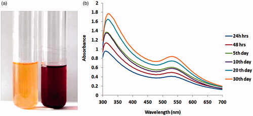
Green synthesized AuNPs were initially confirmed by UV absorbance value of nanoparticles using UV-visible spectroscopy. Chueh et al. (2014) and Geetha et al. (2013) [Citation10,Citation29] reported that green synthesized gold nanoparticles have well-known exact peaks around 540–560 nm. The AuNPs formation was apparent from the conversion of sample color from mild-yellow to ruby-red in addition to the presence of typical plasmon peak that ranged between 525 and 540 nm with a maximum peak absorbance approximately found to be between 527 and 535 nm. The distinct typical plasmon peak characteristic was spherical and oval-shaped AuNPs with 30–50 nm length. The reaction after 24 h to the 30th day as evidenced by the stability of plasmonic peak was found in the reaction kinetics of UV-vis spectroscopy studies (. Spectrophotometrically, AuNPs concentration was studied by Beer-Lambert law with an extinction coefficient ε of 1.8 × 10¹⁰ M−1 cm−1 for a particle diameter of 50 nm. Anisotropic AuNPs is well-known to demonstrate two outstanding absorption bands, (i) wavelength transverse absorption peak was less and (ii) wavelength longitudinal absorption peak was more. The longitudinal plasmon peak is a strong function of the nanoparticle. Through adsorption and desorption biomolecules specifically interacts with the nanoparticle face and the growth tariff of faces is kinetically modulated, which finally controls the nanoparticles structure [Citation30]. It has been well documented that reducing biomolecules mediated controlled reduction of metal salts generally in favor of producing spherical nanoparticles [Citation31].
Figure 1. (a) The synthesis of AuNPs from aqueous leaf extract of Marsdenia tenacissima was confirmed by changes in solution nature from light color to dark color. (b). UV-visible absorption spectrum of synthesized AuNPs.
Transmission electron microscopy and energy dispersive spectroscopy studies
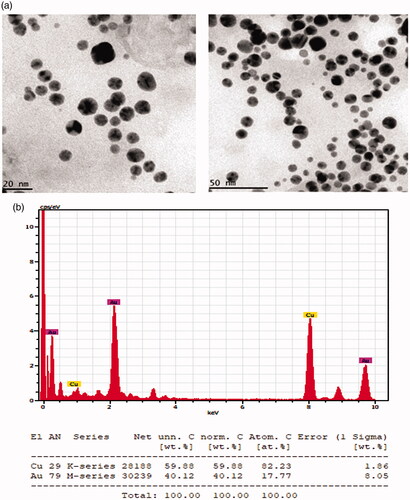
TEM studies were used to investigate the morphology and Au element size distribution profile of the synthesized nanoparticles. The morphology of the AuNPs was analyzed by HR-TEM as shown in . The figure also shows that AuNP particles consist of spherically shaped, having a smooth surface without aggregation. Moreover, anisotropic shapes, i.e triangular and hexagonal platelets which truncate single nanosheets were also noticed. Previous findings also supported the synthesis of AuNPs, which were confirmed by TEM microscopic studies [Citation32]. Moreover, energy dispersive spectroscopy (EDS) was used to determine the crystal elemental analysis of the presence of Au nanoparticles in M. tenacissima extracts (). Meanwhile, the compact sections of the element mapping clearly show the eminent proportion of Au elements in the sample () and significantly diminished Au elements signal. In this study, strong signal was observed in the silver region and it confirmed the synthesis of AuNPs. We also noticed that the strong signals of Au (0.1–0.5 and 2– 2.5 and 9–9.5 keV) at energy characteristic were observed. The typical optical absorption spectrum for the gold crystal is usually shown to be 3 KeV due to surface plasmon resonance [Citation33]. The identical distribution of gold particles was clearly observed in EDS mapping which resulted in monodisperse nanoparticles as confirmed through TEM images.
Figure 2. (a) High resolution transmission electron microscopy (HR-TEM) and (b) Energy dispersive X-ray (EDX) and analysis silver nanoparticles synthesised from Marsdenia tenacissima.
FTIR, SAED, and AFM studies
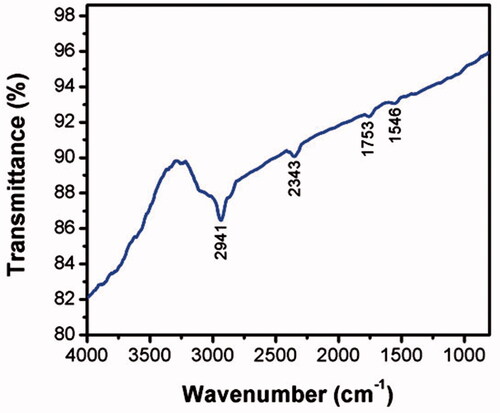
FTIR studies illustrated that more peaks were observed, which indicated the presence of active biomolecules on the surface of gold nanoparticles synthesized from M. tenacissima extracts (. The FTIR spectrum data showed that several absorption peaks were observed such as those placed at ranges of 2941, 2373, 1753, and 1546 cm−1, which indicates stretching for O–H bond, C–H, C–O–C, aldehyde groups, and carboxylic acids. The absorption bands at 2941 and 2373 cm − 1 area, which excludes CeH stretching of the aromatic compound were found. The band ranges at 1556 and 1753 cm–1, which indicated the presence of NeH and CeN (amines) stretch vibration which was found in the amide linkages of the proteins. AuNPs from the evaluation spectrum of M. tenacissima extracts showed high similarity, which indicated capable adsorption of organic molecules on AuNPs. Moreover, time-dependent properties for optical nature was ascribed by the formation of the round shape of AuNPs which aggregates time and formation of anisotropic nanoparticles.
Figure 3. Fourier transforms infrared (FTIR) spectroscopy analysis of gold nanoparticles (AuNPs) synthesized from Marsdenia tenacissima.
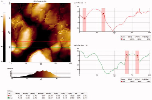
The crystalline nature of gold nanoparticles was also investigated by selected area diffraction patterns (SAED) and we observed bright circular pattern rings present in the image (. This confirmed the crystalline nature of Au present in the synthesized particles from M. tenacissima plant extract. This result was more closely correlated with the HR-TEM and EDX data analysis. Atomic force microscopy (AFM) was used to analyze the three- dimensional effect of size and shape distribution of the synthesized AuNPs (. The AFM images of gold nanoparticles from M. tenacissima extract confirmed the regular distribution of NPs. In this study, the majority of the particles size was shown to be 40–50 nm in diameter and these reports are consistent with the other morphological studies such as EDX and TEM measurements. Moreover, the crystal structure of the gold nanoparticles was additionally evidenced by SAED pattern. In this present work, we noticed that the synthesized gold nanoparticles were completely stable for more than 36 days.
Figure 4. SAED pattern analysis of gold nanoparticles synthesised from Marsdenia tenacissima.
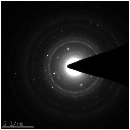
Figure 5. Atomic force microscopyanalysis of gold nanoparticles synthesised from Marsdenia tenacissima.
Anticancer role of AuNPs against lung cancer A549 cell lines
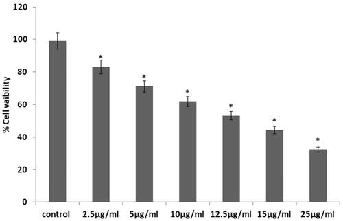
After synthesis, the gold nanoparticles were evaluated for their anticancer activity against lung cancer cell lines. For that purpose, we performed an array of in vitro experiments such as cytotoxicity based assay, apoptotic morphological assay, caspase activity assay, and apoptotic protein expression which were analyzed by Western blotting. The cytotoxic assay exposed the potent anticancer activity of AuNPs against A549 cells depending on the concentration of nanoparticles. The increasing concentration of AuNPs significantly increased cell death in A549 cells (. The IC50 of AuNPs was 15 µg/mL, moreover, 10 and 15 µg/mL concentrations of AuNPs were used for further studies. Generally, AuNPs are considered as non-toxic in normal cells and biocompatible for delivery of nanoparticles among all the metal nanoparticles [Citation32].
Figure 6. Cytotoxic potential of gold from Marsdenia tenacissima in prostate cancer A549 cell.
The role of anticancer activity of AuNPs might be recognized due to the adsorbed active molecules present in the M. tenacissima plant extracts. In our experimental study on the use of AuNPs against anticancer cell lines, the results were significantly higher when compared to previous reports for green synthesized AuNPs. The anticancer activity of M. tenacissima extracts also accounts for the induced cell death in lung cancer cells [Citation18]. Plant-derived compounds and their therapeutic activity against cancer, as well as biocompatible nanoparticles, could be attained. In this study, M. tenacissima extracts caused the synthesis of highly excellent AuNPs that displayed higher activity against cancer cells.
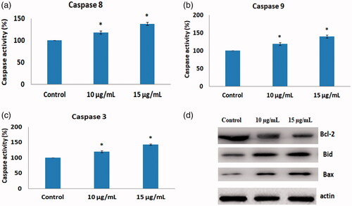
Apoptosis is considered as a usual cell death program and these pathways were regulated by several apoptotic proteins such as proapoptotic protein Bax, caspases, and antiapoptotic protein Bcl-2, Bid [Citation34]. In our current study, AuNPs mediated apoptotic morphological changes were observed by A549 cells stained with acridine orange and ethidium bromide and illustrated in . Acridine orange was used to stain A549 cells and the control cells appear uniformly green, whereas early apoptotic cell debris was yellow or yellowish green. Interestingly, treatment of AuNPs-treated cells showed the presence of ethidium-bromide-stained cells appearing as condensed nuclei, membrane blebbing and apoptotic bodies (. Moreover, apoptotic proteins such as Bax, Bcl-2, caspase 8, caspase 9 and caspase-3 are involved in apoptosis. In addition, AuNPs mediated apoptosis makers such as Bax, Bcl-2, Bid, Caspase-8, 9 and 3 were analyzed by caspase assay kit and Western blotting. As shown in , treatment with AuNPs synthesized from M. tenacissima down-regulated the expression of Bcl-2, Bid, and up-regulated the expression of Bax, caspase-8, caspase-9, and caspase 3 in lung cancer A549 cell lines. These reports are closely related with our acridine orange/ETBR staining. Previously, AuNPs synthesized from different plant extracts induced apoptosis through inhibiting apoptotic protein expressions in different cell lines [Citation32,Citation35]. Based on these studies, AuNPs synthesized from M. tenacissima effectively induces apoptosis by regulating intrinsic apoptotic pathway.
Figure 7. AuNPs mediated apoptotic morphological studied by AO/PI staining.
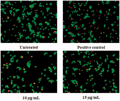
Figure 8. (a-c) Colorimetric assay for caspase-8,9 and 3 activity expression in AuNPs treated A549 cells. (d). Western blotting images for Bax, Bid, and Bcl-2 protein expression in AuNPs treated A549 cells.
Conclusion
In the present work, it is concluded that the synthesis of gold nanoparticles from M. tenacissima leaf extract was confirmed by numerous characterization studies such as UV-absorbance, FTIR, EDS, AFM, and TEM. These nanoparticles inhibit cell proliferation and induce apoptosis in a dose-dependent manner. Furthermore, AuNPs synthesized from M. tenacissima induces apoptosis through the modulation of Bax/Bcl-2 protein levels in A549 lung cancer cell line. In the future, M. tenacissima AuNPs may be applied for the treatment of cancers, which may overcome adverse effects associated with chemotherapy.
Disclosure statement
Authors stated that there are no conflicts of interests.
Related Research Data
References
- Plummer M, de Martel C, Vignat J, et al. Global burden of cancers attributable to infections in 2012: a synthetic analysis. Lancet Glob Health. 2016;4:e609–e616.
- Haga A, Takahashi W, Aoki S, et al. Classification of early stage non-small cell lung cancers on computed tomographic images into histological types using radiomic features: interobserver delineation variability analysis. Radiol Phys Technol. 2018;11:27–35.
- Mokhtari RB, Homayouni TS, Baluch N, et al. Combination therapy in combating cancer. Oncotarget 2017;8:38022.
- Chakraborty S, Rahman T. The difficulties in cancer treatment. Ecancermedicalscience. 2012;6:ed16.
- Kleinstreuer C, Feng Y, Childress E. Drug-targeting methodologies with applications: a review. World J Clin Cases. 2014;2:742–756.
- Jha RK, Jha PK, Chaudhury K, et al. An emerging interface between life science and nanotechnology: present status and prospects of reproductive healthcare aided by nano-biotechnology. Nano Rev 2014;5:10.
- Mody VV, Siwale R, Singh A, et al. Introduction to metallic nanoparticles. J Pharm Bioallied Sci. 2010;2:282–289.
- Shankar SS, Rai A, Ahmad A, et al. Rapid synthesis of Au, Ag, and bimetallic Au core–Ag shell nanoparticles using Neem (Azadirachta indica) leaf broth. J Colloid Interface Sci. 2004;275:496–502.
- Cai W, Gao T, Hong H, et al. Applications of gold nanoparticles in cancer nanotechnology. Nanotechnol Sci Appl. 2008;1:17–32.
- Geetha R, Ashokkumar T, Tamilselvan S, et al. Green synthesis of gold nanoparticles and their anticancer activity. Cancer Nano. 2013;4:91–98.
- Sharma D, Kanchi S, Bisetty K. Biogenic synthesis of nanoparticles: a review. Arab J Chem. 2015; https://doi.org/10.1016/j.arabjc.2015.11.002
- Huang Z, Lin H, Wang Y, et al. Studies on the anti-angiogenic effect of Marsdenia tenacissima extract in vitro and in vivo. Oncol Lett. 2013;5:917–922.
- Shukla R, Bansal V, Chaudhary M, et al. Biocompatibility of gold nanoparticles and their endocytotic fate inside the cellular compartment: a microscopic overview. Langmuir. 2005;21:10644–10654.
- Ikram S. Synthesis of gold nanoparticles using plant extract: an overview. Archivos De Medicina. 2015;1:5.
- Krishnaraj C, Muthukumaran P, Ramachandran R, et al. Acalypha indica Linn: biogenic synthesis of silver and gold nanoparticles and their cytotoxic effects against MDA-MB-231, human breast cancer cells. Biotechnol Rep (Amst). 2014;4:42–49.
- Bhau BS, Ghosh S, Puri S, et al. Green synthesis of gold nanoparticles from the leaf extract of Nepenthes khasiana and antimicrobial assay. Adv Mater Lett. 2015;6:55–58.
- Kumar VG, Gokavarapu SD, Rajeswari A, et al. Facile green synthesis of gold nanoparticles using leaf extract of antidiabetic potent Cassia auriculata. Colloids Surf B Biointerfaces. 2011;187:159–163.
- Jiao YN, Wu LN, Xue D, et al. Marsdenia tenacissima extract induces apoptosis and suppresses autophagy through ERK activation in lung cancer cells. Cancer Cell Int. 2018;18:149.
- Li MQ, Shen JH, Xu B, et al. The mechanism of laboratory research for xiaoaiping treating SGC-7901 gastric carcinoma cellular strains. J Interven Radiol. 2001;10:228–231.
- Li D, Li C, Song Y, et al. Marsdenia tenacssima extract and its functional components inhibits proliferation and induces apoptosis of human Burkitt leukemia/lymphoma cells in vitro and in vivo. Leuk Lymphoma. 2016;57:419–428.
- Fan W, Sun L, Zhou JQ, et al. Marsdenia tenacissima extract induces G0/G1 cell cycle arrest in human esophageal carcinoma cells by inhibiting mitogen-activated protein kinase (MAPK) signaling pathway. Chin J Nat Med. 2015;13:428–437.
- Balupillai A, Nagarajan RP, Ramasamy K, et al. Caffeic acid prevents UVB radiation induced photocarcinogenesis through regulation of PTEN signaling in human dermal fibroblasts and mouse skin. Toxicol Appl Pharmacol. 2018;352:87–96.
- Gunaseelan S, Balupillai A, Govindasamy K, et al. Linalool prevents oxidative stress activated protein kinases in single UVB-exposed human skin cells. PLoS One. 2017;12:e0176699.
- Dhas TS, Kumar VG, Abraham LS, et al. Sargassum myriocystum mediated biosynthesis of gold nanoparticles. Spectrochim Acta A Mol Biomol Spectrosc. 2012;99:97–101.
- Arvizo R, Bhattacharya R, Mukherjee P. Gold nanoparticles: opportunities and challenges in nanomedicine. Expert Opin Drug Deliv. 2010;7:753–763.
- Wang X, Yan Y, Chen X, et al. The antitumor activities of Marsdenia tenacissima. Front Oncol. 2018;8:473.
- Jung J, Park S, Hong S, et al. Synthesis of gold nanoparticles with glycosides: synthetic trends based on the structures of glycones and aglycones. Carbohydr Res. 2014;386:57–61.
- Chen EY, Liu WF, Megido, et al. Understanding and utilizing the biomolecule/nanosystems interface.In: Vuk Uskoković, Dragan P. Uskoković, editors. Micro and Nano Technologies, Nanotechnologies in Preventive and Regenerative Medicine. Elsevier;2018. Chapter 3, p. 207–297, https://doi.org/10.1016/B978-0-323-48063-5.00003-4
- Chueh PJ, Liang RY, Lee YH, et al. Differential cytotoxic effects of gold nanoparticles in different mammalian cell lines. J Hazard Mater. 2014;264:303–312.
- Jo MR, Yu J, Kim HJ, et al. Titanium dioxide nanoparticle-biomolecule interactions influence oral absorption. Nanomaterials (Basel). 2016;6(12):225. doi:10.3390/nano6120225
- Mohamad NA, Arham NA, Jai J, et al. Plant extract as reducing agent in synthesis of metallic nanoparticles: a review. AMR. 2013;832:350–355.
- Vijayan R, Siby J, Beena M. Indigofera tinctoria leaf extract mediated green synthesis of silver and gold nanoparticles and assessment of their anticancer, antimicrobial, antioxidant and catalytic properties. Artif Cells Nanomed Biotechnol. 2018;46:861–871.
- Kaviya S, Santhanalakshmi J, Viswanathan B, et al. Biosynthesis of silver nanoparticles using citrus sinensis peel extract and its antibacterial activity. Spectrochim Acta A Mol Biomol Spectrosc. 2011;79:594–598.
- Britto SM, Shanthakumari D, Agilan B, et al. Apigenin prevents ultraviolet-B radiation induced cyclobutane pyrimidine dimers formation in human dermal fibroblasts. Mutat Res. 2017;821:28–35.
- Sarvamangala D, Kantipriya K, Murthy USN, et al. Green synthesis of AgNP’S using Alternanthera Sessilis leaf extract [a natural source for ocular therapy]. Int J Innov Res Sci Eng Technol. 2014;3:15000–15010.