
Abstract
Nanotechnology has been materialized as a proficient technology for the development of anticancer nanoparticles all the way through an environment-friendly approach. Conventionally, nanoparticles have been assembled by dissimilar methods, but regrettably rely on the negative impact on the natural environment. Amalgamation of nanoparticles by means of plant extract is alternate conservative methods. In the present study, we equipped gold nanoparticles (AuNPs) from Strychni semen; displayed as a less toxic and environment-friendly. Integration of AuNPs was famed by UV-absorbance which displays peak values. Moreover, high-resolution transmission electron microscopy (HR-TEM), energy dispersive X-ray analysis (EDX) and atomic force microscopy (AFM) substantiate the shape of the AuNPs in the combined materials. FTIR results exhibit the active molecules positioned in the flat surface of the AuNPs. Similarly, the anticancer effectiveness of AuNPs is considered in KMCH-1 cells. Also, AuNPs successfully aggravate cytotoxicity and apoptosis by conjugating apoptotic gene expressions in KMCH-1 cells. Eventually, our results confirm the synthesis of AuNPs from Strychni Semen shows anticancer effects with environment-friendly manner.
Introduction
Challenges in anti-tumor tormented drug delivery system fabrication are to extend the appropriate transporter tools. In recent times, nanotechnology has happened to be a well-liked term, which represents the effectiveness of existing science and technology. When compared to other major contributions, nanomedicine field of research is an important area of nanotechnology which is also specific in many medical interventions at the molecular level for diagnosis and management of diseases [Citation1]. The interdisciplinary occurrence of nanolevels deals with partition, classification and purpose of resources [Citation2]. Diseases such as cancer are the leading causes of transience worldwide and their occurrence is still mounting because of the increasing and aging worldwide population. Nanotechnology has transfigured conventional therapies by improving the precision of diagnosis and efficacy of drug delivery, therapeutics and regeneration of tissue due to the similar sizes of nanomaterials to subordinal arrangements of human tissues at the nanolevel. Various nanoparticles have been constructed to extend their total circulation half-life after general administration, scatter from blood into tumor region with improved blood vessel passage, choose and bind to tumor cells, envisage their position and tumor border and discharge antitumor drugs for enhanced cancer treatment [Citation3].
Abundant chemicals are used to widen as nanoparticles for the hopeful effects. Evenly, those chemicals are destructive to the environment and extremely costly but conversely, innate metal nanoparticles are premeditated as low cost and conserved [Citation4]. Numerous nanoparticles drug delivery systems have been predictable to attain the incessantly sprouting need of the essential and medical fields. During recent years, green composite metallic nanohybrids encumbered with bioactive complexes reveal numerous possible applications which include medical and pharmaceutical field [Citation5,Citation6]. Gold is an outstanding metal of option in the field of organic system, existing organism and medicinal field [Citation7]. The powdered form of gold (bhasma) used as an immunological agent to treat many deficiencies like male impotency and numerous diseases especially in South Asian countries like China and India [Citation8]. Primary reason for choosing gold as a candidate metal is when contrast to other inspected metallic particles, gold nanoparticles (AuNPs) are nontoxic in nature, possess many related medical applications and acquire biological properties [Citation9]. Remarkable plane Plasmon resonance displayed by gold nanoparticles surface in conditional size which results in powerful obliteration of burning clarification. This unique property of gold is lost in other substantial materials [Citation10].
Cholangiocarcinoma (CCA) is referred to as bile duct cancer instigated from the biliary tract epithelium. CCA is extremely hard to identify awaiting the disease development to the highly developed phase [Citation11]. CCA is the second universal most important liver cancer worldwide, annually 1500 mortality cases due to this prime liver cancer in the United Kingdom. Conspicuously, the occurrence of CCA is mounting globally for unidentified reasons [Citation12]. Too much of smoking, consumption of alcohol, exposure to fatty foods which results in obesity have not been time after time exposed to augmented menace, even though a small involvement cannot subsist to lined away [Citation13]. Routine exploitation of persuaded phytochemicals, able to diminish the danger and improvement of specific cancers [Citation14]. Strychni Semen, the seed of S. nux-vomica (Loganiaceae), generally known as “Ma qian zi” in China and used as folk medicine. Strychni Semen extracts are employed as a traditional medicine for the treatment of lung, stomach and esophagal cancers in Korea [Citation15].
Numerous researches like pharmacological effects and chemical studies have been carried out in Strychni Semen [Citation16–19]. Especially, Strychni Semen showed some actions against the growth of HepG2 (human hepatoma) cells [Citation20]. In this present study, we shaped gold nanoparticles (AuNPs) from ethanolic extract of Strychni Semen and it was established by numerous studies such as UV-visible absorbance spectrum, resolution-transmission electron microscope (HR-TEM), energy dispersive X-ray analysis (EDX), FTIR with selected area diffraction, elevated size of AuNPs resoluted by atomic force microscopy image (AFM). Additionally, contrived AuNPs conquered the anticancer eventual studies by apoptotic induction and regulation by RT-PCR findings. Finally, intracellular ROS production and cytotoxic efficiency study is done in cholangiocarcinoma cells (KMCH-1).
Materials and methods
Chemicals and antibodies
Antibodies such as Caspase-3, Caspase-9, Bax, Bcl-2, Bid, and goat anti-mouse IgG-HRP polyclonal antibody were procured from Santacruz, USA. Dulbecco’s Modified Eagles Medium (DMEM), fetal bovine serum (FBS), trypsin-EDTA was acquired from Himedia, Mumbai, India. 3–(4, 5-dimethyl-2-thiaozolyl)-2, 5-diphenyl-2H tetrazolium bromide (MTT), acridine orange (AO), propidium iodide (PI) and 2, 7-diacetyl dichlorofluorescein (DCFH-DA) were procured from Sigma chemical, MO, USA.
Gold nanoparticles (AuNPs) from Strychni semen
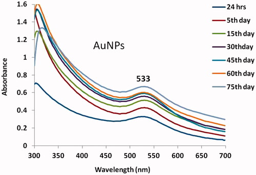
Strychni Semen dried seeds were decayed to make the aqueous extract. Dried seeds of Strychni Semen were weighing 15–20 g are hollowed with double distilled water. Then, it was allowed to dry and compressed into 100 ml disinfected distilled water, then it was filtered by Whatman No.1 filter paper. The gold particles have been equipped by Frens method [Citation21] where the color of the exterior solution for AuNPs was pinkish red and with the UV-vis spectra, the observed maximum colloidal gold was in the range of 533 nm (. Consequently, colloidal nanoparticles were equipped by adding 1 ml of 1% sodium citrate solution to 50 ml of a 0.01% chloroauric acid (boiling condition). The mixture was sustained at boiling point for 10 min and then stimulated for another 10 min after eliminating the heat source. Warmth plays a crucial role in devouring the size allocation of the gold colloids.
Figure 1. UV–visible spectrum absorption pattern of gold nanoparticles synthesised from Strychni semen.
Categorization of AuNPs synthesized from Strychni semen
AuNPs were categorized by various experiments which recognize the size, shape and morphological individuality of nanoparticles. The formed AuNPs was embedded by ultraviolet-visible absorbance spectrum revised at wavelength ranges of 300–700 nm. Morphology and size of AuNPs were recognized and distinguished by high-resolution transmission electron microscopy (HR-TEM) and atomic force microscopy (AFM) images. Firmness of nanoparticles was observed by energy dispersive X-ray (EDX). Presence of active compounds in nanoparticles was studied by FTIR spectroscopy proportions. The FTIR spectra of synthesized nanoparticles were renowned in the arrangement of 1000 and 4000 cm−1 in potassium bromide pellets by FTIR spectrophotometer.
Cell culture
In the present study, cholangiocarcinoma cells (KMCH-1) were used. Human cholangiocarcinoma cell lines (KMCH-1 were purchased from the Cell Bank of Chinese Academy of Sciences (Shanghai, China). Cells were sealed with DMEM medium enhanced with fetal bovine serum (10% FBS) and 1% antibiotics at 37 °C in a humidified incubator at 5% CO2. After 96 h, the culture was vanquished approximately 80% confluent and the cells were harvested with trypsin-EDTA. Further studies are done with the prepared cells.
Cell viability assay
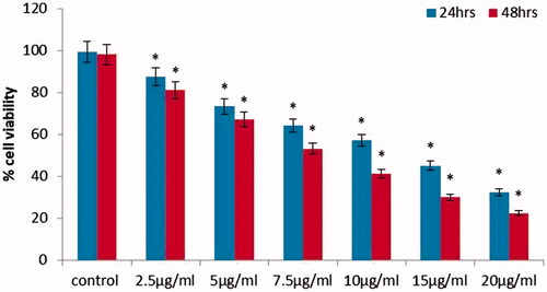
After assembling cholangiocarcinoma cells, nearly 10,000 cells were added in 96-well plates and it was reared for 24 h at 37 °C. Then, combined AuNPs particles from Strychni Semen was indulged with diverse concentrations (20–120 µg/ml) and kept in incubation for 24 and 48 h at 37 °C. After development, the cells were exposed with MTT reagent (1 mg/mL) in all the wells. Finally, culture plates were incubated for 4 h at 37 °C to form purple formazan crystals. About 200 μL of DMSO was supplemented to each well to hang up formazan crystals and then, the optical density was premeditated at 600 nm in microplate reader.
Measurement of intracellular ROS
AuNPs interceded ROS generation in KMCH-1 cells was deliberate by fluorescent probe, 2,7-diacetyl dichlorofluorescein (DCFH-DA) staining. Suddenly, an aliquot of isolated KMCH-1 cells (1 × 10⁶ cells/ml) was equipped and overloaded in 6 well plates. Then, the cells were indulged with 15 and 20 µg/ml absorption of AuNPs at 24 h incubation. DCFH-DA (1 mg/mL) was added to 6 wells and kept in an incubator for 37 °C in a dark room. The fluorescent intensity was calculated by spectrofluorimeter with emission (490 ± 10 nm) and excitation (540 ± 12 nm).
Apoptotic studies for acridine orange/propidium iodide
AuNPs mediated apoptotic changes in KMCH-1 cells were assessed by acridine orange and propidium iodide (AO/PI) staining. In a while, 1 × 10⁶ cells were seeded in 6-well plate and then cultured cells were indulged with 15 and 20 µg/ml concentrations of AuNPs incubation at 24 h. When incubation was over, the cells were sanitized with ice-cold phosphate buffer solution and then discolored with 20 µL of AO/PI staining solution at 37 °C for 20 min. The stained apoptotic and viable cells were scrutinized by a fluorescent microscope.
RT-PCR studies
Apoptotic markers such as Bax, Bcl-2, Bid, caspase-3& 9 were evaluated by RT-PCR studies. mRNA expression of Bax, Bcl-2, Bid, caspase-3&9 genes concerned in apoptosis pathway was examined in KMCH-1 cells. RNA extraction of the cells was done according to RNX-Plus protocol. Eminence of RNA was qualified by agarose gel electrophoresis. Deliberation of extracted RNA was appraised by optical density measurement with NanoDrop 1000 Spectrophotometer (Wilmington, De, USA). Then, the reaction tubes were incubated for 60 min at 45 °C. Real-time PCR was carried out using the SYBR green-based PCR Master Mix thermocycler (QIAGEN Corbett products, Hilden, Germany). Primers for cDNA amplification designed by Eacon Designer software, Boston, MA, USA. The total volume of amplification reactions was 25 µL and each well was included with SYBR Green PCR Master Mix (12 µl) 1 µl of cDNA, 900 nM of both forward and reverse primers. Thermal cycler steps were integrated with 95 °C for 10 min, 45 PCR cycles of 95 °C for denaturation step, annealing temperature 45 °C, and extension carried out at 72 °C, respectively. Lastly, the completion of amplifications done at 72 °C for 10 min.
Statistical analysis
All experiments were agreed out in three self-directed trials and the outcome results were expressed as the mean ± standard deviation (mean ± SD) by means of one-way examination of variance (ANOVA). The p values < .05 were considered significant.
Results
Size, morphology and absorption of synthesized AuNPs
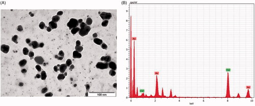
In the current study, we have established that the preparation of gold nanoparticles from Strychni Semen and absorption pattern is illustrated by UV-visible absorbance spectrum analysis in . UV-spectroscopy was used at dissimilar time proposition while the absorption of the Strychni Semen and the gold aqueous solution. The ocular sign of the colours with over and the formation of pinkish red colour precised the development of the AuNPs that express the pattern of gold nanoparticles (). Size, the ultrastructure and morphological uniqueness of AuNPs fashioned from Strychni Semen were additionally précised and premeditated by HR-TEM (high resolution-transmission electron microscope) in . High resolution TEM image exemplifies the morphological array of AuNPs which are scrutinized with different size variations like spherical, hexagonal, oval and triangular with standard sizes between 80 and 10 nm.
Figure 2. HR-Transmission electron microscopy (TEM) & Energy dispersive X-ray analysis (EDX) of gold nanoparticles synthesised from Strychni semen.
Characterization of AuNPs merged from Strychni semen
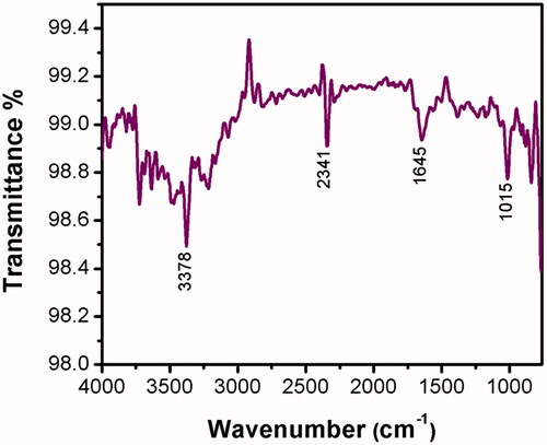
The characterization of AuNPs from Strychni Semen is determined by energy dispersive studies to evaluate the shape of the particles formed from Strychni Semen extract (). Movement, size and force of the synthesized AuNPs from Strychni Semen determined by atomic force microscopy analysis (AFM) (. In addition, FTIR is used to differentiate the numerous functional groups which are present in the synthesized AuNPs from Strychni Semen extract by obtaining different spectrum values. In the present study, FTIR showed values of 3907 cm1, 3745 cm1, 3500 cm1, 3378 cm1, 3210 cm1, 2341 cm1, 1725 cm1, 1645 cm1, 1459 cm1 and 1015 cm1 which recognizes the aromatic, hydroxyl, carboxyl alkyl and amide groups which are nearby to the surface of AuNPs in .
Figure 3. Fourier-transform infrared spectroscopy analysis and SAED pattern of gold nanoparticles synthesised from Strychni semen.
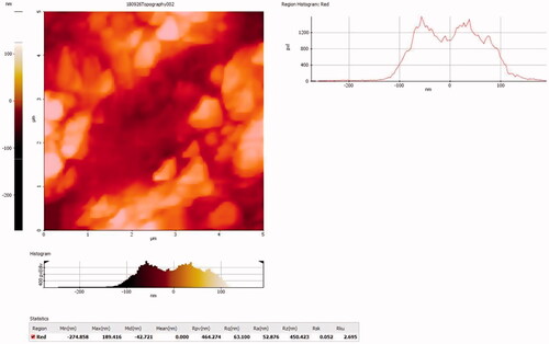
Figure 4. Atomic force microscopyanalysis of gold nanoparticles synthesised from Strychni semen.
Anticancer potential of AuNPs from strychni semen in KMCH-1 cells
AuNPs were knowledgeable for their anticancer proceedings against KMCH-1 cells. Consequently, the anticancer probability of AuNPs was intended by cytotoxicity assay (MTT), in which high AuNPs concentration considerably kept back the KMCH-1 cells expansion in both 24 and 48 h incubation in . Therefore, we establish that 15 µg/ml concentrations of AuNPs exposed outstanding cell death in KMCH-1 cells. Therefore, we have chosen 15 µg/ml concentrations of AuNPs for further apoptotic studies.
Figure 5. Cytotoxicity effect of gold nanoparticles synthesised from Strychni semen in cholangiocarcinoma cell (KMCH-1). The number of viable cells after treatment is expressed as a percentage of the vehicle-only control. This experiment was repeated thrice and the bars in the graph represent S.E. (*p < .05).
Apoptotic studies for acridine orange/propidium iodide
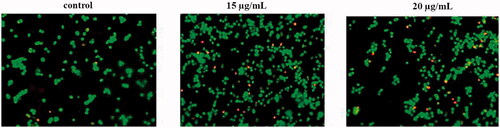
Acridine orange/propidium iodide (AO/PI) double staining is done for cell morphological measurement. Morphological alterations in KMCH-1 cells pretreated with synthesized AuNPs from Strychni Semen for 24 h followed by succeeding revelation to 15 and 20 µg/ml concentrations of AuNPs. This was depicted in untreated cells (control); (B) 15 µg/ml, (C) 20 µg/ml. Feasible cells are blemished green by the presence of acridine orange; necrotic and late apoptotic cells are blemished orange and red by the presence of propidium iodide.
Figure 6. Apoptotic effect of gold nanoparticles synthesised from Strychni semen in KMCH-1 Cells.
ROS, caspase-9 and 3 activity assay
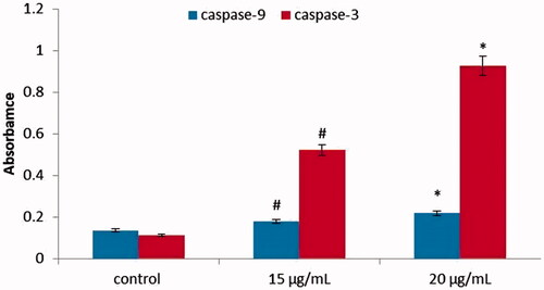
Consequently, AuNPs mediated ROS production that was analyzed by DCFH-DA fluorescent assay. In the present study, we recognized that AuNPs manufactured from Strychni Semen aggravate ROS production in KMCH-1 cells (. Caspase-9 and 3 activities were premeditated by caspase assay kit, as per manufacturer’s direction, the methodology was chased. ELISA technique was used for evaluation of caspase-3 and 9 protein levels. Levels of caspase-3 and 9 were significantly increased in 15 and 20 µg/ml concentrations of AuNPs compared with control cells. There were significant differences in the levels of caspase-3 and caspase-9 is depicted in .
Figure 7. Intracellular production of ROS levels in KMCH-1 cells. This experiment was repeated thrice and the bars in the graph represent S.E. (*p < .05).
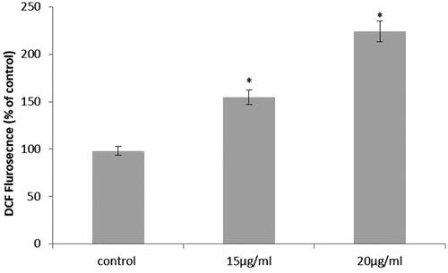
Figure 8. Caspase-3, Caspase-9 activity in KMCH-1 cells. This experiment was repeated thrice and the bars in the graph represent S.E. (*p < .05, #p < .01).
Real-time quantitative RT-PCR
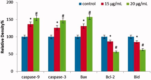
AuNPs intruded apoptosis markers such as Bax, Bcl-2, Bid, caspase-3&9 were analyzed by RT-PCR. illustrates treatment with AuNPs produced from Strychni Semen with the concentration of 15 and 20 µg/ml and diminished the expression of Bcl-2 and Bid at the same time elevated the expression of caspase -3 and 9 in KMCH-1 cells. Finally, with these findings, AuNPs produced Strychni Semen efficiently, persuaded apoptosis by regulating the apoptotic pathway.
Figure 9. Anticancer effect of gold nanoparticles synthesised from Strychni semen on apoptotic gene expression in KMCH-1 Cells. This experiment was repeated thrice and the bars in the graph represent S.E. (*p < .05, #p < .01).
Discussion
Even though there has been enormous innovation in indicative technique, surgical treatment and the development of new anti-cancer agent, the upshot of patients with bile duct cancer has improved discreetly in the past. In contrast to other metals, gold is environmentally friendly and low cost. Production of nanoparticles from plant extracts is an unsophisticated practice, in which metal salt is departed with plant extract and finishing point of reaction takes place within a short period. Specifically, gold nanoparticles (AuNPs) are non-toxic when compared to other metallic nanoparticles [Citation22,Citation23]. Nanoparticles separation in plants can be flattering over all other organic technique by eradicating the complicated process of preserving cell cultures [Citation24]. In the present study, we recognized the Strychni Semen arbitrates the produced AuNPs by UV-visible absorbance. A mounting reflection of Strychni Semen seed extracts direct towards the improved intensity of absorption.
The UV-visible spectra distinguished with unrelated time intervals such as 24 h, 5th day, 15th day and 30th day from the beginning of the reaction with various amounts of seed extracts. Present study exemplify the fashioned AuNPs reveal highest absorbance was found to be in the ranges of 533 nm at 60th day. This result matches with the prior studies in which UV-Vis spectroscopy tattered to examine the shape and size of the constrained nanomaterials in the aqueous suspensions [Citation25]. This connected data were previously reputable by a number of investigations on a combination of gold nanoparticles [Citation26]. When compared with the other metal nanoparticles, mainly AuNPs, enfold free electrons which are positioned in SPR (surface plasma resonance) absorption bands due to the united trembling of electrons of metal nanoparticles in consequence with lightwave [Citation27]. In the current energy dispersive x-ray analysis study, tough and strong signal were recognized in the gold section which validates the AuNPs pattern. In the present study, we obtained the powerful signals of Au (i.e. 1.5–2.5 and 2.5– 3.5 keV), in which previous surface resonance findings were very near to our present results with the unique optical absorption for metallic crystal typically found to be 3 KeV [Citation28].
Gold nanoparticles normally display typically strapping ocular assimilation peak was experienced due to surface plasmon resonance [Citation29]. The reinforcement of importance in a transmission electron microscope (TEM) pursue incessant modifications in examining latest diseases or supplementary features of aged diseases, new remedial drugs and their potential toxic effects, side effects upsetting the liver. In our findings, the development of gold nanostructures definite in TEM figures, in which morphological individuality provides high-density AuNPs shaped by the Strychni Semen extract. Previous studies confirm the size and shape of gold nanoparticles performed by TEM studies in Hep-2 cells, where the TEM examination of all gold nanoparticles was reliable with the ascending like 3, 11, 27, and 51 nm [Citation30]. Many studies support our data like the TEM investigated the phase and contrivance of the cellular incorporations of protein-covered gold nanoparticles by means of the HeLa cell lines [Citation31].
FTIR is used to distinguish the various functional groups which are embedded in the synthesized AuNPs from Strychni Semen extract. Present finding acquaintances with previous studies in which fabrication of AuNPs from various plants extracts have a number of functional groups [Citation32]. Prior FTIR findings chains our data in which gold nanoparticles propose that the biological molecules like enzymes and proteins might perhaps implement the utility for the arrangement and consistency of the AuNPs using the Enterococcus species [Citation33]. Atomic force microscopy images (AFM) corroborate the size of the formed nanoparticles. In (), précised AFM images have enough pampered control through the progress of the AuNPs which matched with the previous findings [Citation34]. A further AFM prior study shows a large number of AuNPs at the cell membrane after incubation of 1 h, in contrast where after 2 h, AuNPs are not noticeable [Citation30]. Intracellular reactive oxygen species (ROS) in a system association influence oxidative stress which results in apoptosis [Citation35]. In addition, prominent ROS directs to the disintegration of nucleus and mitochondria depolarization which effects in oxidative stress intervened apoptosis [Citation36,Citation37].
The propidium iodide/acridine orange stain (PI/AO stain) is a feasibility stain that notices the apoptotic cells. Apoptotic swots were executed with a staining method employing AO and PI according to the previous studies [Citation38]. This was further correlated with prior studies in which vanillin, a flavonoid compound induces apoptosis and cell cycle detains in human colorectal cancer cell line HT-29 [Citation39]. Caspases engage in recreation of crucial role in the extrinsic pathway of apoptosis via commencement of terminal caspases which were in effect [Citation40,Citation41]. RT-PCR investigation in cultured HA22 T, HepG2, and Huh7 human liver cancer cells in which the appearance of estrogen receptor and neurofibromin NF2 gene was assessed [Citation42]. Development and sequence of tumor and alteration to numerous oncologic remedial execution consequences are regularly scarce to apoptotic stimuli. Apoptosis is an essential watchdog in tissue preservation and maintenance [Citation43]. Consequently, apoptosis is a scrupulous and important passageway for cancer treatment. Previous studies support our current findings in which gold nanoparticles induce apoptosis in MCF-7 (human breast cancer cells) [Citation44].
Conclusion
Finally, we demonstrate a simple and rapid technique with reproducibility for the environment-friendly fabrication of AuNPs, which lacks elegant reducing agents. The effectiveness of AuNPs produced from Strychni Semen was well established. The physiochemical distinctiveness of manufactured AuNPs from Strychni Semen is documented by UV-absorbance, EDX, FTIR, AFM and HR-TEM. AuNPs successfully convince cytotoxicity and apoptosis by inflecting intrinsic apoptotic gene expressions in KMCH-1 cells. So, finally, our study confirms the synthesized AuNPs from Strychni Semen, which displays anticancer effects.
Acknowledgment
This work was supported by the grants from the National Natural Science Foundation of China [grant numbers 81660399, 81860423]; the Innovative Research Team Project of Yunnan Province [grant number 2015HC033]; the Yunnan Provincial Academician Workstation of Xiaoping Chen [grant number 2017IC018]; the Breeding Program for Major Scientific and Technological Achievements of Kunming Medical University [grant number CGYP201607]; the Medical Leading Talent Project of Yunnan Province [grant number L201622]; and Yunnan Provincial Clinical Center of Hepato-biliary-pancreatic Diseases to L. W.
Disclosure statement
All the authors declared that there are no conflicts of interest.
Related Research Data
References
- NIH Roadmap Initiatives. Available from: http://nihroadmap.nih.gov/initiatives.asp.
- Sepeur S. Nanotechnology: technical basics and applications. Hannover: Vincentz; 2008.
- Jin G, Zhao X, Xu F. Therapeutic nanomaterials for cancer therapy and tissue regeneration. Drug Discov Today. 2017;22:1285–1287.
- Sharma HS, Ali SF, Hussain SM, et al. Influence of engineered nanoparticles from metals on the blood-brain barrier permeability, cerebral blood flow, brain edema and neurotoxicity. An experimental study in the rat and mice using biochemical and morphological approaches. J Nanosci Nanotechnol. 2009;9:5055–5072.
- Kayalvizhi T, Ravikumar S, Venkatachalam P. Green synthesis of metallic silver nanoparticles using Curculigo orchioides rhizome extracts and evaluation of its antibacterial, larvicidal, and anticancer activity. J Environ Eng. 2016;142:C4016002.
- Venkatachalam P, Kayalvizhi T, Udayabanu J, et al. Enhanced antibacterial and cytotoxic activity of phytochemical loaded-silver nanoparticles using Curculigo orchioides leaf extracts with different extraction techniques. J Clust Sci. 2017;28:607–619.
- Prasannaraj G, Venkatachalam P. Enhanced antibacterial, anti-biofilm and antioxidant (ROS) activities of biomolecules engineered silver nanoparticles against clinically isolated gram positive and gram negative microbial pathogens. J Clust Sci. 2017;28:645–664.
- Astruc D, Daniel MC, Ruiz J. Dendrimers and gold nanoparticles as exo-receptors sensing biologically important anions. Chem Commun. 2004;23:2637–2649.
- Tripathi RM, Shrivastav A, Shrivastav BR. Biogenic gold nanoparticles: as a potential candidate for brain tumor directed drug delivery. Artif Cell Nanomed B. 2014;43:1–7.
- Pattnaik P. Surface plasmon resonance: applications in understanding receptor-ligand interaction. Appl Biochem Biotechnol. 2005;126:79–92.
- Patel T. Cholangiocarcinoma-controversies and challenges. Nat Rev Gastroenterol Hepatol. 2011;8:189–200.
- Khan SA, Emadossadaty S, Ladep NG, et al. Rising trends in cholangiocarcinoma: is the ICD classification system misleading us? J Hepatol. 2012;56:848–854.
- El-Serag HB, Tyson GL. Risk factors for cholangiocarcinoma. Hepatology. 2011;54:173–184.
- Mehta RG, Murillo G, Naithani R, et al. Cancer chemoprevention by natural products: how far have we come? Pharm Res. 2010;27:950–961.
- Lee SM, Kwon JI, Choi YH, et al. Induction of G2/M arrest and apoptosis by water extract of Strychni Semen in human gastric carcinoma AGS cells. Phytother Res. 2008;22:752–758.
- Bisset NG, Datta De B. Alkaloids of Strychnos ignatii. Planta Med. 1990;56:133.
- Yuan LM, Zi M, Ai P, et al. Versatile two-phase solvent system for alkaloid separation by high-speed counter-current chromatography. J Chromatogr A. 2001;927:91–96.
- Shi Y, Zhang H, Du X. A double blind observation for therapeutic effects of the tong luo kai bi tablets on rheumatoid arthritis. J Tradit Chin Med. 1999;19:166–172
- Tsui S, Wong S, Kwan S. Analysis of proprietary Chinese medicines for the presence of toxic ingredients by LC/MS/MS. J Pharm Biomed Anal. 2002;30:161–170.
- Deng XK, Yin W, Li WD. The anti-tumor effects of alkaloids from the seeds of Strychnos nux-vomica on HepG2 cells and its possible mechanism. J Ethnopharmacol. 2006;106:179–186.
- Basu S, Ghosh SK, Kundu S, et al. Biomolecule induced nanoparticle aggregation: effect of particle size on interparticle coupling. J Colloid Interface Sci. 2007;313:724.
- Rai M, Yadav A, Gade A. Current [Corrected] trends in phytosynthesis of metal nanoparticles. Crit Rev Biotechnol. 2008;28:277–284.
- Mittal AK, Chisti Y, Banerjee UC. Synthesis of metallic nanoparticles using plant extracts. Biotechnol Adv. 2013;31: 346–356.
- Kumar V, Yadav SK. Plant‐mediated synthesis of silver and gold nanoparticles and their applications. Journal of Chemical Technology & Biotechnology: International Research in Process, Environmental & Clean Technology. 2009 Feb;84(2):151-7.
- Zhang XF, Liu ZG, Shen W, et al. Silver nanoparticles: synthesis, characterization, properties, applications, and therapeutic approaches. IJMS. 2016;17:1534.
- Aljabali A, Akkam Y, Al Zoubi M, et al. Synthesis of gold nanoparticles using leaf extract of Ziziphus zizyphus and their antimicrobial activity. Nanomaterials. 2018;8:174.
- Eustis S, El-Sayed MA. Why gold nanoparticles are more precious than pretty gold: noble metal surface plasmon resonance and its enhancement of the radiative and nonradiative properties of nanocrystals of different shapes. Chem Soc Rev. 2006;35:209–217.
- Kaviya S, Santhanalakshmi J, Viswanathan B, et al. Biosynthesis of silver nanoparticles using citrus sinensis peel extract and its antibacterial activity. Spectrochim Acta A Mol Biomol Spectrosc. 2011;79:594–598.
- Magudapathy P, Gangopadhyay P, Panigrahi BK, et al. Electrical transport studies of Ag nanoclusters embedded in glass matrix. Physica B. 2001;299:142.
- BoyogluHe C, He Q, Willing G, et al. Microscopic studies of various sizes of gold nanoparticles and their cellular localizations. ISRN Nanotechnol. 2013;2013:13.
- Chithrani BD, Chan WC. Elucidating the mechanism of cellular uptake and removal of protein-coated gold nanoparticles of different sizes and shapes. Nano Lett. 2007;7:1542–1550.
- Kuppusamy P, Yusoff MM, Maniam GP, Govindan N. Biosynthesis of metallic nanoparticles using plant derivatives and their new avenues in pharmacological applications – an updated report. Saudi Pharm J. 2016;24:473–484.
- Fayaz MA, Girilal M, Venkatesan R, et al. Biosynthesis of anisotropic gold nanoparticles using Maduca longifolia extract and their potential in infrared absorption. Colloids Surf B Biointerfaces. 2011;88:287–291.
- Darwich S, Mougin K, Rao A, et al. Manipulation of gold colloidal nanoparticles with atomic force microscopy in dynamic mode: influence of particle-substrate chemistry and morphology, and of operating conditions. Beilstein J Nanotechnol. 2011;2:85–98.
- Ivanova D, Zhelev Z, Aoki I, Bakalova R, Higashi T. Overproduction of reactive oxygen species – obligatory or not for induction of apoptosis by anticancer drugs. Chin J Cancer Res. 2016;28:383–396.
- Guo C, Sun L, Chen X, et al. Oxidative stress, mitochondrial damage and neurodegenerative diseases. Neural Regen Res. 2013;8:2003–2014.
- Zorov DB, Juhaszova M, Sollott SJ. Mitochondrial reactive oxygen species (ROS) and ROS-induced ROS release. Physiol Rev. 2014;94:909–950.
- Lakshmi S, Dhanya GS, Joy B, et al. Inhibitory effect of an extract of Curcuma zedoariae on human cervical carcinoma cells. Med Chem Res. 2008;17:335–344.
- Ho KLi, Yazan LS, Ismail N, et al. Apoptosis and cell cycle arrest of human colorectal cancer cell line HT-29 induced by vanillin. Cancer Epidemiol. 2009;33:155–160.
- Wachmann K, Pop C, van Raam BJ, et al. Activation and specificity of human caspase-10. Biochemistry. 2010;49:8307–8315.
- Schultz DR, Harringto WJ. Apoptosis: programmed cell death at a molecular level. Semin Arthritis Rheum. 2003;32:345–369.
- Letizia C, Vitale M, Orazia M G, et al. Merlin, the product of NF2 gene, is associated with aromatase expression and estrogen formation in human liver tissues and liver cancer cells. J Steroid Biochem Mol Biol. 2017;23:222–230.
- Igney FH, Krammer PH. Death and anti-death: tumour resistance to apoptosis. Nat Rev Cancer. 2002;2:277–288.
- Shyur LF, Chen CH, Lo CP, et al. Induction of apoptosis in MCF-7 human breast cancer cells by phytochemicals from Anoectochilus formosanus. J Biomed Sci. 2004;11:928–939.