
 ?Mathematical formulae have been encoded as MathML and are displayed in this HTML version using MathJax in order to improve their display. Uncheck the box to turn MathJax off. This feature requires Javascript. Click on a formula to zoom.
Abstract
Nanotechnology has been materialized as a proficient technology for the development of anticancer nanoparticles all the way through an environment-friendly approach. Conventionally, nanoparticles have been assembled by dissimilar methods, but regrettably rely on the negative impact on the natural environment. Amalgamation of nanoparticles by means of plant extract is alternate conservative methods. Scutellaria barbata species was used majorly as food or as medicines against various diseases, and extensive research was conducted for their therapeutic properties. The present research was mainly focused on the synthesis of gold nanoparticles from the Scutellaria barbata by green route method and evaluation of its anticancer activity against pancreatic cancer cell lines (PANC-1). The gold nanoparticles have been characterized by UV–visible spectroscopy, TEM, SAED, AFM, and FTIR analysis. The synthesized gold nanoparticles (AuNPs) possessed effective anticancer activity against pancreatic cancer cell lines (PANC-1). Hence, further research on this plant may lead to the development of novel anticancer drugs which can be used to combat pancreatic cancer.
Introduction
Nanotechnology is one of the important thriving and rapidly developing field of science and technology. In history, people of the 16th century are known to use splendid gold coated materials for many purposes including the medical field. It was confirmed that they used materials coated with gold particles for oral medicine, pharmaceuticals and tissue and organs implantation [Citation1]. Plant parts contain several bioactive constituents such as flavonoids, alkaloids, terpenoids, phenolics, amino acids and steroids which act as reducing agents for the formation of nanoparticles. In the field of nanotechnology, the plant-mediated synthesis (green route method) of nanoparticles is an emerging area due to the active plant constituents which act as a capping and reducing agent [Citation2].
Green synthesis by plant extracts can be comparable to some conventional methods, however, they did not give details about nanoparticle yields, stability, and ions reduction mechanism which are not considered environmentally friendly. To overcome this problem, researchers began to use more environmentally benign methods that are collectively called green synthesis [Citation3]. Plant extracts have shown to be effective in the reduction of Au ions to Au NPs. gold nanoparticles play a vital role in nanotechnology as biomedicine because of their convenient surface bioconjugation with biomolecular probes and remarkable plasmon-resonant optical parameters [Citation4].
Green synthesis of AuNPs using plant extract was reported in its range of biological applications such as antibacterial [Citation5], antiparasitic[Citation6], antioxidant [Citation7] and anticancer activities [Citation8,Citation9]. The synthesis of the metal nanoparticle using plant extracts is known as enamors extent because of the comparatively easier synthesis for the control of monodispersed nanoparticles because of its non-toxic, eco-friendly, and biocompatibility characters [Citation10]. Recently, Divakaran et al. (2019) [Citation11] reported that AuNPs are more significant in the research field owing to their non-toxic effects; self-assembled natural and improved drug delivery.
Cancer is one of the major diseases responsible for the majority of human deaths worldwide. Globally, there has been a constant fight against cancer with a lot of progressions in the treatment and preventive methods, however, the expected successful treatment rate is yet to be achieved. Cancer is distinguished as cells which constantly divide and multiply rapidly, with the inability to stop or control the progression of cell multiplication. The continually developing malignant or cancer cells have the potential to be metastatic [Citation12].
Pancreatic cancer is a type of cancer with the successful and lifesaving treatment limited by the belligerent neoplasm. There have been many common chemotherapy and/or radiotherapy employed for the treatment of pancreatic cancer. However, studies show that pancreatic cancer cells are highly resistant phenotypes. Annually, the occurrence of pancreatic cancer is constantly increasing worldwide. Pancreatic cancer has become the fourth main common reason for cancer-related deaths globally. Statistics have it that the new pancreatic cancer patient rate in 2008 was 277,000 and it increased to 338,000 in 2012. The death rate due to pancreatic cancer was recorded as 266,000 in 2008 and it increased to 331,000 in 2012. It is estimated that this could increase by 3-fold from 2018 to 2019 [Citation13].
Most of the patients with pancreatic cancer are diagnosed at the very late stage and this reduces the chances of exploring available treatment options. In the last six years, medical records showed that the relative survival rate of pancreatic cancer patients was relatively low and it has been estimated that the median survival rate of pancreatic cancer patients will be approximately 6 months, even with intensive medical care. The desire to attain a novel approach to combat cancer led to the improvement of preventive and effective treatment strategies; however, many research efforts have focused on better understanding of the mechanisms (in molecular level) underlying the progression of pancreatic cancer in humans [Citation14,Citation15].
The present treatment options include chemotherapy, radiotherapy, and drugs derived from chemicals which potentially cause severe side effects. The use of chemotherapy and radiotherapy place the patient under a lot of pain, strain, and additional deterioration of the patient’s health. Hence, researches have been ongoing to seek new alternative treatment and therapeutic methods against pancreatic cancer [Citation16].
Scutellaria barbata is one of the important species of the Scutellaria genus. It is commonly called “Ban Zhi Lian” in Chinese. Historically, this species was used widespread as food or medicines for the treatment of various diseases and it was extensively researched for its therapeutic properties. In the traditional Chinese system of medicine, this plant is widely used to treat various human ailments due to its high medicinal value. Hence, its name “the miracle root for the preservation of life”. Scutellaria barbata has a potent antitumor activity in breast cancer [Citation17], colorectal cancer [Citation18], hepatocarcinoma [Citation19], skin cancer [Citation20], lung cancer [Citation21], and ovarian cancer [Citation22]. Moreover, Lee et al., (2017) [Citation23] reported that the active fractions of Scutellaria barbata have anti-inflammatory activity in BV-2 cells. The present research study was mainly focused on the synthesis of gold nanoparticles from the Scutellaria barbata by green route method and evaluation of its anticancer activity against pancreatic cancer cell lines (PANC-1).
Materials and methods
Chemicals and antibodies
Dulbecco’s modified eagle’s medium (DMEM)/nutrient mixture F-12 Ham, bovine serum albumin (BSA), dimethyl sulfoxide (DMSO), antibiotic/antimycotic (100X) solution (penicillin/streptomycin/amphotericin), ampicillin, trypsin-EDTA (0.25%) and fetal bovine serum (FBS) were purchased from GIBCO (Carlsbad, CA, USA). Protein estimation kit, acrylamide, APS, TEMED, and pre-stained sodium dodecyl sulfate-polyacrylamide gel electrophoresis (SDS-PAGE) standard were from Bio-Rad (Irvine, CA, USA) and enhanced chemiluminescence (ECL) kit was procured from Thermo Scientific (Madison, WI, USA). Protease inhibitor cocktail was purchased from Pierce Biotechnology Inc. (Rockford, IL, USA). Polyvinylidene difluoride (PVDF) membrane was procured from Millipore (Millipore, USA). All primary antibodies used in this study were purchased from Cell Signalling Technologies (Beverly, MA, USA).
Preparation of extract
The healthy and matured plant of Scutellaria barbata (Lamiaceae family) were collected, shade dried, and finely powdered plant was weighed and mixed with 100 ml of deionized water. This suspension was allowed to stay for 10 min with occasional shaking, followed by boiling at 90˚C for 40 min to obtain the major bioactive constituents. After boiling, the suspension was allowed to cool to room temperature and filtered using a Whatman No.1 filter paper. After this process, the resulted crude aqueous extract was used for the synthesis of the gold nanoparticles.
Green synthesis of gold nanoparticles
About 10 ml of aqueous extract of the plant was added to 50 ml of 0.1 M of an aqueous solution of chloroauric acid (HAuCl4). Then, this reaction mixture was constantly stirred in a magnetic stirrer for the reduction of gold ions. After 15–20 min, the reaction mixture turned from brown to reddish yellow and the suspension was constantly stirred for 3 h. The suspension was allowed to incubate in a cool and dark place. Following this stage, the suspension was centrifuged at 10,000 rpm for 15 min to obtain the synthesized gold nanoparticles. Then, the pelleted gold nanoparticles were used for further characterization and analytical purposes.
Characterization of synthesized gold nanoparticles
UV-Visible spectroscopy analysis
The analysis through UV-Visible spectroscopy is one of the widely used techniques for the characterization of gold nanoparticles which are synthesized biologically. It is necessary to measure the optical density by using the UV-Visible spectroscopy in order to confirm the formation of gold nanoparticles. The reduction of gold (Au) ions into gold nanoparticles in the reaction medium were determined by the measurement of the spectrum of UV-visible spectroscopy at the 540 nm range.
Size distribution analysis of synthesized gold nanoparticles
The average size of the synthesized gold nanoparticles was measured by using Dynamic Light Scattering Analysis. It is a method of multiple light scattering and laser diffraction techniques. The synthesized gold nanoparticles from the aqueous extract of Scutellaria barbata were dissolved in deionized water followed by ultra sonification. The prepared suspension was centrifuged at 7000 rpm for 25 min at 26˚C. After the centrifugation process, the supernatant was separated and used for the analysis. The separated supernatant was discrete for six more times to carry outsize distribution analysis. Then, the average size of the synthesized gold nanoparticles in the liquid suspension was analyzed using the computer-aided nanoparticles size analyzer.
TEM analysis of synthesized gold nanoparticles
Transmission electron microscopic (TEM) analysis of synthesized gold nanoparticles from the Scutellaria barbata was done to detect the average size and shape of the gold nanoparticles. For this purpose, synthesized gold nanoparticles were collected in a copper grid coated with carbon and then it was preserved overnight under vacuum desiccators to ensure proper drying. After overnight incubation, the carbon coated copper grid was supplemented with the synthesized gold nanoparticles and placed on a specimen holder. The micrograph of the synthesized gold nanoparticles was taken by using JOEL JSM 100CX TEM apparatus operating at an accelerating voltage of 200 kV.
FT-IR analysis of synthesized gold nanoparticles
Fourier Transform Infrared Spectroscopy (FT-IR) analysis was done to identify the specific biomolecules mainly present on the surface of the gold nanoparticles. About 100 ml suspension of synthesized gold nanoparticles were centrifuged at 10,000 rpm for 20 min at room temperature. Then, the solution was dissolved in 10 ml of sterile deionized water. The purified solution was freeze-dried to obtain the dried powder. Finally, the dried gold nanoparticles were analyzed using FT-IR spectrometry.
Atomic force microscope (AFM) analysis of synthesized gold nanoparticles
The synthesized gold nanoparticles were diluted with deionized water (0.05 mg per ml) and then spread over zinc (Zn) substrate for analysis using AFM. The images of the synthesized gold nanoparticles were obtained at a set of 10 nm using a scan rate of 1 μm per second.
Cytotoxicity assay
The MTT cytotoxicity assay was performed according to the previously described method to assess the survival of the pancreatic cancer cells by assessing the mitochondrial activity inside the cells before and after the treatment with synthesized gold nanoparticles. To test the cytotoxicity of the synthesized gold nanoparticles, the PANC-1 cell lines (purchased from the Institute of Biochemistry and Cell Biology, Chinese Academy of Sciences, Shanghai, China) were treated with different concentrations of gold nanoparticles. The gold nanoparticles treated PANC-1 cell lines were placed in DMEM medium and incubated for 24 h. After incubation, the cell lines were taken in the ELISA plate reader. 10 μl MTT (3–(4, 5-dimethyl-thiazol-2-yl)-2, 5-diphenyl tetrazolium bromide) dye was added in each well and set aside for 2 h. The MTT dye reacted with the oxidoreductase enzyme present in the mitochondria and then converted it into crystals of formazan. This process occurred only in viable cells. Further, the formazan crystals were dissolved with dimethyl sulfoxide (DMSO) and then its absorbance was measured using a plate reader at 570 nm. Finally, the percentage of viability of PANC-1 cell lines treated with the synthesized gold nanoparticles was calculated.
Acridine orange/propidium iodide (Ao/Pi) staining
To assess the actively proliferating cells, acridine orange and propidium iodide staining were performed. For this, the PANC-1 cell lines were cultured on their growth at the density of 5 × 10³ cells per well. After growth, the medium was discarded and the obtained cells were washed with phosphate buffer. Then, the cells were trypsinized and then placed on the surface of a clean glass slide. The cells were then stained using acridine orange and propidium iodide; the stained cells were observed under a fluorescence microscope (Carl Zeiss, Jena, Germany).
Estimation of reactive oxygen species (ROS) level
The generation of ROS was quantitatively estimated by the H2DCFDA staining method. For this, the PANC-1 cell lines treated with synthesized gold nanoparticles were fixed with 70% ice-cold methanol. Then, incubation was done using 10 μl of H2DCFDA for 30 min at room temperature. The fluorescence intensity of the cells was measured with the excitation wavelength of 480 nm and the emission wavelength of 530 nm by using a flow cytometer. The obtained data were analyzed using cyflogic software.
Analysis of apoptotic protein expression by Western blotting
To assess the apoptotic protein expressions, the PANC-1 cell lines were cultured in DMEM medium. Then, the cultured cell lines were washed with a phosphate solution and then separated from the culture plates by treating with trypsin/EDTA solution. The obtained cell suspension was centrifuged at 2000 rpm for 10 min. After centrifugation, the pellets were lysed with ice-cold protease inhibitor-containing RIPA buffer. Then, the cell lysate was cleared by centrifugation at 13,000 rpm for 15 min at 4 °C. Finally, the resulted supernatant was used to estimate the protein concentration by using the Lowry protein estimation method (Lowry, 1951).
Quantitative analysis of apoptotic gene expressions by qRT-PCR
For this, the total RNA was extracted from the PANC-1 cell lines by using the RNeasy mini test kit (Qiagen, Hilden, Germany) according to the manufacturer’s instructions. The expressions of mRNA of Caspase3, Caspase9, Bax, Bcl-2, and Bid genes present in the PANC-1 cells were measured by using the quantitative Real-Time PCR (qRT-PCR). The RNA purification and the quantity were analyzed spectromatically by measuring the optical density at 260 nm. The primer pairs supplied with the RNeasy Test Kit was used to measure the expression of mRNA. For the synthesis of cDNA or complementary DNA, the cyclic conditions of PCR were used as 10 min at 25˚C, 50 min at 42˚C, and 15 min at 75˚C, respectively. The PCR cyclic conditions used for the amplification of cDNA were 2 s at 95˚C, 15 s for 55˚C, and 20 s for 68˚C, respectively as mentioned and recommended by the manufacturer. The level of expression of genes was normalized to the level of expression of the mRNA GADPH on each sample. Gene expression levels were calculated using the cyclic threshold method (Ct method). The mean of the Ct values from the triplicate was used to calculate the expression level of the target gene by using the 2-ΔΔCt formula
Results
Synthesis of gold nanoparticles from Scutellaria barbata
The gold nanoparticles were synthesized from the aqueous extract of Scutellaria barbata by using green route method. The formation of gold nanoparticles in the reaction medium was evidenced by observing the color change before and after incubation. The reaction mixture containing the aqueous extract of Scutellaria barbata and an aqueous solution of 0.1 M chloroauric acid changed color from brown to reddish yellow. This reaction demonstrates the formation of gold nanoparticles (AuNPs) in the reaction medium.
UV-visible spectroscopic analysis of synthesized gold nanoparticles
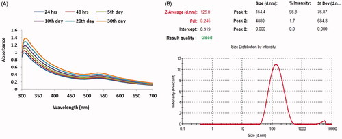
The aqueous solution of gold nanoparticles (AuNPs) synthesized from the aqueous extract of Scutellaria barbata may contain complimentary electrons. These electrons may provide the absorption band of the plasma resonance due to the combined vibration of metallic gold nanoparticles (AuNPs). The gold nanoparticles synthesized from the aqueous extract of Scutellaria barbata exhibited the spectrum of surface plasma resonance at the range of 525 nm (). The surface plasma resonance of the synthesized gold nanoparticles (AuNPs) at the increasing concentration was observed and also color changes were observed in the synthesized gold nanoparticles.
Figure 1. UV–visible spectrum absorption pattern and dynamic light scattering of gold nanoparticles synthesised from Scutellaria barbata. (A). UV-visible absorption spectrum of synthesized AuNPs. (B). Dynamic light scattering (DLS) images of AuNPs
Size distribution analysis of the synthesized gold nanoparticles
The characteristic structure of the gold nanoparticles synthesized from the aqueous extract of Scutellaria barbata was determined using the method of dynamic light scattering (DLS). The observed histogram of the dynamic light scattering clearly demonstrates that the synthesized gold nanoparticles may have an average size range of 154 nm ().
Transmission electron microscopic analysis of the synthesized gold nanoparticles
TEM analysis was carried out to determine the average size and shape of the synthesized gold nanoparticles from the aqueous extract of Scutellaria barbata by green synthesis method. The TEM photographs revealed the presence of the gold nanoparticles as well as the presence of the aggregates of the nanoparticles. The images of the TEM analysis showed that the gold nanoparticles (AuNPs) possibly possessed spherical shape and the typical size of the nanoparticles may be in the range of 0.4 μm to 1 μm ().
Figure 2. HR-transmission electron microscopy (HR-TEM) and energy dispersive X-ray analysis (EDX) of gold nanoparticles synthesised from Scutellaria barbata. (A) Transmission electron microscopy (HR-TEM) and (B) energy dispersive X-ray (EDX) and analysis AuNPs synthesised from Scutellaria barbata.

Energy dispersive X-ray crystallographic analysis of synthesized gold nanoparticles
The synthesized gold nanoparticles from the aqueous extract of the Scutellaria barbata was analyzed through the energy dispersive X-ray crystallography for the quantitative estimation of the major elements present in the reaction medium which may be responsible for the formation of the gold nanoparticles (AuNPs) from the gold (Au) ions. The EDX elemental analysis clearly demonstrated the presence of the major portion of gold (Au) followed by copper (Cu) at the 3 keV (). This result confirms the presence of the gold nanoparticles in the reaction medium.
Fourier transform – infrared (FT-IR) analysis of synthesized gold nanoparticles
Fourier transform infrared analysis (FT-IR) was used to determine the profile of the biomolecules specifically found on the surface of the synthesized gold nanoparticles (AuNPs). The spectrum of the FT-IR analysis demonstrated the presence of carboxylic acid group, hydroxyl group, alkanes group, and carbonyl group of the exterior of the synthesized gold nanoparticles. The peak regions of 3313 cm−1 demonstrated the strong band of oxygen and hydrogen (O–H) stretches of carboxylic bands. The peak region of 2161 cm−1 showed the presence of stretching bands of the carbonyl group. The peak region of 1635 cm−1 may be due to the presence of alkane group on the exterior of the synthesized gold nanoparticles ().
Figure 3. Fourier transform infrared (FTIR) spectroscopy analysis and SAED pattern of AuNPs synthesized from Scutellaria barbata.
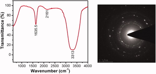
Analysis of selected area electron diffraction (SAED) pattern of synthesized gold nanoparticles
The obtained results from the analysis of selected area electron diffraction (SAED) demonstrated the presence of concentric diffraction rings with a 480 mm optical distance (. The result clearly showed that the synthesized gold nanoparticles possessed polycrystalline nature. The lighter and darker regions observed in the high-resolution transmission electron microscope indicated the different planes and symmetry of the synthesized gold nanoparticles. The SAED analysis revealed the polyhedral morphology of the synthesized gold nanoparticles.
Atomic force microscopic analysis of the synthesized gold nanoparticles
Atomic force microscope analysis was done to detect the presence and distribution of the synthesized gold nanoparticles (AuNPs). In the tapping mode, the scanning area of 1 × 1 of the nanoparticles was taken (). The AFM images confirmed the presence of gold nanoparticles (AuNPs) which were uniformly distributed. The majority of the gold nanoparticles were approximately 30–40 nm in diameter with a spherical topology ().
Figure 4. Atomic force microscopy analysis of gold nanoparticles synthesised from Scutellaria barbata.
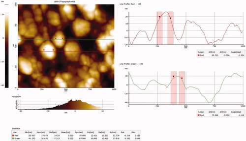
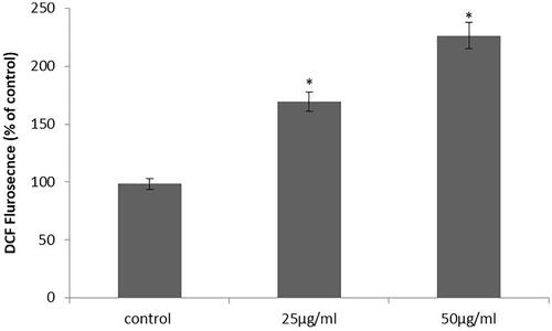
Figure 5. Effect of synthesized gold nanoparticles on intracellular ROS generation. This experiment was repeated thrice and the bars in the graph represent S.E. (**p < .05, ***p < .01).
MTT cytotoxic activity of synthesized gold nanoparticles from Scutellaria barbata
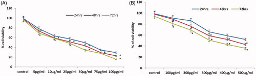
Cytotoxic potential of the synthesized gold nanoparticles against the pancreatic cancer cell lines (PANC-1) was evaluated by using MTT cytotoxicity assay. The synthesized gold nanoparticles showed a significant (p < .001) cytotoxic activity against the pancreatic cancer cell lines. When the cells were treated with synthesized gold nanoparticles, the viability of the cells was constantly decreased in time and dose-dependent manner. The cytotoxicity level of the synthesized gold nanoparticles against the pancreatic cancer cell lines (PANC-1) at the different dose and time interval was carefully noted and the result is presented in .
Figure 6. Cytotoxic activity of the synthesized gold nanoparticles and Scutellaria barbata extract in 24, 48 and 72 h. (A) MTT assay of gold nanoparticles. (B) MTT assay of Scutellaria barbata extract. This experiment was repeated thrice and the bars in the graph represent S.E. (**p < .05, ***p < .01).
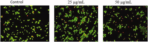
Staining of cancer cell morphology by acridine orange/propidium iodide (Ao/Pi)
The fluorescent staining method was carried out to assess the cytotoxic potential and the mode of cell death provoked by the synthesized gold nanoparticles from the Scutellaria barbata at the dose of 25 μg/ml and 50 μg/ml. Acridine orange and propidium iodide stain was used to carry out the analysis. The condensed nucleus of the apoptotic cells was selectively taken using only propidium iodide (Pi) stain while the nucleus of the control cells were taken by using only the acridine orange (Ao) stain. The viable cells with the intact DNA and the nucleus exhibited green colour and round nucleus. The fragmented DNA containing early apoptotic cells exhibited some green with orange-coloured nucleus while the DNA of the late apoptotic and necrotic cells exhibited orange and red colours. The fluorescence staining of the cancer cells by using acridine orange (Ao) and propidium iodide (Pi) demonstrated the condensation of chromatin, blebbing of the membrane and apoptotic cells which clearly exhibited the apoptotic properties of the synthesized gold nanoparticles (AuNPs) at the dose of 25 μg/ml and 50 μg/ml (.
Figure 7. Effect of synthesized gold nanoparticles on morphological assessment of PANC-1 cells stained with acridine orange (green) and propidium iodide (red).
Estimation of accumulation of reactive oxygen species (ROS)
The ROS production levels of pancreatic cancer cell lines (PANC-1) were estimated by using H2DCFDA staining method. The intracellular ROS production level in the synthesized gold nanoparticles from Scutellaria barbata treated cell lines significantly (p<.001) increased the ROS generation (). When the pancreatic cancer cell lines (PANC-1) were treated with synthesized gold nanoparticles, it demonstrated significant (p<.001) increase in the intracellular ROS production which is in contrast to the control cell lines. Though the treatment with 50 μg/ml concentration of synthesized gold nanoparticles from Scutellaria barbata exhibited more ROS production than 25 μg/ml treatment (). This effect may be responsible for the apoptosis induction properties of gold nanoparticles of Scutellaria barbata plant.
Figure 8. Effect of synthesized gold nanoparticles on apoptosis related protein expressions. PANC-1 cells were treated with gold nanoparticles (25 and 50 µg) for 24 h and dose-dependent changes in the expressions of Bid, Bcl-2, and Bax were monitored by Western blotting. β-actin was used as a loading control.
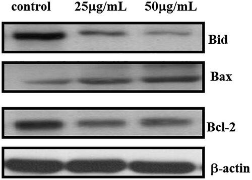
Effect of synthesized gold nanoparticles on apoptosis-related protein expression
The pancreatic cancer cell lines (PANC-1) were used to determine the apoptosis associated protein levels. The expression of Bcl-2 protein was slightly decreased when the dose of the synthesized gold nanoparticles from Scutellaria barbata was increased. The expression of Bax and β-actin proteins was up-regulated on the treatment of the synthesized gold nanoparticles (AuNPs) (). The observed results clearly demonstrated that the synthesized gold nanoparticles from Scutellaria barbata promoted the cancer cell apoptosis by increasing the apoptosis-related protein expressions in time and dose-dependent mode.
Quantitative analysis of the apoptotic gene expressions by qRT-PCR
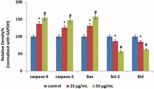
The apoptotic-related genes expression of the pancreatic cancer cells (PANC-1) induced by the synthesized gold nanoparticles from Scutellaria barbata was determined using the qRT-PCR method. The gene expression and mRNA level of Caspase3, Caspase9, Bax, Bid, and Bcl-2 on the cancer cells was determined by using qRT-PCR. The synthesized gold nanoparticles from Scutellaria barbata treated pancreatic cancer cell lines (PANC-1) demonstrated up-regulated expression level of Caspase3, Caspase9, and Bax at the doses of 25 μg/ml and 50 μg/ml when compared to the control cells (). Treatment with the synthesized gold nanoparticles slightly down-regulated the expression of Bid and Bcl-2 at the dose of 50 μg/ml compared to the 25 μg/ml in contrast to the control cells.
Figure 9. Effect of synthesized gold nanoparticles on apoptotic gene expressions by qRT-PCR. This experiment was repeated thrice and the bars in the graph represent S.E. (**p < .05, ***p < .01).
Statistical analysis
All experiments were carried out in three self-determined experiments and the results were articulated as the mean ± standard deviation (mean ± SD) by using one-way analysis of variance (ANOVA). Values of p<.05 were indicative of significant differences.
Discussion
Nanotechnology is becoming the most important and rapidly progressive field in science and technology. Gold nanoparticles are gradually taking the forefront in the area of nanotechnology and there is expansion in its research among researchers due to its unique properties and various applications in the medical field. The exclusive nano size-dependent properties and mode of action make them indispensable in the field of medicine. The nanomaterials and nanoparticles coated materials are constantly receiving attention and recommendation due to their potential of targeting specific and crucial actions and also their unique properties in the area of biomedicine and pharmacology [Citation24,Citation25].
In nanotechnology, for the synthesis of nanoparticles, advances are being made in the development of good physical shape without causing any side effects and a request for improved environmentally friendly processes which do not require the use of toxic and hazardous chemical substances. The synthesis of metallic nanoparticles by using physical and chemical methods are not considered as good or health beneficial because of the usage of capping or reducing agents required for the synthesis of nanoparticles. The chemicals are extremely toxic to the environment and unhealthy for human consumption. In green route method or green synthesis of gold nanoparticles, microbes are used as the reducing or capping agents such as bacteria, fungi, algae, and various plants and their parts. Among these sources, plants are commonly known as the “factories of bio-nanoparticles”. Moreover, they have been considered as being eco-friendly, reasonably cheap to produce, having a unique structure and having the highest capacity of uptake of metallic ions [Citation26–28].
The improvement of a green route method for the synthesis of the gold nanoparticles is constantly gaining attention and recommendations in the field of nanotechnology. The green route method for the synthesis of gold nanoparticles is considered as a suitable method because it excludes the use of toxic chemicals and toxic containing by-products. The green route method of nanoparticle synthesis by using plant and other biological materials is trusted and accepted because it is a cost-effective method, friendly to the environment, and can be easily scaled up for large scale synthesis of gold nanoparticles [Citation29]. Hence, the green synthesis of gold nanoparticles by using biological materials is gaining much interest in the field of pharmaceuticals.
Scutellaria barbata is known as an important traditional medicinal plant that is well described in the traditional Chinese system of medicine. Unlike other medicinal plants, Scutellaria barbata has high-therapeutic values and curative effects for various human diseases. The traditional system of Chinese medicine and pharmacopeia recommends and describes this plant as the most efficient medicinal herb to promote the circulation of blood in the human body. This plant can treat all kinds of human diseases which are directly associated with the blood [Citation30].
The medicinal plants and the plant products are accepted and appreciated among the global populations and also the plant-based products have made a positive impact on the health of the world populations. For this, medicinal plants and plant-based products are playing a major role on the global population healthcare system. Moreover, a number of medicinal plants worldwide, especially in China and India, are recommended and are being used for the treatment and prevention of cancer while a few number of medicinal plants have only received attention for scientific research on the development of therapeutic products for cancer or tumour treatment.
Plant extracts may act both as reducing agents and stabilizing agents in the synthesis of nanoparticles. The source of the plant extract is known to influence the characteristics of the nanoparticles. In green synthesis of gold nanoparticles from plant materials, the reduction of gold ions (Au) to gold atoms may involve the binding of atoms to the surface of cells to form the gold nanoparticles in the reaction medium [Citation31,Citation32]. The initial step of characterization of synthesized gold nanoparticles begins with the visual observation of colour change of the reaction medium due to the effect of surface Plasmon resonance. This colour change develops when the size of the nanoparticles is increased. The variation of the colour change of the reaction medium may be due to the demonstrated surface Plasmon resonance. The absorbance of the changes of the colour is measured by using UV visible spectroscopy [Citation33]. The ideal optical properties demonstrated by the synthesized metallic nanoparticles may be due to the oscillations of the conduction band of the electrons’ nanoparticle surfaces.
The transmission electron microscopic analysis of the synthesized gold nanoparticles gives the precise data on the morphology, shape and size of the nanoparticles [Citation34]. Whereas, the selected area electron diffraction (SAED) pattern analysis confirms the metallic nature of the synthesized gold nanoparticles [Citation35]. The energy dispersive X-ray crystallographic (EDX) analysis is the common technique used to analyze the nature of element or chemical. This technique is usually employed for the determination of the total number and size of the synthesized gold nanoparticles by using biological materials as a source, such as plants [Citation36]. Fourier Transform Infra-red (FT-IR) was employed to determine and identify the presence of possible biomolecules on the surface of the synthesized gold nanoparticles [Citation37]. The crystalline nature of the synthesized gold nanoparticles was analyzed using an X-ray diffraction method [Citation38].
Cancer is commonly described as a class of disease in which the cells proliferate uncontrollably due to the various physical and chemical causes. It is one of the deadliest diseases with a very low survival rate. Pancreatic cancer is a type of belligerent condition of malignancy usually associated with the very low or poor rate of survival [Citation39]. There are various chemotherapeutic and radioactive treatments employed for the treatment of pancreatic cancer, however, the survival rate is minimal and the prognosis is really not clear [Citation40].
The MTT cytotoxicity assay is commonly employed in vitro technique to assess the viability of cancer cell lines. This technique is usually used to determine the in vitro cytotoxicity of the anticancer drugs. In the present study, MTT cytotoxicity assay was used to determine the cytotoxicity effects of synthesized gold nanoparticles from Scutellaria barbata. The results of previous research suggest that the various kinds of cytotoxicity assays may produce varying results based on the samples and test agents used for the study. In the present study, the synthesized gold nanoparticles exhibited significant (p<.001) cytotoxicity activity against human pancreatic cancer cell lines (PANC-1) in a dose-dependent manner. When the concentration of synthesized gold nanoparticles increased, the percentage of viability of the cancer cell lines (PANC-1) was significantly (p<.001) decreased. It clearly demonstrates that the synthesized gold nanoparticles have the potential cytotoxic activity against the human pancreatic cancer cell (PANC-1) lines.
Apoptosis is commonly described as the physiological process of programmed cell death which may be directed by the various molecules and genes [Citation41]. The apoptotic process is very necessary for the removal of excess cells found in the normal tissue structure. For this reason, the process of apoptosis may be as the therapeutic objective for cancer cells whereas the programmed cell death induced by any kind of harmful stimuli should be prevented in the normal cells. Apoptosis is distinct from the cellular necrosis both biochemically and morphologically. The process of apoptosis is involved in the shrinkage of the cells, condensation of chromatin, fragmentation of DNA, blebbing of the plasma membrane and the membrane-bound apoptotic bodies [Citation42]. Hence, apoptosis induction has become a key role for the development of anticancer drugs. The drugs with the capability of induction of apoptosis in cancer cells may become perfect anticancer drugs. The quantitative real-time PCR (qRT-PCR) novel technique is usually employed for the precise quantification of levels of apoptotic-related gene expression. In the present study, the synthesized gold nanoparticles from the aqueous extract of the Scutellaria barbata increased the expression of apoptotic-related protein expressions. Hence, it clearly demonstrates that the gold nanoparticles of the Scutellaria barbata may become an effective anticancer drug after the crucial evaluation. One of the reasons cancer cells can develop resistance to cancer drugs may be directly related to the tendency to resist apoptosis. So, it is necessary to develop novel drugs with the effectiveness of the induction of apoptosis on the cancer cells.
Free radicals are classified as molecules or fragment of molecules that usually contain single or many unpaired electrons on the outermost orbital of the molecule. The sources for the production of free radicals in our body can be exogenous or endogenous in nature. The free radicals such as reactive oxygen species (ROS) are extremely reactive in nature and may be involved in various kinds of pathological conditions [Citation43]. The reactive oxygen species are nothing but by-products of usual cellular metabolism and play a major role in the cell signalling pathways, such as signal transduction between cells, cellular metabolism, cell proliferation and cell apoptosis. In the present research, the gold nanoparticles synthesized from the aqueous extract of the Scutellaria barbata exhibited significantly increased production of intracellular reactive oxygen species (ROS) in the pancreatic cancer cell lines (PANC-1) at the dose of 25 μg/ml and 50 μg/ml in contrast to the control cell lines. This effect may be the key factor of the anticancer activity of synthesized gold nanoparticles from Scutellaria barbata plant.
Medicinal plants play a vital role in the treatment of various kinds of human diseases and the development of novel therapeutic drugs. It is estimated that above 60% of the anticancer drugs which are currently used for cancer treatment are isolated from medicinal plants and their parts and almost 3000 medicinal plants worldwide have been reported to have anticancer properties. However, scientific research on the anticancer properties of medicinal plants is limited. The present research work was aimed to screen the anticancer properties of the synthesized gold nanoparticles from the Scutellaria barbata plant. The synthesized gold nanoparticles (AuNPs) possessed the effective anticancer activity against the pancreatic cancer cell lines (PANC-1). Hence, further research on this plant may lead to the development of the novel anticancer drugs to combat pancreatic cancer.
Conclusion
Based on the above findings, it can be concluded that Scutellaria barbata plant is a major source for the large scale production of gold nanoparticles. The synthesized gold nanoparticles showed effective anticancer activity against the human pancreatic cancer cell lines (PANC-1). Therefore, further scientific research on this plant could lead to the development of the novel anticancer drugs for the treatment of human pancreatic cancer.
Disclosure statement
No potential conflict of interest was reported by the authors.
Additional information
Funding
References
- Sane N, Hungund B, Ayachit N. Biosynthesis and characterization of gold nanoparticles using plant extracts. Int Conf Adv Nanomater Emerg Eng Technol. 2013;12082:295–299.
- Rai M, Yadav A. Plants as potential synthesiser of precious metal nanoparticles: progress and prospects. IET Nanobiotechnol. 2013;7:117–124.
- Vaghela HM, Pathan AA, Shah RH. The biogenic synthesis of Au, Pd and Pt nanoparticles and its medicinal applications: a review. UK: Cambridge Scholars Publishing; 2018.
- Peralta-Videa JR, Huang Y, Parsons JG, et al. Plant-based green synthesis of metallic nanoparticles: scientific curiosity or a realistic alternative to chemical synthesis? Nanotechnol Environ Eng. 2016;11:4.
- Soni N, Prakash S. Green nanoparticles for mosquito control. Sci World J. 2014;2014:1.
- Lakshmana A, Umamaheswari C, Nagarajan NS. A facile phytomediated synthesis of gold nanoparticles using aqueous extract of Momordica cochinchinensis rhizome and their biological activities. J Nanosci Technol. 2016;2:76–80.
- Patil MP, Kim GD. Eco-friendly approach for nanoparticles synthesis and mechanism behind antibacterial activity of silver and anticancer activity of gold nanoparticles. Appl Microbiol Biotechnol. 2017;101:79–92.
- Patil MP, Ngabire D, Thi HHP, et al. Eco-friendly synthesis of gold nanoparticles and evaluation of their cytotoxic activity on cancer cells. J Clust Sci. 2017;28:119–132.
- Anand K, Gengan RM, Phulukdaree A, et al. Agroforestry waste Moringa oleifera petals mediated green synthesis of gold nanoparticles and their anti-cancer and catalytic activity. J Ind Eng Chem. 2015;21:1105–1111.
- Abid Ali Khan MM, Jain DC, Bhakuni RS. Occurrence of some antiviral sterols in Artemisia annua. Plant Sci. 1991;75:161–165.
- Divakaran D, Lakkakula JR, Thakur M, et al. Dragon fruit extract capped gold nanoparticles: synthesis and their differential cytotoxicity effect on breast cancer cells. Mater Lett. 2019;236:498–502.
- Ribas A, Wolchok JD. Cancer immunotherapy using checkpoint blockade. Science. 2018;359:1350–1355.
- Ferlay J, Soerjomataram I, Dikshit R, et al. Cancer incidence and mortality worldwide: sources, methods and major patterns in GLOBOCAN 2012. Int J Cancer. 2014;136:359–386.
- Loc WS, Smith JP, Matters G, et al. Novel strategies for managing pancreatic cancer. WJG. 2014;20:14717–14725.
- Carrato A, Falcone A, Ducreux M, et al. A systematic review of the burden of pancreatic cancer in Europe: real-world impact on survival, quality of life and costs. J Gastrointest Cancer. 2015;46:201–211.
- Ochwang DO, Kimwele CN, Oduma JA, et al. Medicinal plants used in treatment and management of cancer in Kakamega County Kenya. J Ethnopharmacol. 2014;151:1040–1055.
- Marconett CN, Morgenstern TJ, San Roman AK, et al. BZL101, a phytochemical extract from the Scutellaria barbata plant, disrupts proliferation of human breast and prostate cancer cells through distinct mechanisms dependent on the cancer cell phenotype. Cancer Biol Ther. 2010;10:397–405.
- Yang N, Zhao Y, Wang Z, et al. Scutellarin suppresses growth and causes apoptosis of human colorectal cancer cells by regulating the p53 pathway. Mol Med Rep. 2017;15:929–935.
- Kan X, Zhang W, You R, et al. Scutellaria barbata D. Don extract inhibits the tumor growth through down-regulating of treg cells and manipulating Th1/Th17 immune response in hepatoma H22-bearing mice. BMC Complement Altern Med. 2017;17:41.
- Suh SJ, Yoon JW, Lee TK, et al. Chemoprevention of Scutellaria bardata on human cancer cells and tumorigenesis in skin cancer. Phytother Res. 2007;21:135–141.
- Chen CC, Kao CP, Chiu MM, et al. The anti-cancer effects and mechanisms of Scutellaria barbata D. Don on CL1-5 lung cancer cells. Oncotarget. 2017;8:109340.
- Zhang L, Ren B, Zhang J, et al. Anti-tumor effect of Scutellaria barbata D. Don extracts on ovarian cancer and its phytochemicals characterisation. J Ethnopharmacol. 2017;206:184–192.
- Lee SR, Kim MS, Kim S, et al. Constituents from Scutellaria barbata inhibiting nitric oxide production in LPS‐stimulated microglial cells. Chem Biodiversity. 2017;14:e1700231.
- Pal S, Tak YK, Song JM. Does the antibacterial activity of silver nanoparticles depend on the shape of the nanoparticle? A study of the gram-negative bacterium Escherichia coli. Applied and Environmental Microbiology. 2007;73:1712–1720.
- Salata OV. Applications of nanoparticles in biology and medicine. J Nanobiotechnology. 2004;2:3.
- Sarkar J, Ray S, Chattopadhyay D, et al. Mycogenesis of gold nanoparticles using a phytopathogen Alternaria alternate. Bioprocess Biosyst Eng. 2012;35:637–643.
- Honary S, Fathabad EG, Paji ZK, et al. A novel biological synthe- sis of gold nanoparticle by Enterobacteriaceae family. Trop J Pharm Res. 2012;11:887–891.
- Dumur F, Guerlin A, Dumas E, et al. Controlled spontaneous generation of gold nanoparticles assisted by dual reducing and capping agents. Gold Bull. 2011;44:119–137.
- Roopan SM, Madhumitha G, Rahuman AA, et al. Low-cost and eco-friendly phyto-synthesis of silver nanoparticles using Cocosnucifera coir extract and its larvicidal activity. Ind Crops Prod. 2013;43:631–635.
- Sun S, Wang CZ, Tong R, et al. Effects of steaming the root of Panaxnotoginseng on chemical composition and anticancer activities. Food Chem. 2010;118:307–314.
- Yu KF, Kelly KL, Sakai N, et al. Morphologies and surface plasmon resonance properties of monodisperse bumpy gold nanoparticles. Langmuir. 2008;24:5849–5854.
- Brolossy TAE, Abdallah T, Mohamed MB, et al. Shape and size dependence of the surface plasmon resonance of gold nanoparticles studied by Photoacoustic technique. Eur Phys J Spec Top. 2008;153:361–364.
- Zhang X, Qu Y, Shen W, et al. Biogenic synthesis of gold nanoparticles by yeast MagnusiomycesingensLH-F1 for catalytic reduction of nitrophenols. Colloids Surf A Physicochem Eng Asp. 2016;497:280–285.
- Montes MO, Mayoral A, Deepak FL, et al. Anisotropic gold nanoparticles and gold plates biosynthesis using alfalfa extracts. J Nanopart Res. 2011;13:3113–3121.
- Nejad SM, Bonjar SG, Khaleghi N. Biosynthesis of gold nanoparticles using Streptomyces fulvissimusisolate. Nanomed J. 2015;2:153–159.
- Rajeshkumar S, Malarkodi C, Vanaja M, et al. Antibacterial activity of algae mediated synthesis of gold nanoparticles from Turbinariaconoides. Der PharmaChemica. 2013;5:224–229.
- Elavazhagan T, Arunachalam KD. Memecylon edule leaf extract mediated green synthesis of silver and gold nanoparticles. Int J Nanomedicine. 2011;6:1265–1278.
- Bennur T, Khan Z, Kshirsagar R, et al. Biogenic gold nanoparticles from the ActinomyceteGordoniaamarae: application in rapid sensing of copper ions. Sens Actuators B Chem B. 2016;233:684–690.
- Siegel R, Naishadham D, Jemal A. Cancer statistics. CA Cancer J Clin. 2012;62:10–29.
- Thota R, Pauff JM, Berlin JD. Treatment of metastatic pancreatic adenocarcinoma: a review. Oncology. 2014;28:70–74.
- Ulukaya E, Acilan C, Yilmaz Y. Apoptosis: why and how does it occur in biology? Cell Biochem Funct. 2011;29:468–480.
- Kerr JF, Winterford CM, Harmon BV. Apoptosis. Its significance in cancer and cancer therapy. Cancer. 1994;73:2013–2026.
- Rajendran P, Nandakumar N, Rengarajan T, et al. Antioxidants and human diseases. Clin Chim Acta. 2014;436:332–347.