
Abstract
A novel nerve conductor made out of polypyrrole (PPY), collagen (Coll) and nano-strontium substituted bioactive glass (n-Sr@BG) (PPY/Coll/n-Sr@BG) was fabricated by electrospinning. SEM demonstrated that the mean distances across of the pores in the nerve channels were under 15 mm and more prominent than 2 mm. These biocomposite films had biomimetic morphology, bigger porosity and moderately higher surface territory than customary nerve channels, consequently not just allowing the transportation of nerve development factor and glucose yet, in addition, hindering the section of lymphatic tissue and fibroblasts. The consistent filaments of the nerve can copy the characteristic ECM, which is valuable to cell bond, cell multiplication, and cell movement. PPY/Coll/n-Sr@BG demonstrated great cell fondness rate, which is useful for neurilemma cell cells bond, relocation and expansion. Its great viability empowers its wellbeing animal models. Sciatic nerve deformity was crossed over an animal model with PPY/Coll/n-Sr@BG in rodents. PPY/Coll and autotransplants were utilized as control gatherings. Contrasted with PPY/Coll and PPY/Coll/n-Sr@BG accomplished fundamentally increasingly viable recovery of sciatic nerve wounds following 24 weeks implantation and the mean distance across of muscle fibres occasions bigger than that in PPY/Coll/n-Sr@BG, and it was nearer to that in control. The rejuvenated nerve filaments in PPY/Coll/n-Sr@BG had an increasingly standard round shape, the thickness of neuro-filaments in c was more than those in PPY/Coll, and was near that in control.
Introduction
Nerve tissue engineering is a promising methodology that can possibly address this need with engineered nerve channels. There has been a huge exertion devoted to creating manufactured nerve courses that have brought about empowering recovery and some level of utilitarian recuperation of fringe nerve defects [Citation1–5]. Though, more materials should be done to improve their adequacy contrasted with standard nerve grafts. A significant part of engineered nerve unites is their capacity to direct power. Electrical incitement can essentially advance the recovery of fringe nerve wounds in the wake of looking at an extensive number of creature tests [Citation6]. In this manner, there has been an extensive spotlight on the utilization of conductive substances in neural tissue designing. Scientists want to build up a novel substance that can fulfil both the conductivity needs of nerve tissue and the necessities of tissue designing all in all. Preferably, this course ought to be degradable, cyto-compatible and give a fitting electrical condition.
As a way to accomplish this, analysts have considered electroconducting macromolecules, for example, polyphosphazene, polyaniline and polypyrrole. They have been researched as of late for various applications in the field of biosensor and clinical and have been appeared to have phenomenal electrical properties [Citation7,Citation8]. For clinical applications, polypyrrole has been the most generally contemplated [Citation9,Citation10]. In addition to the fact that it has the astounding electrical characteristic, biological examinations have demonstrated it to have great cell and tissue similarity. Moreover, polypyrrole is anything but difficult to blend, has a promptly adjustable exterior, and is economical, which are all inconceivably engaging for tissue building purposes. In this way, polypyrrole has been perceived as a promising platform substance for neural prostheses and nerve tissue designing [Citation11]. Consequent investigations have concentrated on advancing the macromolecule platforms by fusing different prompts, for example, cell glue atoms, geographical highlights and neurotrophins, underscoring the significance of numerous sign for increased adjustment of neuronal reactions [Citation12,Citation13].
Though, most of the works done on polypyrrole include biocompatibility assessment. Thinking about its downsides, including its pitiable dissolvability, additional investigate should be done animal model to affirm the reasonability of polypyrrole as a platform substance [Citation14,Citation15]. The reason for this examination was to explore a conceivable dealing for fixing harmed nerves and to beat the present weaknesses polypyrrole has in tissue building. Collagen (Coll) is broadly utilized in nerve tissue building because of its great viability, non-biodegradable, and tensile strength [Citation16–19].
In this paper, a composite made out of PPY/Coll, and PPY/Coll/n-Sr@BG was fabricated via electro-spinning. These composites were moved up and adhered to make the nerve courses. The conductors were dissected as far as the physicochemical character utilizing SEM. We utilized the viability assay to survey the composite poisonous quality. So, as to contemplate the possibility of this composite conductor, sciatic nerve imperfection was crossed over with PPY/Coll/n-Sr@BG courses in rodents. PPY/Coll and PPY/Coll/n-Sr@BG were position and autotransplants were utilized as control gatherings. The recovered nerves were dissected by H&E and resistant fluorescence re-colouring.
Experimental section
The electrospinning (PPY/Coll/n-Sr@BG composite) arrangement was stacked into a syringe with an end spout constrained by a syringe siphon at a sustaining [Citation20,Citation21]. The catapulted fibres were gathered on a gatherer secured with aluminium foil. The separation between the spout and the gatherer was 10 cm. In the wake of electrospinning, the as-got polymer mats were put in a vacuum drying stove for 3 days at RT to evacuate remaining dissolvable. These mats were stuck into courses with Methylene chloride. Every one of these courses was cleaned with ethylene oxide and further illuminated with a bright light before further appraisals to defeat the ecological and different components that could influence the test results. The analyses were stringent as far as a logical activity to keep up wellbeing amid the procedure of the test. We additionally set countless gatherings in order to guarantee the objectivity of the analysis.
Characterization
The chemical structure examination of the nanomats was performed in the FTIR spectrometer (Bruker instrument) [Citation22]. The phase analyses of fabricated nanomats were obtained through XRD studies using an X’PertPRO; PANalytical [Citation23]. The morphological features of the fabricated nanomats were dissected utilizing FESEM utilizing a quickening voltage of 10 kV. The nanomats were sputter covered with platinum utilizing sputter coater [Citation24].
Viability assay
For viability tests, cells were seeded onto nanomats covered coverslips put in 96-culture plates and permitted to develop for one day before investigation as depicted before [Citation25]. The suitability of the dermal fibroblasts on the nanomats was resolved utilizing the colourimetric Solution Cell Proliferation Assay (MTT examine) as per maker's directions. Quickly, cells developed on the nanomats were hatched with 500 µL of DMEM for one day and 100 µL of MTT reagent were included for 12 min at RT before the examination. Each test was performed in triplicates.
Walking track analysis
To assess engine recuperation in each exploratory rodent at 12 and 24 weeks post-task, strolling track examination was led by two free analysts who were blinded to tests. Singular impressions were recorded in the white paper after the rodent strolled through a glass box [Citation26].
Thermal hyperalgesia test
This examination was performed by a solitary agent who was blinded to the analysis to assess the tactile utilitarian recuperation by estimating the rodent's capacity to defeat and tremble its rear paw on the hot plate each week following medication organization [Citation27]. Prior to testing, each gathering was housed in entity cages to oblige the earth for one day. At that point, the rodents were put on a hot plate. Subsequently, the reaction time at whatever points the creatures shacked their paws was evidenced. The discontinue time was set at twenty seconds to limit skin damage. All tests were rehashed multiple times with five minutes interims. In the event that no rear paw pull back was seen after twenty seconds, the reaction time was evidenced as twenty seconds.
In vivo
All creatures were housed in explicit pathogen free circumstances, and the exploratory methods including creatures were as per NIH Guidelines for the Care and Use of Laboratory Animals and under the endorsement of the Administration Committee of Experimental Animals, China. 24 weeks subsequent to operation, the rodents were relinquished with an overdose of ketamine, at that point the distal segment of the sciatic nerves was expelled and promptly fixed in a cushioned formalin, dried out in a reviewed arrangement of ethyl alcohol arrangements lastly inserted in paraffin, recoloured with H&E for microscopy examination. In re-coloured areas, recovered nerve tissues in the channels were analyzed [Citation28].
Results and discussion
Functional group and phase analysis
FTIR spectra of PPY/Coll and PPY/Coll/n-Sr@BG appear in . The FTIR spectrum of PPY/Coll portrays the peaks at 3320, 1649, 1551 and 1239 cm−1 relating to hydroxyl, amide I–III gatherings, individually [Citation29]. The FTIR spectra of n-Sr@BG portray the peaks at groups at 468, 666, 1459, 1728, 2670 and 3429 cm−1 relating to P–O in PO4, Si–O–Si, C–H in CH3, C = O, and O–H adsorbed water molecules, respectively [Citation30]. Additionally, a few bands are moved, which gives more motivation to trust that the carbonyls bunch partakes in the communication with bioglass. In this manner, it appears that there is decent security among nanobioglass and PPY/Coll in the interface of composite stages [Citation31].
Figure 1. (a) FTIR spectrum of PPY/Coll, n-Sr@BG and PPY/Coll/n-Sr@BG. (b) XRD spectrum of PPY/Coll and PPY/Coll/n-Sr@BG nanomats.
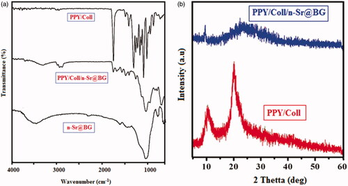
The XRD spectrum of PPY/Coll and PPY/Coll/n-Sr@BG appear in . As can be seen, the PPY/Coll composite display wide planes at two-theta = 24, which might be because of the undefined idea of collagen particles [Citation19]. Similarly, the PPY/Coll/n-Sr@BG (demonstrates a wide planes at two-theta = 24) [Citation32]. It might be because of the formless idea of PPy and the dissipating from PPy chains at the interplanar dividing [Citation33]. The characteristic diffraction planes for PPY and Coll were smothered by the undefined pinnacle of n-Sr@BG inside the scope of 20–40 scale.
Morphology analysis
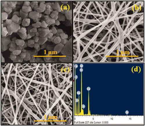
Scanning electron microscopy pictures of the nanomats specimens demonstrated the nearness of flat nano-fibrous surface with no signs of structural deformities (). This exhibits no stage partition of the bioglass amid electrospinning progression which prompts homogeneous conveyance of the mixes all through the fibres. The normal mats breadths of PPY/Coll and PPY/Coll/n-Sr@BG were in the scope of 276 ± 25 and 241 ± 17 nm separately. It has been accounted for that the distance across of the electrospun nanofibers diminished by diminishing the thickness and expanding the conductivity of dope arrangement [Citation32, Citation34]. The exceptional peak of Ca, Sr, P, O and Si affirms the statement of PPY/Coll/n-Sr@BG.
Figure 2. SEM images of (a) n-Sr@BG, PPY/Coll, PPY/Coll/n-Sr@BG and EDX spectrum of PPY/Coll/n-Sr@BG composite.
Biocompatibility assay
Subsequently, we examined the in vitro viability of the PPY/Coll and PPY/Coll/n-Sr@BG nanomats using MTT suitability measure and cytoskeleton recolouring following one-day post seeding of human dermal fibroblasts. MTT measure showed that none of the nanofibers showed any cytotoxic impact for human dermal fibroblasts, recommending that cells held their metabolic movement (). In any case, when contrasted with control, the human dermal fibroblasts development supposedly was fundamentally higher for PPY/Coll and PPY/Coll/n-Sr@BG nanomats frameworks. Accordingly, the MTT results demonstrated higher human dermal fibroblasts metabolic movement and expansion on PPY/Coll mats containing n-Sr@BG [Citation35].
Figure 3. (a) Viability assay of human dermal fibroblasts cell on different nano-mats using MTT assay. CLSM picture of human dermal fibroblasts cell incubated on (b) control, (c) PPY/Coll and (d) PPY/Coll/n-Sr@BG.
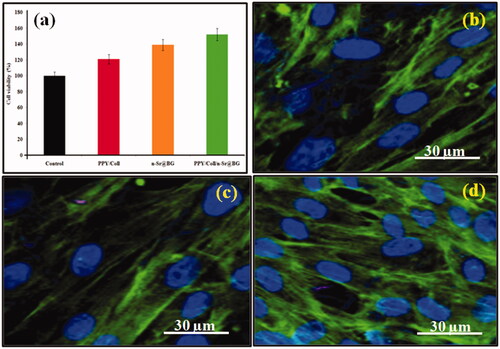
The human dermal fibroblasts phenotype was seen by picturing cell cytoskeleton under microscopy. From demonstrated that fibroblasts presented to both PPY/Coll and PPY/Coll/n-Sr@BG nanomats showed flawless cytoplasmic filamentous conveyances of Factin with a noticeable core and absence of any auxiliary variations from the norm. Strangely, a more prominent thickness of cells was seen when seeded on to PPY/Coll and PPY/Coll/n-Sr@BG nanomats, authenticating the outcomes got from MTT test. The human dermal fibroblasts were joined on all the nanomats and showed ordinary cell structure. These perceptions showed that the PPY/Coll stacked n-Sr@BG nanomats were biocompatibility for human dermal fibroblasts and advanced cell multiplication [Citation36].
Sciatic functional index analysis
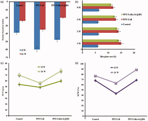
shows the normal sciatic functional index values and the harmed foot strolling example of all investigation bunches at 12 and 24-week post medical procedure. The strolling track investigation indicated better utilitarian recuperation results in the control gathering. The control, as the highest quality level of nerve crossing over, demonstrated improved sciatic functional index from toward the finish of the fourth week to and toward the finish of eighth and twelfth weeks, separately. There was a critical contrast between the PPY/Coll and PPY/Coll/n-Sr@BG bunches with the control bunch toward the finish of the main month. Results showed the two gatherings achieved a comparable dimension of engine recuperation as the control. After the 12th week, the request of sciatic functional index esteem from high to low was controlled bunch PPY/Coll/n-Sr@BG and PPY/Coll gathering. There was a clear contrast between the PPY/Coll/n-Sr@BG and PPY/Coll bunches with control gathering and in the midst of 12 W and 24 W, showing the control bunch had next to no improvement and minimal engine recuperation contrasted and nerve courses alone and channel stacked with n-Sr@BG [Citation37].
Figure 4. (a) SFI of all rats groups. (b) Hot plate response of all rats groups. (c) Nerve conduction velocities and (d) recovery rate of triceps weight after implantation.
Hot plate latency test
The aftereffects of the hotplate test are demonstrated in . A month after careful transaction, most rodents react in the hotplate analysis by licking their rear paws and the withdrawal reflex latency dropped to 12 s [Citation38]. This defers coordinated the time required to see the crossing over of the recovering nerve axons over a hole, with a surmised mean rate of recovery every day. Toward the finish of the 12 W, huge contrasts were found between gathering's PPY/Coll PPY/Coll/n-Sr@BG contrasted and control, gathering. Additionally, PPY/Coll/n-Sr@BG treated rodents had essentially preferred responsiveness over the control gathering. A short time later, the withdrawal reflex latency consistently improved amid the 24 W recuperation time. 24 weeks after damage, the removal reaction showed better tangible recuperation results in gathering's PPY/Coll/n-Sr@BG and control. Though, no factually noteworthy contrasts were found between gathering's PPY/Coll/n-Sr@BG with together and contrasted and control gathering.
shows that the muscle weight proportion of the PPY/Coll/n-Sr@BG bunches was essentially higher than that of PPY/Coll gathering, whereas the TA (triceps surae) muscle weight proportion of control bunches was fundamentally not quite the same as that of PPY/Coll/n-Sr@BG. The improvement in muscle weight demonstrates that there was a huge reinnervation of the objective muscle.
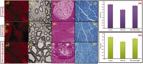
Figure 5. (a–c) Intraoperative photographs of samples 24 weeks postoperatively of all rats group. (d–f) TEM images of regenerated axons and myelin sheath. (g–i) Rejuvenated sciatic nerve stained with H&E staining in different groups at 24 weeks postoperation. (j–l) Rejuvenated sciatic nerve stained with MB staining in different groups at 24 weeks postoperation. (m) The density of myelinated fibres and (n) thickness of myelin sheath in all groups rats.
In vivo rejuvenated sciatic nerve
TEM was utilized to watch the ultra-structural morphology of recovered nerves. Fascicles with various sizes, each comprises of a few layers of axons, fibroblasts and enclosed SCs in the majority of the gatherings (). Myelin sheath thickness was fundamentally more noteworthy in the fibroblasts and enclosed SCs bunch than in the PPY/Coll/n-Sr@BG gathering. While the thicknesses of the control and PPY/Coll gatherings were comparative, they were unmistakably not exactly those seen in the other two gatherings [Citation39]. To more readily assess the recuperation of harmed nerves, the centre fragment of recovered nerves was coloured with H&E re-coloring 24 weeks after the careful activity. All rodents were healthy and no passings were accounted for; wound disease and extreme tissue responses occurred amid the recuperation. Veins existed in the composite course gatherings (), which demonstrated that the veins produce and give supplements to harmed nerves amid the procedure of recuperation. Neurilemma cell existed in the three gatherings and advanced the development of harmed nerves. The myelinated fibre in 5 g and 5 h were appropriated in course of action with slender myelin cover in a manner and most unpredictable connective tissues in development. The fibre of recovered nerves in is increasingly roundabout fit as a fiddle, better-proportioned in size and orchestrated more thickly than in . There are an expansive number of fascicular structures dispersed all through nerves in . In light of the structural perception, the myelinated fibres in is more prominent in number and more concentrated than those in 24 weeks after implantation. Nerves were in more prominent extent in , and this extent was near what was available in .
Immunofluorescent assay
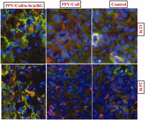
demonstrates blue and green shading in each of the three gatherings, which shows that the recovered sciatic nerves indicated the development of neuro-filaments amid the recuperation of harmed nerves. CLSM image shows that the cells were stout and the neuro-filaments were speck like in exterior in all gatherings at 3 months subsequent to a medical procedure. The thickness of cells in all gatherings was high while the thickness of recovered neuro-filaments in PPY/Coll/n-Sr@BG was more than that in PPY/Coll, yet not as much as that in control. The structure of recovered neuro-filaments in PPY/Coll/n-Sr@BG was more roundabout and stuffed in game plan than those in PPY/Coll, while it was nearer in incentive to that in control. Cells develop around the recovered neuro-filaments, which improve nerve recovery through emitted nerve development feature, cerebrum determined the neurotrophic issue, etc. [Citation40].
Figure 6. Rejuvenated sciatic nerve stained with immunofluorescence staining in different groups at 12 and 24 weeks post operation.
Electrophysiological assessment
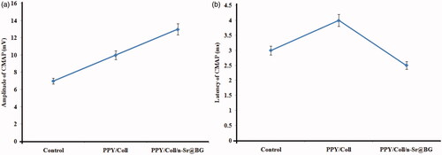
Electromyography was executed to assess conduction from recovered nerves to the gastrocnemius muscle (). The compound muscle action potentials pinnacle and inactivity periods were determined and their centrality examined. Contrasted and the control gathering, best recuperation records were seen in PPY/Coll and PPY/Coll/n-Sr@BG gatherings; however, the improvement was undeniably increasingly sensational in the PPY/Coll/n-Sr@BG gathering. Furthermore, there were no critical contrasts between the control and PPY/Coll gatherings [Citation41,Citation42].
Figure 7. Electro-physiological assessment. The peaks (a) and latencies (b) of the action potentials were documentation and statistically examined.
Conclusion
Nerve tissue designing offers an imaginative and promising way to deal with treating nerve surrenders. Conductive polymers have extraordinary potential as clinical to create manufactured nerve courses. We have effectively fabricated PPY/Coll/n-Sr@BG via the electrospinning technique and researched its cytocompatibility and fringe nerve recovery. PPY, Coll and n-Sr@BG exist in PPY/Coll/n-Sr@BG course through the pertinent ingestion tops appeared from XRD and FTIR spectra. The scanning electron microscopy picture demonstrates that the composite of PPY/Coll/n-Sr@BG has a bio-mimic morphology that cannot just allow the transportation of nerve development factor and glucose yet additionally obstruct the section of lymphatic and fibroblasts. Following a half year, the rodents with the PPY/Coll/n-Sr@BG demonstrated practical recuperation like that of the best quality level control nerve join and essentially superior to that of the PPY/Coll channels. Whereas accomplishing recuperation on the dimension of sound nerve is a definitive objective of nerve tissue recovery, making a unite that gives the equivalent practical recuperation level as a control join is energizing in light of the fact that such a channel takes out the disadvantages related with utilizing a control unite, including constrained benefactor source, giver site dreariness, numerous medical procedure locales, and conceivable size confound. The conductivity nano-structure and of the nerve direction course were valuable to the bond and managing expansion of neurons. This work gave significant data to the nerve fix and the plan of bio-mimic gadgets equipped for animal model applications.
Disclosure statement
No potential conflict of interest was reported by the authors.
References
- Subramanian A, Krishnan UM, Sethuraman S. Development of biomaterial scaffold for nerve tissue engineering: biomaterial mediated neural regeneration. J Biomed Sci. 2009;16:108–119.
- Mobini S, Spearman BS, Lacko CS, et al. Recent advances in strategies for peripheral nerve tissue engineering. Curr Opin Biomed Eng. 2017;4:134–142.
- Sensharma P, Madhumathi G, Jayant RD, et al. Biomaterials and cells for neural tissue engineering: current choices. Mater Sci Eng C. 2017;77:1302–1315.
- Heidari M, Bahrami SH, Ranjbar-Mohammadi M, et al. Smart electrospun nanofibers containing PCL/gelatin/graphene oxide for application in nerve tissue engineering. Mater Sci Eng C. 2019;103:109768.
- Salehi M, Bagher Z, Kamrava SK, et al. Alginate/chitosan hydrogel containing olfactory ectomesenchymal stem cells for sciatic nerve tissue engineering. J Cell Physiol. 2019;234:1–12.
- Lee B, Koripalli MK, Jia Y, et al. An implantable peripheral nerve recording and stimulation system for experiments on freely moving animal subjects. Sci Rep. 2018;8:6115–6127.
- Anderson M, Shelke NB, Manoukian OS, et al. Peripheral nerve regeneration strategies: electrically stimulating polymer based nerve growth conduits. Crit Rev Biomed Eng. 2015;43:131–159.
- Ghasemi-Mobarakeh L, Prabhakaran MP, Morshed M, et al. Application of conductive polymers, scaffolds and electrical stimulation for nerve tissue engineering. J Tissue Eng Regen Med. 2011;5:e17–e35.
- Bober P, Capáková Z, Acharya U, et al. Highly conducting and biocompatible polypyrrole/poly(vinyl alcohol) cryogels. Synth Met. 2019;252:122–126.
- Maruthapandi M, Nagvenkar AP, Perelshtein I, et al. Carbon-dot initiated synthesis of polypyrrole and polypyrrole@cuo micro/nanoparticles with enhanced antibacterial activity. ACS Appl Polym Mater. 2019;1:1181–1186.
- Massoumi B, Hatamzadeh M, Firouzi N, et al. Electrically conductive nanofibrous scaffold composed of poly(ethylene glycol)-modified polypyrrole and poly(ε-caprolactone) for tissue engineering applications. Mater Sci Eng C. 2019;98:300–310.
- Tsai N‐C, She J-W, Wu J-G, et al. Poly(3,4‐ethylenedioxythiophene) polymer composite bioelectrodes with designed chemical and topographical cues to manipulate the behavior of pc12 neuronal cells. Adv Mater Interfaces. 2019;6:1801576.
- Ferreira CL, Valente CA, Zanini ML, et al. Biocompatible PCL/PLGA/polypyrrole composites for regenerating nerves. Macromol Symp. 2019;383:1800028.
- Su D, Zhou J, Ahmed KS, et al. Fabrication and characterization of collagen-heparin-polypyrrole composite conductive film for neural scaffold. Int J Biol Macromol. 2019;129:895–903.
- Chakraborty R, Seesal VS, Manna JS, et al. Synthesis, characterization and cytocompatibility assessment of hydroxyapatite-polypyrrole composite coating synthesized through pulsed reverse electrochemical deposition. Mater Sci Eng C. 2019;94:597–607.
- Khan MA, Cantù E, Tonello S, et al. A review on biomaterials for 3d conductive scaffolds for stimulating and monitoring cellular activities. Appl Sci. 2019;9:961.
- Heo DN, Hospodiuk M, Ozbolat IT. Synergistic interplay between human MSCs and HUVECs in 3D spheroids laden in collagen/fibrin hydrogels for bone tissue engineering. Acta Biomater. 2019. doi: 10.1016/j.actbio.2019.02.046
- Goodarzi H, Jadidi K, Pourmotabed S, et al. Preparation and in vitro characterization of cross-linked collagen–gelatin hydrogel using EDC/NHS for corneal tissue engineering applications. Int J Biol Macromol. 2019;126:620–632.
- Lin K, Zhang D, Macedo MH, et al. Advanced collagen-based biomaterials for regenerative biomedicine. Adv Funct Mater. 2018;29:1804943.
- Lee Y-S, Arinzeh TL. Electrospun nanofibrous materials for neural tissue engineering. Polymers. 2011;3:413–426.
- Liu J, Rawlinson SCF, Hill RG, et al. Strontium-substituted bioactive glasses in vitro osteogenic and antibacterial effects. Dent Mater. 2016;32:412–422.
- Fan S, Huang Zhang ZY, et al. Magnetic chitosan-hydroxyapatite composite microspheres: Preparation, characterization, and application for the adsorption of phenolic substances. Bioresour Technol. 2019;274:48–55.
- Yang CR, Wang YJ, Chen XF. Preparation and evaluation of biomimetric nano-hydroxyapatite-based composite scaffolds for bone-tissue engineering. Chin Sci Bull. 2012;57:2787.
- Gnaneshwar PV, Sudakaran SV, Abisegapriyan S, et al. Ramification of zinc oxide doped hydroxyapatite biocomposites for the mineralization of osteoblasts. Mater Sci Eng C. 2019;96:337–346.
- Samadian H, Salehi M, Farzamfar S, et al. In vitro and in vivo evaluation of electrospun cellulose acetate/gelatin/hydroxyapatite nanocomposite mats for wound dressing applications. Artif Cells Nanomed Biotechnol. 2018;46:964–S974.
- Brown CJ, Mackinnon SE, Evans PJ, et al. Self‐evaluation of walking‐track measurement using a sciatic function index. Microsurgery. 1989;10:226–235.
- Liu C, Fan L, Xing J, et al. Inhibition of astrocytic differentiation of transplanted neural stem cells by chondroitin sulfate methacrylate hydrogels for the repair of injured spinal cord. Biomater Sci. 2019;7:1995–2008.
- Soucy JR, Sani ES, Lara RP, et al. Photocrosslinkable gelatin/tropoelastin hydrogel adhesives for peripheral nerve repair. Tissue Eng A. 2018;24:1393–1405.
- Babu A, Prasanth KG, Balaji B. Effect of curcumin in mice model of vincristine induced neuropathy. Pharm Biol. 2015;53:838–848.
- Mekonnen BT, Ragothaman M, Kalirajan C, et al. Palanisamy, conducting collagen-polypyrrole hybrid aerogels made from animal skin wastes. RSC Adv. 2016;6:63071–63077.
- Koutsopoulos S. Synthesis and characterization of hydroxyapatite crystals: a review study on the analytical methods. J Biomed Mater Res. 2002;62:600–612.
- Oryan A, Eslaminejad MB, Kamali A, et al. Synergistic effect of strontium, bioactive glass and nano-hydroxyapatite promotes bone regeneration of critical-sized radial bone defects. J Biomed Mater Res. 2019;107B:50–64.
- Manivasagan P, Bui NQ, Bharathiraja S, et al. Multifunctional biocompatible chitosan-polypyrrole nanocomposites as novel agents for photoacoustic imaging-guided photothermal ablation of cancer. Sci Rep. 2017;7:4359.
- Yeow ML, Liu YT, Li K. Morphological study of poly(vinylidene fluoride) asymmetric membranes: effects of the solvent, additive, and dope temperature. J Appl Polym Sci. 2004;92:1782–1789.
- Yu H, Peng J, Xu Y, et al. Bioglass activated skin tissue engineering constructs for wound healing. ACS Appl Mater Interfaces. 2016;81:703–715.
- Naseri S, Lepry WC, Nazhat SN. Bioactive glasses in wound healing: hope or hype? J Mater Chem B. 2017;5:6167–6174.
- Ma Y, Dong L, Zhou D, et al. Extracellular vesicles from human umbilical cord mesenchymal stem cells improve nerve regeneration after sciatic nerve transection in rats. J Cell Mol Med. 2019;23:2822–2835.
- Salehi M, Naseri-Nosar M, Ebrahimi-Barough S, et al. Regeneration of sciatic nerve crush injury by a hydroxyapatite nanoparticle-containing collagen type I hydrogel. J Physiol Sci. 2018;68:579.
- Breshah MN, Sadakah AA, Eldrieny EA, et al. Functional and histological evaluation of rat sciatic nerve anastomosis using cyanoacrylate and fibrin glue. Tanta Dent J. 2013;10:67–74.
- Dubový P, Klusáková I, Hradilová-Svízenská I, et al. A conditioning sciatic nerve lesion triggers a pro-regenerative state in primary sensory neurons also of dorsal root ganglia non-associated with the damaged nerve. Front Cell Neurosci. 2019;13:1–16.
- Dai L-G, Huang G-S, Hsu S-H. Sciatic nerve regeneration by cocultured Schwann cells and stem cells on microporous nerve conduits. Cell Transplant. 2013;22:2029–2039.
- Wu D, Raafat A, Pak E, et al. Dicer-microRNA pathway is critical for peripheral nerve regeneration and functional recovery in vivo and regenerative axonogenesis in vitro. Exp Neurol. 2012;233:555–565.