
 ?Mathematical formulae have been encoded as MathML and are displayed in this HTML version using MathJax in order to improve their display. Uncheck the box to turn MathJax off. This feature requires Javascript. Click on a formula to zoom.
Abstract
The extensive relevance of nanoparticles arouses the requirement for manufacturing although the predictable technique are frequently perilous and energy saving. In the current study, zinc oxide nanoparticles manufactured from Allium cepa avert UVB radiation interceded irritation in human epidermal keratinocytes (HaCaT cells). In the current study, the zinc oxide nanoparticles (ZnO-NPs) was synthesized from the extract of A. cepa. The optimized ZnO-NPs hence attained and was enumerated and exemplified by UV visible spectroscopy, X-ray diffraction, Fourier transform infrared spectroscopy (FT-IR), transmission electron microscopy (TEM), scanning electron microscope (SEM) and EDAX impending analysis. In addition, amalgamated ZnO-NPs were experienced for cell viability (MTT), formation of reactive oxygen species (ROS), apoptosis, and antioxidant and lipid peroxidation (TBARS) levels. Also, we explored the effect of A. cepa ZnO-NPs in molecular level by evaluating the inflammatory and apoptotic markers, in which ZnO-NPs reinstated the interleukins 6, 10 and related signaling molecules like iNOS, COX-2 levels. Ultimately, ZnO-NPs induce apoptotic markers (Bax, Bcl-2) and also recommended that ZnO-NPs might aggravate cancer cell apoptosis in HaCaT cells.
Introduction
Nanotechnology is swiftly mounting with the incessant progression of nanomaterial-related user products and their industrialized purpose. Nano levels of carbon-based resources, metal oxides and biopolymers are life form worn out in numerous industries which includes disease diagnosis, drug discovery and delivery, perfumes, sunscreens, food materials, paints, electronic materials and sports [Citation1]. Zinc oxide nanoparticles (ZnO-NPs) encompass numerous usages like the different biological ground, such as perfumes, cosmetics, as food flavor, as drug deliverance, cellular blueprint, and cancer treatments [Citation2,Citation3]. ZnO-NPs are supplementary to cosmetic and facial rubs to supply guard adjacent to skin damage by UVA irradiation [Citation4,Citation5]. Nano level range bestowed them inimitable substantial, element, and biological possessions plus performance. A consideration of actions and inherited property of nanoparticles in existing systems is imperative for both the intent of function and relevance of nanomaterial-related preparation for remedy previously in usage [Citation6].
ZnO-NPs, as a novel type of the inexpensive and low-toxic nanomaterial, has concerned marvelous attention in a range of biomedical research, which includes antibacterial, antioxidant, antidiabetic, anticancer and anti-inflammatory properties, in addition to drug liberation and bioimaging purpose [Citation7,Citation8]. The relevance of ZnO-NPs among an extent of 100 nm is normally documented as secure by the USA Food and Drug Management because of their improved bioagreeable when compared to lesser ZnO-NPs and size can be transported addicted to cells and can mount up in various associate cellular organelles [Citation7]. Human epidermal keratinocytes (HaCaT) cells are nontumor epidermal keratinocytes that are accounted to enclose usual p53 action, and due to this reason, it has been extensively worn out to learn biological sequence modification and process implicated in nanoparticles provoked noxious [Citation9–11].
Preceding studies demonstrates nanoparticles postponed the cell cycle next to the gap/mitotic checkpoint that was tracked by compacted HaCaT cell feasibility. Elevated methylation of histone, lessened acetylation of histone and reduced chromatin were experimental via Western and immunological blemishing examination escorted by the increased altitude of mRNA and abridged appearance of the acetyltransferase genes (CBP, P300 and GCN5) after management with ZnO-NPs [Citation12]. The production of oxygen species was established to be augmented, and DNA smash up was experimental subsequent management with ZnO-NPs. In addition transmission electron microscopy deep-rooted that ZnO-NPs were engrossed into the cell. Apoptotic studies (HaCaT) illustrates the phrase of the proapoptotic genes Bax, Puma and Noxa was originated to augment considerably, while the expression of the antiapoptotic gene Bcl-xl was diminished, behind disclosure to ZnO-NPs [Citation6]. Expected compounds from the herbals are ahead of reputation since of numerous compensation such as frequently having smaller amount side effects, enhanced patient lenience, less expensive and adequate owing to an extended narration of use [Citation13]. In addition natural medicines afford balanced resources for the management of numerous diseases that are stubborn and untreatable in further classification of medicine. Due of this, a number of plants have been explored for the handling of skin diseases sorted from itching to skin melanoma. Until now, 31 plants have been accounted to be effectual in different skin diseases throughout the precedent 17 years that is from 1995 to 2012 [Citation13].
Allium genus has been deliberated by a number of botanists of the world beneath diverse families, viz., Alliaceae, Liliaceae, or Amaryldiaceae [Citation14]. Allium genus is inexpensively significant because it comprises of numerous imperative chronological vegetable crops like onion, garlic and lots of wild decorative species and it is affirmed to be instigated in the Near East and Central Asia [Citation15]. Allium cepa has the capability to progress the manifestation of scars subsequent to excision and illustrated that extracted gel enhanced blemish faintness, ruddiness, surface and inclusive look at the removal site at study weeks 4, 6, 8 and 10 as evaluated by the unsighted examiner [Citation16]. So, in the present study, we explored the effect of ZnO-NPs amalgamated from A. cepa which might avert UVB radiation interceded TBARS, antioxidant levels, ROS, inflammatory response and apoptosis in HaCaT cells. Ultrastructure from TEM, SEM, and EDX confirms that ZnO-NPs were immersed into the cell. Finally, we confirm that nano synthesized A. cepa averted skin cancer in HaCaT cells.
Materials and methods
Chemicals
Fetal bovine serum (FBS), Dulbecco's modified Eagle's medium (DMEM), trypsin-EDTA were acquired from Invitrogen (Carlsbad, San Diego, CA, USA). Sodium citrate, sodium borohydride, tribasic dehydrates, 2', 7'-dichlorofluorescein diacetate (DCFH-DA), zinc-oxide, Tris-buffered saline and Tween 20 were procured from Sigma-Aldrich (St. Louis, MO, USA). Subsequent antibodies were used: Bax, Bcl-2, COX-2, iNOS were obtained from Kamiya Biomedicinal Company, Seattle, Washington. All particular horseradish peroxidase-conjugated secondary antibodies were acquired from Santa Cruz Biotechnology (Rockland antibodies, Dallas, TX, USA). All the other chemicals used in this experiment were of analytical grade.
Amalgamation of zinc oxide nanoparticles
The onion bulbs were rinsed with disinfected distilled water and the external casing of the bulb was physically shed off and the fleshy part of the onion was again washed with germ-free distilled water. The onion bulb was slashed into minute pieces and 10 gm of onion bulb was positioned using pestle and mortar with distilled water. The pulled out extraction was filtered by means of muslin cloth and then again filtered by Whatman No.1 filter paper which is used as dropping agent and as a preservative. Zinc nitrate was worn out as a predecessor for the production of zinc oxide nanoparticles. For ZnO-NPs mixture, the reaction mixture was equipped by mixing of 10 ml of leaf extract and 90 ml of 1 mM zinc nitrate in a 250 ml conical flask. Then, the combination was nurtured in the darkroom at 37 °C. Finally, the combination was centrifuged at 10,000 rpm for 15 min. After centrifugation, the pellet was allowed to dry [Citation17]. Then, the pellet was used for further studies.
Purification of ZnO-NPs
The nanoparticles thus acquired were cleansed by frequent centrifugation nearly at 10000 rpm at 25 °C for 15 min. Then, it was pursued by re-diffusion of the pellet in deionized water to throw away any clumsy organic molecules. The procedure of centrifugation and re-diffusion were repetitive with disinfected distilled water to make sure a better partition of remaining particles from the nanoparticles [Citation17].
Characterization of ZnO-NPs
The manufactured nanoparticles were exemplified by UV-visible spectroscopy, X-ray diffraction (XRD), transmission electron microscopy (TEM), scanning electron microscopy (SEM), Fourier transform infrared spectroscopy (FTIR), energy dispersive X-ray spectroscopy analysis (EDX).
UV-visible spectroscopy examination
The decline of untainted metal ions was established by determining the incorporation of the reaction mixture by UV visible Spectrophotometer (UV Model 1700 Shimadzu) from 300 to 800 nm].
X-ray diffraction analysis
To resolve the character and size of the ZnO nanoparticles, X-ray diffraction (XRD) was executed. The sample pellets were suspended in sterile water (deionized) and the cleaning process repeated for three times by the same solution followed by centrifugation. The pellet was preserved and allowed to dry. The powder commencing from the sample was covered on the XRD grid, the spectra were documented 40 kV and a current of 30 mA with K alpha and K beta radiation by means of XRD (Model PW1050/37 Philips). The diluted intensities were verified from 20 to 80° C at 2 theta angles. The crystalline environment of blended nanoparticles was premeditated from the breadth of the XRD crest, by means of Debye–Scherrer formula.
Transmission electron microscopy
Disseminated sections are divided into carbon covered copper lattice and observed under an electron microscope (HIH-8100 Model, Hitachi) at an escalated volt of 200 kV. Photomicrographs of transmission electron microscopy were explored by ImageJ (NIH) to evaluate the position of ZnO-NPs in human epidermal keratinocytes cells and the connoted size of ZnO-NPs which was cumulative.
Scanning microscopy studies
The produced nanoparticles were scattered in water and the consequential deferment were pasteurized by ultrasonicator for about 2 h. Few drops of the nanoparticles suspension were positioned on a section of micro glass slide fond of to a metal grid covered with carbon coat and dehydrated steadily at 37 °C. The section was then spit coated and visualized with a JSM-6480 LV (JEOL) scanning electron microscope to evaluate the size, shape and percentage of the particle.
Energy dispersive X-Ray spectroscopy (EDAX) analysis
Few drops of the nanoparticles postponement were located on a part of microglass slide fonded to a metal crisscross covered with carbon film and allowed to dry it slowly at room temperature. X-ray spectrometer (EDAX) activated at an increasing voltage for about 10KeV. The section was then gasp coated with gold and imagined with an analytical instrument (BRUKER) to review the mass, figure and proportion of manufactured particles.
Fourier transform infrared (FTIR) analysis
FTIR has been converted into a significant tool for the consideration of the contribution of functional groups in communications between metal particles and biological molecules. FTIR spectra were verified at 1 cm−1 motion by FTIR spectrophotometer (Model-8400S Shimadzu) using potassium bromide pellet procedure. The frequency array was deliberated as wave figures characteristically over the range 4500–400 cm−1.
Cell viability
MTT assay
The mitochondrial motion was evaluated using the MTT assay [Citation18]. The cells were nurtured for a dissimilar point in time with ZnO-NPs and for 4 h with MTT [3–(4, 5-dimethylthiazoyl-2-yl)-2,5-diphenyltetrazolium bromide] dye. The intermediate from each well was redundant and the resultant formazan crystals were solubilized by adding up 200 µl of DMSO (dimethylsulphoxide) and enumerated by determining the absorbance at 540 nm in a SYNERGY- HTX plate reader, BioTek instruments (Winooski, USA). The CC50 value of ZnO-NPs at 24 h was intended from MTT assay by means of the method of Reed and Muench [Citation19].
Measurement of intracellular ROS
The intracellular ROS production was predictable by the regular method [Citation20] using 2, 7-dichlorofluorescein diacetate (DCFDA; Sigma, St Louis, MO, USA) dye. Cells were sowed in a 96-well bottom plate (106 cells/well). After 24 h, the cells were exposed to ZnO-NPs for 6 h. Subsequent to disclosure, the cells were cleansed more than one time with phosphate buffer saline and nurtured with DCFDA dye (20 µM) for 30 min at room temperature. The reaction combination was then restored by 200 µl of phosphate buffer saline and fluorescence strength was calculated in a Multiwell plate reader, (Winooski, USA) at emission and excitation wavelengths of 495 and 538 nm, correspondingly. The conformity investigation of ROS production was prepared by means of a microscope with an attached fluorescence (Leica DMLB epi-fluorescence microscope, Wetzla, Germany).
Mitochondrial membrane potential assessment
Mitochondrial membrane potential was perceived with the fluorescent probe 5,5′,6,6′-tetrachloro-1,1′,3,3′-tetraethyl benzimidazolcarbocyanine iodide. Cells uncovered to ZnO-NPs were collected by treatment with trypsin and bathed with phosphate buffer saline twice. The cells were developed with 10 µM mitochondrial membrane potential dye (JC-1) for 15 min at room temperature and then washed with PBS again re-suspended in PBS at a deliberation of 106 cells/ml. For qualitative analysis, the cells were imaged for green and red fluorescence using a microscope with a fluorescence attachment (Leica Phase Contrast Fluorescence Microscope, Germany).
ETBR/AO double staining
HaCaT cells were placed in a 12-well plate in exposure with the concentrations of 15 and 20 μg/ml ZnO-NPs for 24 h. Following the achievement of the disclosure period, cells were treated with phospate buffer saline. adding together 300 μl phospate buffer saline which holds 100 μg/ml acridine orange and 100 μg/ml ethidium bromide, we scrutinized dyeing consequences using a fluorescence microscope (Nikon Eclipse Shinjuku, Tokyo, Japan).
Anti-oxidant assay
SOD activity was deliberated by Superoxide Dismutase Assay Kit, Cayman Chemical, Michigan, USA as per manufacturer′s directions. This method makes use of tetrazolium salt to identify superoxide radicals produced from xanthine oxidase and hypoxanthine. CAT activity was measured by the same above-mentioned assay kit (Cayman Company) as per producer directions. In this assay, CAT enzyme retort with methanol in the occurrence of H2O2 to fabricate formaldehyde which was precised spectrophotometrically along with chromogen. When coming to the procedure Cells were collected and centrifuged at 400 rpm for 5 min at 4 °C. The supernatant was detached. The suspension was cleaned with subsequent centrifugation for about two times with cold Phosphate Buffer Saline to eliminate all traces of the medium. The final pellet was sonicated at 300 W to attain the cell lysate. GPx and GSH activity was also measured using Cayman′s kit. The regular group was 20 μM GSH suspended in GSH buffer solution. The uniform group was restored by phospate buffer saline. The absorbance was read at 420 nm by means of a microplate reader. GPx speeds up the decline of hydroperoxides, together with H2O2, by reduced glutathione.
Determination of TBARS
The culture media was resoluted using TBARS assay. Culture media for about 1 ml was accumulated to which 200 L of 10% SDS was further added and spun forcefully. 2 ml of newly equipped thiobarbituric acid (TBA) was supplemented to the above mix and was nurtured at 95°C for 60 min. It was allowed to cool to room temperature and was centrifuged at 3000 rpm for 15 min. The assimilation range of the supernatant was documented at a range of 530 nm to appraise the development of TBARS. All examinations were conceded out in triplicate for further confirmation.
Quantification of serum cytokine
The plane of the cytokines (IL-6, 10 and tumor necrosis factor (TNF-α), Caspase 3 & 9) in serum was determined using ELISA kit (Bio-Rad Lab, CA, USA) [Citation21]. Premixed blob covered among the intended antibodies was supplemented to a well, and brewed with the section in a 96-well microtiter cover to respond with exact analytes. Then, mixed noticed antibodies are supplemented to sample wells pursued by a fluorescent tagged blogged molecule that exclusively attaches to the analyte. Every step obliges precise interval time, with shuddering at 37 °C and followed by washing. Again washing the wells was performed using a Bio-Plex (Bio-Rad Lab) wash position. Lastly, the section was then analyzed by the same assortment reader, and data attainment and examination were conceded out by means of the advertent technique.
Western blot studies
HaCaT Cells treated with ZnO-NPs were pelleted and lysed using cell lysis reagent (Sigma, MO, USA) in the presence of protease inhibitor cocktail. The whole protein attentiveness was deliberated by the standard procedure and process [Citation22]. Proteins were determined by the sodium dodecyl sulfate and polyacrylamide gel electrophoresis and reassigned to nitrocellulose covering. The covering membranes were sterilized by means of non-plump milk (5%) for 2 h at 37 °C and explored with anti-human primary antibodies against -actin, Bcl2, Bax, COX-2, and iNOS for 1 h at room temperature subsequent by 12 h incubation at 4 °C. Next day, the membrane was again incubated for 1 h at 37 °C with secondary anti-primary antibody conjugated to horseradish peroxidase (Calbiochem Research Biochemicals, St. Louis, MO, USA). The protein bands were envisaged by improved chemiluminescence (Chemiluminescent Western reagent, Rockford, IL, USA) and densitometry was ended using the Scion Image (Scion Corporation, Fredrick, Maryland, USA).
-actin is treated as loading control.
Statistics
Outcome was articulated as the mean ± SEM and data were evaluated using one-way analysis of variance (ANOVA) with the Dunnett post hoc test to conclude connotation comparative to the unexposed control using SPSS v. 16.0 software (SPSS Inc., IL, USA). In all cases, p < .05 was measured noteworthy.
Results
X-Ray diffraction analysis
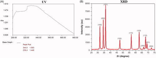
shows the different peaks in the XRD prototype could be dispensed to the crystalline zinc oxide with the hexagonal structure with the lattice parameters. The XRD pattern demonstrated dissimilar concentration peaks in the entire band of 2θ values ranging from 25° to 75° for the onion. The A. cepa digs out arbitrated synthesized ZnO-NPs were indexed as 100, 002, 101, 102, 110, 103, 200, 112, 201 and 004. The zinc was indexed and excited at 101 and 100. The standard crystal formed size was resoluted by means of Scherer’s equation. In addition, no extra peaks have been noticed in the sample and established the biosynthesis of ZnO-NPs at a high limpidness level. Biological synthesis of ZnO-NPs from A. cepa extract showed strong diffraction peaks which was a significant agreement with the JCPDS folder No: 36145 [Citation23]. High purity and crystal form of the manufactured ZnO-NPs were also confirmed.
Figure 1. UV-visible spectrum absorption pattern and X-ray diffraction analysis of ZnO-NPs synthesized from Allium cepa. UV-VIS absorption spectra of ZnO-NPs. The peak values for UV-VIS, plotted between ZnO-NPs/absorbance ratios. The highest absorbance peak is about 344.00. All solutions were in water. The XRD pattern showed dissimilar concentration peaks in the entire band of 2θ values between range from 25° to 75° for the A. cepa extract. The A. cepa synthesized ZnO-NPs were indexed as 100, 002, 101, 102, 110, 103, 200, 112, 201 and 004. The zinc was indexed and excited at 101 and 100.
UV spectroscopical investigation
The diminution of zinc oxide ions in the onion extract was supplementary long-established by UV-Vis Spectrophotometer. UV-Vis absorption spectra of nanoparticles were shown in (. The UV-Vis absorption range of ZnO-NPs was at the peak maximum of 344 nm which is equivalent to the absorbent range of zinc oxide nanoparticles. Following incubation, the colour was distorted since the excitation of Surface Plasmon ambiance in the ZnO-NPs. The decrease of zinc was issued to the examination by using the UV-Vis spectrophotometer. Absorption spectra of ZnO-NPs shaped which has a peak at 300 nm, enlargement of peak specifies that the elements are dispersed. The regularity and breadth of the surface Plasmon absorption relies on the volume and form of the metal nanoparticles and also on the dielectric invariable of the metal itself and the neighboring intermediate [Citation24]. Allium cepa extract and zinc nitrate changed the color of the ZnO-NPs. The highest intensity at 238 nm was experimental which indicates the whole decline of zinc ions. The UV-Vis inclusion spectrum of ZnO-NPs demonstrated a pointed absorbance at 344 nm, which designate an approximate consistent mass of the nanoparticles. The UV-Vis absorption range of ZnO-NPs with flower extract of Nyctanthes arbor-tristis illustrated a prickly absorbance at 369 nm [Citation25]. Nevertheless, alter in particle volume or form, a small swing in the incorporation was previously experimental [Citation26].
ZnO-NPs absorption in HaCaT cells
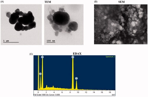
Nanoparticles emerge to put forth diverse cytotoxicity, which relies on cellular intake, positioning, and relocation [Citation27,Citation28]. In the present study, we assessed Nanoparticles intake and position in cells with TEM (Transmission Electron Microscopy). TEM descriptions displayed that ZnO-NPs be not originated in the protoplasm and nucleus of the cells (), however, ZnO-NPs were noticed as cumulative detached within the cytoplasm as a substitute of a single particle.
Figure 2. TEM, SEM and EDAX analysis of ZnO-NPs synthesized from Allium cepa. Nanoparticles position in HaCaT cells using TEM. TEM images displayed ZnO-NPs were not originated in the cytoplasm and nucleus of the cells (), but ZnO-NPs were noticed as cumulative disconnected within the cytoplasm as a replacement of single particle after management with 15 μg/mL ZnO-NPs. Allium cepa extract ZnO-NPs were found as nano-shaped (. The organization, segment and form of amalgamated product were examined by the SEM study. The EDAX examination validates peaks for ZnO-NPs. Production and categorization of ZnO nanoparticles using the leaf extract of A. cepa, the EDS spectrum displays the high value of zinc (82.2%) and oxygen (19.68%).
Scanning electron microscope examination
displays the exterior morphology of the nanoparticles was distinguished using Scanning Electron Microscopy. Allium cepa extract arbitrated ZnO-NPs were found as nano shaped (). The natural move toward using A. cepa extract used for the initial time as a dropping material and also as exterior alleviating agent for the production of minute sphere shaped ZnO-NPs. The organization, segment and form of the amalgamated product were examined by the SEM study [Citation29]. Preceding SEM results also denotes ZnO-NPs produced with Ocimum tenuiflorum leaf extract [Citation18]. In the present study, SEM outcome illustrated the cylindrical nanoparticle fashioned with a width range of about 50–100 nm.
EDAX analysis
EDAX analysis is an alternative corroboration of attendance of nanoparticles. The EDAX examination explained the validating peaks for ZnO-NPs. In the present study, the ZnO nanoparticles were long-established by distinctive inclusion peak at 1KeV (). The gathering of particles should have been instigated from the huge explicit exterior area and elevated surface energy of ZnO nanoparticles. Production and categorization of nanoparticles via the leaf extract of A. cepa EDS spectrum display the high value of zinc (82.2%) and oxygen (19.68%).
Fourier transform infrared spectroscopy analysis
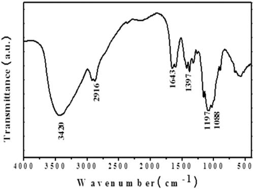
FTIR analysis was agreed to recognize the biomolecules which were accountable for the declining of metal ions into ZnO-NPs in the company of Allium cepa extract (. The phytochemical originated in the A. cepa extract were accountable for the configuration of a variety of nanoparticles. The FTIR range of A. cepa illustrated numerous assimilation peaks ranged from 3420 cm−1 to 500 cm−1. shows the representative FT-IR spectra obtained from A. cepa. The frequency ranges from 3420 cm−1 peaks are symbolized the O–H stretching tremor, and also attendance of carbohydrate and amino acids. The frequencies are 2916 cm−1 peaks were signifies the C–H stretching especially, lipids. Incidence ranges from 1643 cm−1 peaks are the Amide I: C = O elongating that is proteins. Then, the frequency ranges from 1397 cm−1 peak represents sulfur compounds. The frequency ranges from 1197 cm−1 peaks shows the C–N stretching: amino acids [Citation30].
Figure 3. FTIR spectra of ZnO-NPs synthesised from Allium cepa. Allium cepa extract were answerable for the configuration of a variety of nanoparticles. The FTIR range of A. cepa illustrated several assimilation peaks ranged from 3420cm−1 to 500cm−1. shows the representative FT-IR spectra obtained from A. cepa.
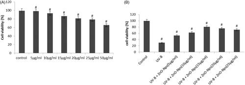
Cytotoxicity results of ZnO-NPs
The cytotoxicity of ZnO-NPs in HaCaT cells was appraised by MTT assays. demonstrates the cell viability assay for normal HaCaT cells were first analyzed in a concentration-dependent manner (5–50 μg/ml) in which the increased viability was normalized in 15 μg/ml. Subsequently, HaCaT cells were uncovered to ZnO-NPs (0.9–20 lg/ml) for 12 h. The MTT results established as concentration-dependent cytotoxicity following exposure to ZnO-NPs (). The proportion mitochondrial commotion experimental after 12 h disclosure at concentrations of 10 and 15 lg/ml was 95 and 85%, The CC50 value of ZnO-NPs at 12 h was 15 lg/ml as intended from the MTT assay.
Figure 4. Cytotoxicity results of ZnO-NPs synthesized from Allium cepa. The cell viability assay for normal HaCaT cells were first analyzed in concentration dependent manner (5–50 μg/ml) in which the increased viability was normalized in 15 μg/ml. Subsequently, HaCaT cells were uncovered to ZnO-NPs (0.9–20 lg/ml) for 12 h. The MTT results established as concentration dependent cytotoxicity following exposure to ZnO-NPs ().
Measurement of intracellular ROS
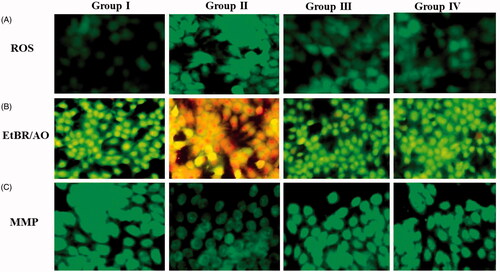
The cells uncovered with ZnO nanoparticles (15 μg/ml) for 6 h illustrated a noteworthy decline in the ROS generation. This was evident that DCF fluorescence when measured qualitatively as well as quantitatively (). Group I normal shows no reactive oxygen species formation. UVB irradiated (180 mJ/cm2) group II shows elevated ROS formation. ZnO nanoparticles (15 μg/ml) along with pretreatment of UVB illustrate declined ROS. Treatment of standard drug (paraamino benzoic acid) (15 µM/ml) along with UVB showed much regular normalized ROS formation.
Figure 5. (A) Inside the cell ROS generation was précised by DCFH-DA: (B)Fluorescence images of apoptotic morphology by dual staining (AO/EtBr): (C) Effect of Allium cepa on mitochondrial membrane potential (MMP). DCF fluorescence when measured qualitatively as well as quantitatively (). Group I normal shows no reactive oxygen species formation. UVB irradiated (180 mJ/cm2) group II shows elevated ROS formation. ZnO nanoparticles (15 μg/ml) along with pretreatment of UVB illustrates declined ROS. ZnO-NPs results in a dose-dependent decline in the number of viable cells and also augments in early apoptotic, late apoptotic, and necrotic cells (). Control group is near normal and does not show much colour change. Group II exposed with UVB radiation displayed declined viable cells that too necrotic cells. In addition, group III UVB+ Allium cepa extract of 15 μg/ml ZnO-NPs normalized number of viable cells which was comparable with the control. The control cells discharge high concentration of green fluorescence which represents polarized mitochondria membrane (). On the other hand, ZnO-NPs treated HaCaT cells were demonstrated noteworthy modification of ΔΨM which was frequently diminished green fluorescence shows in .
Acridine orange and ethidium bromide staining results
The lethality of ZnO-NPs results in a dose-dependent decline in the number of viable cells and also augments in early apoptotic, late apoptotic, and necrotic cells (). The AO/EB assay is appropriate for ZnO nanoparticles in accordance with cell membrane destabilization probable. Control group is near normal does not show much colour change. Group II exposed with UVB radiation displayed declined viable cells that too necrotic cells. In addition group III UVB+ A. cepa extract of 15 μg/ml ZnO-NPs normalized number of viable cells which was comparable with the control, with an associated augment in the number of early apoptotic cells. Cells uncovered to attentiveness of standard drug also (15 μg/ml) showed that late apoptotic cells and necrotic cells which become the gradual predominant cell type.
Effect of Allium cepa on mitochondrial membrane potential (MMP)
Near the beginning phase of apoptosis is prompted by modification of mitochondrial membrane potential which was evaluated by lipophilic cationic dye, Rho-123 (). The control cells discharge high concentration of green fluorescence which represents polarized mitochondria membrane (). On the other hand, ZnO-NPs treated HaCaT cells were demonstrated a noteworthy modification of ΔΨM which was frequently diminished green fluorescence shows in . Especially UVB alone treated group II displays no fluorescence. Standard drug discharges some fluorescence.
ZnO-NPs of Allium cepa treatment of inflammatory markers in HaCaT cells
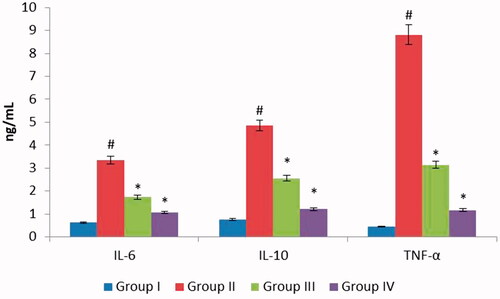
shows the ZnO-NPs of A.cepa treatment of inflammatory markers, in which UVB irradiated group II, illustrates prominent expressions of TNF-α, IL-6, and IL-10. In dissimilarity ZnO-NPs of A. cepa indulged groups III (15 μg/ml) corresponding pretreatment with A. cepa along with UVB demonstrate condensed graph results of TNF-α, IL-6, and IL-10. Group IV which was treated with para-amino benzoic acid displays normal diagram results of which were properly comparable with group 1 control.
Figure 6. ZnO-NPs of Allium cepa treatment of inflammatory markers in HaCaT cells. shows the ZnO-NPs of Allium cepa action of inflammatory markers, in which UVB irradiated group II, demonstrates high-flying expressions of TNF-α, IL-6, and IL-10. In difference, ZnO-NPs of Allium cepa pampered groups III (15 μg/ml) equivalent pre-treatment with Allium cepa along with UVB display reduced graph results of TNF-α, IL-6, and IL-10.
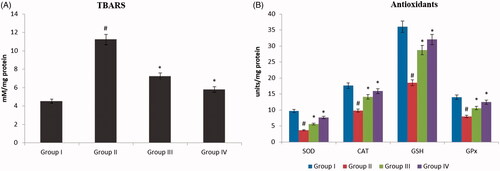
ZnO-NPs augment SOD/CAT/GPx and TBARS activity
Subsequently, we resoluted the consequence of ZnO-NPs pretreatment on the expression of genes encodes for enzymes of most important antioxidant protection system (SOD (superoxide dismutase), CAT (catalase) and GPx (glutathione peroxidase). UVB irradiation somewhat improved the gene expression level of these antioxidant enzymes as a contrast to untreated cells. Conversely, ZnO-NPs pretreated along with UVB radiation cells demonstrated faintly amplified expression of CAT and GPx along with a remarkable elevation in the expression of SOD. On the other hand, treatment with UVB alone cells did not show any momentous outcome on the expression level of these enzymes (). Fascinatingly, when we scrutinized the enzymatic activity of SOD, CAT and GPx, it was declined in UVB-irradiated cells, while elevated in ZnO-NPs pretreated cells with comparative to control cells. Same results were observed in TBARS levels. In control, the thiobarbituric acid reactive substances were normal in the graph. UVB irradiated (alone) group II displayed an elevated level of TBARS. In contrast, UVB irradiated along with ZnO-NPs shows recovered TBARS levels. Group IV standard drug treatment shows a slight increase in TBARS levels.
Figure 7. ZnO-NPs augment SOD/CAT/GPx and TBARS activity. UVB irradiation somewhat improved the gene expression level of these antioxidant enzymes as contrast to untreated cells. Conversely, ZnO-NPs pretreated along with UVB radiation cells demonstrated faintly amplified expression of CAT and GPx along with a remarkable elevation in the expression of SOD. On the other hand, treatment with UVB alone cells did not show any momentous outcome on the expression level of these enzymes (). Thiobarbituric acid reactive substances were normal in graph. UVB irradiated (alone) group II displayed elevated level of TBARS. In, contrast UVB irradiated along with ZnO-NPs shows recovered TBARS levels.
Investigation of apoptotic and signaling proteins
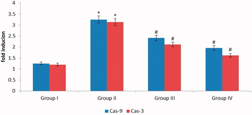
The plane of apoptotic signaling proteins was observed in cells exposed to ZnO-NPs for 9 h by Western blot analysis of important apoptotic proteins blotting (. The cells showed intonation of Bax and Bcl2 protein expression levels with an increase in Bax levels and a corresponding decrease in Bcl2. A significant increase in COX-2 and iNOS at the same time ZnO-NPs along with pretreated UVB recovered the apoptotic and other signaling molecules like COX-2 and iNOS. Likewise, the cascade molecules like caspase-9 & 3 dispaly the same as a comparison with apoptotic and proapoptotic molecules which was depicted in . Beta-actin treated as a standard marker.
Figure 8. Investigation of apoptotic and signaling proteins. showed modulation of Bax and Bcl2 protein expression levels with an augment in Bax levels and an equivalent decline in Bcl2. A noteworthy enhance in COX-2 and iNOS at the same time ZnO-NPs along with pretreated UVB recovered the apoptotic and other signaling molecules like COX-2 and iNOS.
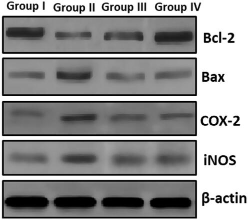
Figure 9. Apoptotic signaling cascade molecules. Similarly, the cascade molecules like caspase-9&3 exhibit the same as contrast with apoptotic signaling cascade molecules which was depicted in . Beta-actin treated as standard marker.
Discussion
Skin cancer vestiges most important health distress, predominantly in the industrial nation [Citation31]. Current study discloses the effects of ZnO nanoparticles on HaCaT cells and offers noteworthy impending into the probable mechanism all the way through which ZnO nanoparticles exert their toxic effects on these cells. Our outcome exhibits the cytotoxic potential of ZnO-NPs in HaCaT cells. Our records also exposed that the form of cell death was apoptosis which was arbitrated by the ROS simultaneously activates mitochondrial pathway as confirmation by a reduction in MMP, the inflection of Bax/Bcl2 ratio and breakage in cascade molecules like Caspase-9&3. ZnO-NPs were accounted to be generally less effective at low concentrations in HaCaT and mouse embryonic fibroblasts cells but displayed improved shielding competence with rising concentrations [Citation32].
UVB radiation is as well notorious to grounds for DNA injure throughout ROS production, which outcome from DNA corrosion [Citation33]. The UV-Vis assimilation spectrum of ZnO nanoparticles by flower extract of Nyctanthes tristis demonstrated a jagged absorbance at 369 nm [Citation34]. FTIR range of ZnO nanoparticles exposed that the peak at 417.52 cm−1 was the distinguishing incorporation of ZnO bond and the wide assimilation of peak at 3438 cm−1 accredited to the distinctive inclusion of hydroxyl group [Citation26]. The peak at 668.29 cm - 1 point out the extending vibrations of ZnO nanoparticle which was reliable with a previous report [Citation35]. Our finding coincides with preceding studies. Further, in XRD results, no additional peaks have been perceived in the sample and completed the macromolecule synthesis of ZnO-NPs pursued to high purity. The represented crystal mass was found to be 21 nm [Citation36].
ZnO-NPs produced by means of Trifolium pratense extract exposed that scanning electron microscope images of the ZnO-NPs were shuffled with an element size arrayed from 100–190 nm [Citation37]. In the present study, our SEM data which is closely related with preceding findings in which scuffled particles size ranges from 50–100 nm. Energy dispersive x-ray spectroscopy analysis of the previous report denotes that the size of the nanoparticle is more than the standard was considered from the Debye–Scherrer formula representing the mismatch of crystallites in ZnO-NPs[Citation38]. The emerald synthesis of zinc oxide nanoparticles using Moringa oleifera leaf extract the tough and fragile peaks are experimental from Zn and O atom and feeble peaks are pragmatic form P, K, Ca elements [Citation39]. Preceding finding shows that examination of the belongings of ZnO-NPs on epidermal cell lines as a replica for uncovered covering at the genomic altitude plus molecular levels. The outcome illustrated that 100 nm ZnO-NPs might modify normal cell cycle development, grounds for cell cycle detain that progress the defeat of feasibility of HaCaT cells, which was related with heritable phenotypic alteration and convoyed through p53 and Bax interceded apoptosis [Citation40].
ZnO-NP revelations have formerly be accounted to consequence in cytology transformation at different segments of the cell sequence [Citation6,Citation41,Citation42]. The uniqueness of ZnO-NPs, the dosage, the dealing time, and the cell line specification chosen might influence the consequences acquired [Citation43,Citation44]. The mRNA plane elevated almost fifty-fold over for methyltransferase gene and virtually fivefold for the methyltransferase gene following disclosure to 50 μg/mL A. cepa, which recommended the lysine workstation tails in histone (H3) may be optionally methylated by G9a and GLP genes [Citation45]. Consequently, this ZnO-NPs induced excess methylation of chromatin may direct the convenience of spoiled sections of DNA to renovate signaling molecules [Citation46].
Slighter elements have an elevated outside region per unit accumulation, resultant in the fabrication of extra ROS in the microenvironment. Reactive species produced by NPs be able to diminish innermost antioxidants, and amend mitochondrial utility, and grounds for oxidative smash up to DNA [Citation47,Citation48]. Also, an important study demonstrates that ROS contented altitude after healing with 20 μg/mL ZnO-NPs for 6 h, signifying the ROS detonation was a premature occurrence in reply to ZnO-NPs related pressure and arose previous to the fecundity of HaCaT cells diminish. The volatile ROS consequently founds DNA injure escorted by histone adjustment and alteration, which resulted in cell series detain. Our outcome also proved that the ROS level fell down to normal levels after 12 h. Nucleus among reduced and limited DNA and protein complex and mitochondria with withered, strong mold, and distended cristae were persuaded by 60–70 nm ZnO-NPs in colon cancer cells in humans [Citation49].
Previous ultrastructural characterization of ZnO-NPs was done by TEM. Though, the size achieved from TEM (100 nm) was slightly elevated than the size measured by dynamic light scattering (70 nm). This dissimilarity in mass is owed to the fact that dissimilar size resolved technique give unusual results based on the procedures engaged [Citation50]. The intervention of a few nanomaterials with frequently used cytotoxicity test scheme has been sound recognized in the researches. Consequently, it has been recommended that the cytotoxicity of nanomaterials ought to be evaluated with a best self-determining method for authenticating the results [Citation39,Citation51]. Previous work appraised the cytotoxicity of ZnO-NPs by MTT assay. The attempt exposed a cytotoxic probable of ZnO-NPs at 14 and 20lg/ml subsequent to 12 as well as 24 h disclosure in HepG2 cells [Citation1]. Oxidative stress is the main conversed pattern for the toxicity of NPs. This has been accredited to their tiny size and thus huge exterior area which is usually thought to manufacture ROS and oxidative trauma [Citation52].
The preceding study illustrates that ZnO-NPs were established to be able to produce intracellular ROS when observed by the cell porous dye DCFH-DA. This examination is dependable with the prior studies which have exposed comparable possessions on mouse embryo fibroblast and human lymphoblastoid cells [Citation53,Citation54]. This previous data coincides with our present data. Similar result established that the ROS fabrication would be considerably provoked by ZnO nanoparticles in HaCaT cells. Again another study illustrates that nanoparticles tempt intracellular oxidative trauma throughout ROS production in different cell lines [Citation55–57]. Numerous facets of ZnO-NPs in vivo, they preferred 5 categories of pro- and anti-inflammatory cytokines. Cytokine examination illustrated the on the whole cytokines (serum) in ZnO-fed animals reduce as a contrast with normal. Among many of cytokines, IL-10 and IL-1 β were outstanding in identifying the cellular kines alter in ZnO-NP animals. The diminished plane of these cytokines in ZnO-NP-fed animals may be recognized to distracted resistant repression [Citation32]. This was correlated with our finding in which UVB irradiated alone group elevated the interleukins 6&10 levels. At the same time, UVB along with ZnO-NPs restored the interleukins and related signaling molecules like iNOS, COX-2 levels. ZnO-NPs have been extensively implemented in cancer treatment and accounted to persuade discerning cytotoxic consequence on cancer cell propagation [Citation50].
The cytotoxicity of ZnO nanoparticles adjacent to coculture C2C12 myoblastoma cancer cells and 3T3-L1 adipocytes. Contrast to 3T3-L1 cells, it appeared that ZnO-NPs inhibited C2C12 cell propagation and grounds for a noticeable apoptosis through ROS arbitrated mitochondrial inherent apoptotic pathway and Bax/Bcl-2 proportion, p53, and caspase-3 alleyway [Citation58] These outcome coincide with our finding in which ZnO-NPs induce apoptotic and proapoptotic markers and also recommended that ZnO-NPs could provoke cancer cell apoptosis, which might additionally worked as a promising entrant for cancer rehabilitation.
Conclusion
The biological fabrication of metal nanoparticles is attractive in the field of chemistry, biology and material science. The hasty biological mixture of zinc nanoparticles using the extract of Allium cepa offers an ecologically friendly, easy and proficient way for the synthesis of nanoparticles. Zinc nanoparticles can survive in ions only in the attendance of strong oxidizing material. The process of ZnO nanoparticles is quiet in its immaturity and more investigation required to be paying attention to the mechanism of nanoparticle formation. The present outcome denotes A. Cepa synthesized from ZnO-NPs which induce apoptotic and proapoptotic markers and also induce cancer cell apoptosis, modification in ultrastructures, cellular cytotoxicity, ROS formation, antioxidants and TBARS levels, modification in interleukin molecules and necrosis factor. This might moreover facilitate as a talented candidate for cancer rehabilitation. The present study further requires in vivo experiments to understand the ZnONPs toxicity and other applications.
Disclosure statement
No potential conflict of interest was reported by the authors.
Correction Statement
This article has been republished with minor changes. These changes do not impact the academic content of the article.
References
- Sharma V, Anderson D, Dhawan A. Zinc oxide nanoparticles induce oxidative DNA damage and ROS-triggered mitochondria mediated apoptosis in human liver cells (HepG2). Apoptosis. 2012;17:852–870.
- Brayner R. The toxicological impact of nanoparticles. Nano Today. 2008;3:48–55.
- Nel A, Xia T, Meng H, et al. Nanomaterial toxicity testing in the 21st century: use of a predictive toxicological approach and high-throughput screening. Acc Chem Res. 2013;46:607–621.
- Nohynek G, Dufour E, Roberts M. Nanotechnology, cosmetics and the skin: is there a health risk? Skin Pharmacol Physiol. 2008;21:136–149
- Nohynek GJ, Lademann J, Ribaud C, et al. Grey goo on the skin? Nanotechnology, cosmetic and sunscreen safety. Crit Rev Toxicol. 2007;37:251–277.
- Gao F, Ma N, Zhou H, et al. Zinc oxide nanoparticles-induced epigenetic change and G2/M arrest are associated with apoptosis in human epidermal keratinocytes. Int J Nanomedicine. 2016;11: 3859–3874.
- Rasmussen JW, Martinez E, Louka P, et al. Zinc oxide nanoparticles for selective destruction of tumor cells and potential for drug delivery applications. Expert Opin Drug Deliv. 2010;7:1063–1077.
- Xiong HM. ZnO nanoparticles applied to bioimaging and drug delivery. Adv Mater Weinheim. 2013;25:5329–5335.
- Boukamp P, Popp S, Altmeyer S, et al. Sustained nontumorigenic phenotype correlates with a largely stable chromosome content during long-term culture of the human keratinocyte line HaCaT. Genes Chromosom Cancer. 1997;19:201–214.
- Yang X, Liu J, He H, et al. SiO2 nanoparticles induce cytotoxicity and protein expression alteration in HaCaT cells. Part Fibre Toxicol. 2010;7:1.
- Gius DR, Ezhevsky SA, Becker-Hapak M, et al. Transduced p16INK4a peptides inhibit hypophosphorylation of the retinoblastoma protein and cell cycle progression prior to activation of Cdk2 complexes in late G1. Cancer Res 1999;59:2577–2580.
- Meyer B, Fabbrizi MR, Raj S, et al. Histone H3 lysine 9 acetylation obstructs ATM activation and promotes ionizing radiation sensitivity in normal stem cells. Stem Cell Reports. 2016;137:1013–1022.
- Tabassum N, Hamdani M. Plants used to treat skin diseases. Phcog Rev. 2014;8:52.
- Shah NC. Status of cultivated and wild Allium species in India: a review. The Scitech Journal 2014;01:28–36.
- Ashalata D, Rakshit K, Sarania B. Ethnobotanical notes on Allium species of Arunachal Pradesh, India. Indian J Tradit Know. 2014;13:606–612.
- Draelos ZD. The ability of onion extracts gel to improve the cosmetic appearance of postsurgical scars. J Cosmet Dermat. 2008;7:101–104.
- Tensingh Baliah N, Lega Priyatharsini S. Biosynthesis and characterization of zinc oxide nanoparticles using onion bulb extract. IJTSRD. 2018;2:36–43.
- Sangeetha G, Rajeshwari S, Venckatesh R. Green synthesis of zinc oxide nanoparticles by Aloe barbadeneis Miller. leaf extract: structure and optical properties. Mat Res Bull. 2011;46:2560–2566.
- Mosmann T. Rapid colorimetric assay for cellular growth and survival: application to proliferation and cytotoxicity assays. J Immunol Methods. 1983;65:55–63.
- Reed LJ, Muench H. A simple method of estimating fifty percent endpoints. Am J Hy. 1938;27:493–497.
- Wan CP, Myung E, Lau BH. (An automated microfluorometric assay for monitoring oxidative burst activity of phagocytes. J Immunol Methods. 1993;159:131–138.
- Kim C-S, Nguyen H-D, Ignacio RM, et al. Immunotoxicity of zinc oxide nanoparticles with different size and electrostatic charge. Int. J Nanomed. 2014;9:195–205.
- Ahmed B, Solanki B, Zaidi A, et al. Bacterial toxicity of biomimetic green zinc oxide nanoantibiotic: insights into ZnONP uptake and nanocolloid–bacteria interface. Toxicol Res. 2019;8:246–261.
- Bradford MM. A rapid and sensitive method for the quantitation of microgram quantities of protein utilizing the principle of protein-dye binding. Anal Biochem. 1976;72:248–254.
- Melvin Joe M, Jayochitra J, Vijayapriaya M. Antimicrobial activity of some common spices against certain human pathogens. J Med Plants Res. 2009;3:1134–1136
- Jamdagni P, Poonam Khatri J, Rana S. Green synthesis of zinc oxide nanoparticles using flower extract of Nyctanthes arbor-tristis and their antifungal activity. J King Saud Univers Sci. 2016;3:417–432.]
- Guo L, Cheng JX, Li X-Y, et al. Synthesis and optical properties of crystalline polymer-capped ZnO nanorods. Mat Sci Eng. 2001;16:123–127.
- Sohaebuddin SK, Thevenot PT, Baker D, et al. Nanomaterial cytotoxicity is composition, size, and cell type dependent. Part Fibre Toxicol. 2010;7:22.
- Zhao F, Zhao Y, Liu Y, et al. Cellular uptake, intracellular trafficking, and cytotoxicity of nanomaterials. Small. 2011;7:1322–1337.
- Ahmed B, Dwivedi S, Abdin MZ, et al. Mitochondrial and chromosomal damage induced by oxidative stress in Zn 2+ ions, ZnO-bulk and ZnO-NPs treated Allium cepa roots. Sci Rep. 2017;7:40685.
- Elumalai K, Velmurugan S. Green synthesis, characterization and antimicrobial activities of zinc oxide nanoparticles from the leaf extract of Azadirachta indica (L.). Appl Surf Sci. 2015;345:329–336.
- Watson M, Thomas CC, Massetti GM, et al. CDC grand rounds: prevention and control of skin cancer. Am. J. Transpl. 2016;16:717–e720.
- Wu MS, Sun DS, Lin YC, et al. Nanodiamonds protect skin from ultraviolet B-induced damage in mice. J. Nanobiotechnology 2015;13:35.
- Narendhirakannan RT, Hannah MA. Oxidative stress and skin cancer: an overview. Indian J Clin Biochem. 2013;28:110–e115.
- Yuan Z, Wang J, Wang Y, et al. P reparation of a poly(acrylic acid) based hydrogel with fast adsorption rate and high adsorption capacity for the removal of cationic dyes. RSC Adv. 2019;9:21075–21085.
- Suresh D, Nethravathi PC, Rajanaika H, et al. Green synthesis of multifunctional zinc oxide (ZnO) nanoparticles using Cassia fistula plant extract and their photodegradative, antioxidant and antibacterial activities. Mater Sci Semicond Process. 2015;31:446–454.
- Zheng Y, Fu L, Han F, et al. Green biosynthesis and characterization of zinc oxide nanoparticles using Corymbia citriodora leaf extract and their photocatalytic activity. Green Chem Lett Rev. 2015;8:59–63.
- Dobrucka R, Długaszewska J. Biosynthesis and antibacterial activity of ZnO nanoparticles using Trifolium pratense flower extract. Saudi J Biol. Sci. 2016;23:517–523.
- Sharma V, Shukla RK, Saxena N, et al. DNA damaging potential of zinc oxide nanoparticles in human epidermal cells. Toxicol Lett. 2009;185:211–218.
- Mishra A, Mishra DK, Bohra NK. Synthesis and characterization of zinc oxide nanoparticles by Azadirachta indica leaves. Annals Arid Zone. 2015;54:43–49.
- Kocbek P, Teskač K, Kreft ME, et al. Toxicological aspects of long-term treatment of keratinocytes with ZnO and TiO2 nanoparticles. Small. 2010; 6:1908–1917.
- Mahmoudi M, Azadmanesh K, Shokrgozar MA, et al. Effect of nanoparticles on the cell life cycle. Chem Rev. 2011;111:3407–3432.
- Valdiglesias V, Costa C, Kiliç G, et al. Neuronal cytotoxicity and genotoxicity induced by zinc oxide nanoparticles. Environ Int. 2013;55:92–100.
- Heng BC, Zhao X, Tan EC, et al. Evaluation of the cytotoxic and inflammatory potential of differentially shaped zinc oxide nanoparticles. Arch Toxicol. 2011;85:1517–1528.
- Pujalté I, Passagne I, Brouillaud B, et al. Cytotoxicity and oxidative stress induced by different metallic nanoparticles on human kidney cells. Part Fibre Toxicol. 2011;8:10–16.
- Huyen Y, Zgheib O, DiTullio RA, Jr, et al. Methylated lysine 79 of histone H3 targets 53BP1 to DNA double-strand breaks. Nature. 2004;432:406–411.
- Sanders SL, Portoso M, Mata J, et al. Methylation of histone H4 lysine 20 controls recruitment of Crb2 to sites of DNA damage. Cell. 2004;119:603–614.
- Li N, Sioutas C, Cho A, et al. Ultrafine particulate pollutants induce oxidative stress and mitochondrial damage. Environ Health Perspect. 2003;111:455.
- Møller P, Jacobsen NR, Folkmann JK, et al. Role of oxidative damage in toxicity of particulates. Free Radic Res. 2010;44:1–46.
- De Berardis B, Civitelli G, Condello M, et al. Exposure to ZnO nanoparticles induces oxidative stress and cytotoxicity in human colon carcinoma cells. Toxicol Appl Pharmacol. 2010;246:116–127.
- Dhawan A, Sharma V. Toxicity assessment of nanomaterials: methods and challenges. Anal Bioanal Chem 2010;398:589–605.
- Monteiro-Riviere NA, Inman AO, Zhang LW. Limitations and relative utility of screening assays to assess engineered nanoparticle toxicity in a human cell line. Toxicol Appl Pharmacol. 2009;234:222–235.
- Xia T, Kovochich M, Brant J, et al. Comparison of the abilities of ambient and manufactured nanoparticles to induce cellular toxicity according to an oxidative stress paradigm. Nano Lett. 2006;6:1794–1807.
- Yang H, Liu C, Yang D, et al. Comparative study of cytotoxicity, oxidative stress and genotoxicity induced by four typical nanomaterials: the role of particle size, shape and composition. J Appl Toxicol. 2009;29:69–78.
- Yin H, Casey PS, McCall MJ, et al. Effects of surface chemistry on cytotoxicity, genotoxicity, and the generation of reactive oxygen species induced by ZnO nanoparticles. Langmuir. 2010;26:15399–15408.
- Xia T, Kovochich M, Liong M, et al. Comparison of the mechanism of toxicity of zinc oxide and cerium oxide nanoparticles based on dissolution and oxidative stress properties. ACS Nano. 2008;2:2121–2134.
- Akhtar MJ, Ahamed M, Kumar S, et al. Nanotoxicity of pure silica mediated through oxidant generation rather than glutathione depletion in human lung epithelial cells. Toxicology. 2010;276:95–102.
- Jiang J, Pi J, Cai J. The advancing of zinc oxide nanoparticles for biomedical applications. Bioinorg Chem Appl. 2018;2018:1062562. https://doi.org/10.1155/2018/1062562.