
Abstract
Nanotechnology is creating a bang in each and every field of life science. Scientists are mounting their interest of research towards gold nanoparticles as they are capable with bigger and advanced properties.Traditionally nanoparticles have been manufactured by various chemical and physical methods but have negative impact on the environment and are also highly toxic. Synthesis of nanoparticles by using plant extracts is substituting the conventional methods and it is eco-friendly too. In the current study, we prepared gold nanoparticles (AuNPs) from Magnolia officinalis, which is identified as an eco-friendly and less toxic method. Incorporation of AuNPs was renowned by UV-absorbance and it shows peak values. Nanoparticle sizes are recognized by dynamic light scattering scrutiny and it shows a value of 128 nm. Besides, high resolution transmission electron microscopy (HR-TEM), energy dispersive X-ray analysis (EDX) and atomic force microscopy (AFM) incorrigibly define the shape of the AuNPs which are present in the complex materials. Fourier-transform infrared spectroscopy (FTIR) findings display that the active molecules are positioned in the plane of the AuNPs. Similarly, anticancer efficacy of AuNPs have been assessed in A549 cells. our study show that AuNPs effectively provoke cytotoxicity, and apoptosis by inflecting apoptotic gene expressions in A549 cells.
Introduction
Most challenging notion in anti-tumour embattled drug delivery system production is to extend the suitable carter tools [Citation1]. Nanotechnology is a multi faceted field that engages the plan and engineering of practical systems at the molecular level. This interdisciplinary approach deals with separation, categorization and application of resources and procedure on nanolevel [Citation2]. Nano rescue systems are capable of meticulous identification of tumour tissues, which sanction the utilization of the enhanced diffusion and preserving effect [Citation3]. Numerous chemicals are used to expand as nanoparticles for the encouraging effects. Equally, those chemicals are harmful to the atmosphere and expensive; at the same time, natural metal nanoparticles are deliberated as ecological and economical [Citation4,Citation5]. Several nanoparticles based drug delivery systems have been anticipated to achieve the continually evolving needs of the fundamental and medical research fields.
In most of the South Asian countries especially in India and China, gold is used as healthiness adjuvant in the form of powdered metal as “bhasma” for dealing with numerous health problems and deficits like escalating crucial power and for male impotency treatments [Citation6]. When compared to other scrutinized metallic particles, gold nanoparticles (AuNPs) are investigated for various nano-related medical applications, bearing in mind their nontoxic nature and distinctive visual, biological properties [Citation7]. Important characteristics of nanomaterials are morphology and size distribution. Size reliant quantum internment performance of gold nanoparticles surface displays an unusual surface Plasmon resonance, resulting in sturdy annihilation of blistering illumination. This sole activity is a special feature of AuNPs, this characteristic is lost in massive substance [Citation8]. AuNPs, are catalytically energetic for CO oxidation reactions with selective and, as well as, for water gas transfer response [Citation9]. Lung cancer is the foremost cause of cancer deaths all over the world. The efficiency of modern treatment is relentlessly partial, with an age-standardized death rate [Citation10,Citation11]. Cigarette smoking and exposure to too much of tobacco is the main cause of the rising lung cancer cases [Citation12,Citation13].
Numerous other factors are also connected with lung cancer development such as exposure to carcinogens such as asbestos, silica, radon, arsenic and air pollution [Citation14,Citation15]. Habitual utilization of conventional phytochemicals is capable of lessening the risk and enlargement of definite cancers [Citation16]. Magnolia officinalis belongs to Magnoliaceae family and is used for many disorders like cough, body pain, nervousness, gastrointestinal disorders and sensitive allergic diseases. Root and stem of Magnolia officinalis was shown to have anti-inflammation and antioxidant properties, muscle relaxation property and effective against cardiovascular diseases [Citation17–20]. Previous studies prove that Magnolol displays anti-cancer activity by reducing differentiation, proliferation, and restrains angiogenesis, counteracting metastasis [Citation21–24].
In addition, Magnolol is effective against human non-small-cell lung carcinoma (NSCLC) cells, which activate apoptosis, inhibits proliferation and induce autophagy [Citation25]. Conversely, Magnolia officinalis’ effectiveness is unknown in A549 lung cancer cells. In this study, we fashioned gold nanoparticles (AuNPs) from ethanolic extract of Magnolia officinalis and it was demonstrated by several studies such as Fourier-transform infrared spectroscopy (FTIR) with selected area diffraction, UV-visible absorbance spectrum dynamic light scattering analysis (DLS), and energy dispersive X-ray analysis (EDX), high resolution-transmission electron microscope (HR-TEM), size of AuNPs was determined by atomic force microscopy image (AFM). Furthermore, manufactured AuNPs subjugated the anticancer prospective studies by apoptotic induction and regulation, cytotoxic efficacy, intracellular ROS production in A549 lung cancer cells.
Materials and methods
Chemicals, solvents and antibodies
Antibodies used in the study are Bax, Beclin-1, Bcl-2, Bid, β-actin, caspase-3 and goat anti-mouse IgG-HRP. Polyclonal antibodies were purchased from the suppliers in Santacruz, USA. Dulbecco’s Modified Eagles Medium (DMEM), antibiotics, trypsin-EDTA and Fetal bovine serum (FBS), was obtained from Himedia, Mumbai, India. 3–(4,5-dimethyl-2-thiaozolyl)-2,5-diphenyl-2H tetrazolium bromide (MTT), 2,7-diacetyl dichlorofluorescein (DCFH-DA), acridine orange (AO), propidium iodide (PI), 4′,6-diamidino-2-phenylindole (DAPI), Terminal dUTP Nick-End Labeling (TUNEL) were purchased from Sigma chemical, USA. Other fine chemicals of analytical grade were used in this study.
Gold nanoparticles (AuNPs) synthesis
Magnolia officinalis leaves were dilapidated to make the aqueous extract. The sterile leaves from Magnolia officinalis weighing about 20–25 g are dipped in double-distilled H2O. Then it was tolerable to dry and compressed into 100 mL sterile distilled water then it was filtered by Whatman No.1 paper. For separation of gold metal, the digestive budding method was used to separate gold nanoparticles from polyscattering nanoparticles using the digestive budding agents. Pursued by warming colloidal suspension at renowned temperatures (∼140 °C) for 3 min followed by dropping of temperature in addition to disulfide or alkanethiols. Temperature plays a crucial role in devising the size allocation of the gold colloids.
Characterization of AuNPs synthesized from Magnolia officinalis
AuNPs were confirmed by several characterization experiments are recognized by the size, shape and morphological uniqueness of nanoparticles. The produced AuNPs was embedded by Ultraviolet-visible absorbance spectrum studies at wavelength ranges of 300–750 nm. Size and morphological distinctiveness of AuNPs were documented and defined by HR-TEM and AFM images. Nanoparticles nature and inflexibility was studied by EDX. FTIR spectroscopy was executed to observe the active compounds present in the produced AuNPs. The FTIR spectra of AuNPs were predictable in the range of 1000 and 4000 cm−1 in KBr pellets by means of FTIR spectrophotometer. Size and surface morphology of the nanoparticles were resolved by DLS.
Cell culture
Lung adenocarcinoma (A549) cells were used for the present study and were obtained from Institute of Biochemistry and Cell Biology, Chinese Academy of Sciences (Shanghai, China). Cells were potted with DMEM medium enhanced with 10% fetal bovine serum (FBS) and 1% antibiotics at 37 °C in a dampened incubator at 5% CO2. After 4 days the culture was subjugated to ∼80% confluent and the cells were reaped with 1 mL of trypsin-EDTA solution. Now the cells were ready for further studies.
Cell viability assay
A549 lung adenocarcinoma cells (5000) were harvested and placed in 96-well plates and it was nurtured for 24 h at 37 °C. Then, merged AuNPs from Magnolia officinalis were treated with different concentrations (20–100 µg/mL) and incubated for another 24 h for examining the cytotoxicity potential at 37 °C. Then, the cells were exposed with MTT reagent (1 mg/mL) and kept for 4 h to ensure the development of purple formazan. DMSO (200 μL) was added into the well to hang up formazan crystals, and then the optical density was deliberated at 600 nm using a microplate reader.
Measurement of intracellular ROS
AuNPs depended production of ROS generation was intended by addidion of 2,7-diacetyl dichlorofluorescein (DCFH-DA) staining in A549 [Citation26]. Then the aliquoted cells (1 × 10⁶ cells/mL) were prepared and placed in 6 well plates. Then the cells were pampered with 15 and 20 µg/mL concentrations of AuNPs at 24 h incubation. Then 1 μL of DCFH-DA (1 mg/mL) was added to all the wells and kept in dark environmental condition. The fluorescent intensity was premeditated by spectrofluorimeter with equal emission and excitation at 490 ± 10 and 540 ± 12 nm, respectively.
TUNEL assay
AuNPs arbitrated nuclear disintegration was scrutinized by Sigma TUNEL assay kit. After exposure of AuNPs, the A549 cells were washed with Phosphate Buffer Saline (PBS) twice, then fixed with 2% paraformaldehyde and incubated for 30 min, and permeabilized with Triton X-100 (0.1%) for 30 min. Then, nurtured with TUNEL reaction buffer at 37 °C for 1 h (dark) and again soaked twice with PBS. At last cells were tainted with DAPI then it was envisioned with a fluorescence microscope (Nikon).
Western blot studies
The protein expression of Bax, Beclin-1, Bcl-2, Bid, and caspase-3 were compared with standard β-actin studied by western blotting analysis. Treated cells are scraped and proteins were extracted by addition of RIPA and protease inhibitor. The extracted protein concentrations were analyzed by the Thermo scientific nanodrop instrument. For, SDS-PAGE, 10% gel were placed to electrophoresis. Afterwards, the separated proteins were transferred to PVDF membranes and it was blocked with an addition of 5% bovine serum albumin for about 2–3 h. The membranes were pampered with an appropriate primary antibody and it set aside for 24 h incubation at 4 °C. Furthermore, it has nurtured with apt conjugated horseradish peroxidase secondary antibodies for 1 h. Bands were recognized by using enhanced chemiluminescence substrates and the intensity of bands were analyzed by Image J Software.
Statistical analysis
All experiments were conducted in triplicates and the results were displayed as the mean ± standard deviation (mean ± SD) by means of one-way investigation of conflict (ANOVA). Values of p < .05 were pinpointing of significant differences.
Results and discussion
Although there have been great developments in diagnostic techniques, surgical treatments and novel anti-cancer agents, the outcome of patients with lung cancer has enhanced discreetly over the earlier period. When compared to other metallic compound gold is eco-friendly and low cost. Synthesis of nanoparticles from plant extracts is an uncomplicated practice, in which metal salt diverges from the plant extract and completion of reaction takes place within a few hours. Specifically, gold nanoparticles (AuNPs), are harmless in contrast to other metallic nanoparticles [Citation27,Citation28]. Nanoparticles severance in plants can be favorable above other organic methods by eliminating the convoluted process of conserving cell cultures [Citation29]. In the current study, we have established that preparation of gold nanoparticles from Magnolia officinalis and it was exemplified by UV-visible absorbance spectrum based DLS, HR-TEM and AFM, in addition to FTIR and EDX. Nuclear fragmentation studies were carried by TUNEL assay. Finally, fabrication of AuNPs subjugates the anticancer prospective studies by apoptotic regulation in Lung adenocarcinoma (A549) cells.
Characterization of AuNPs blended from Magnolia officinalis
The production of AuNPs from Magnolia officinalis was first and foremost indicated by the switch over of colour, and the formation of red colour (ruby) specifies the formation of the AuNPs (. The change in colour was precisely timed to the excitation of surface plasmon resonance blended AuNPs. UV-spectroscopy was used at different time periods while hoaxing the concentration of the Magnolia officinalis and the aqueous solution of gold chloride. The optical sign of the colour switch over and the formation of red colour (ruby) specified the development of the AuNPs; that express the pattern of gold nanoparticles (). This associated data was formerly established by several researches on the amalgamation of gold nanoparticles [Citation30]. Furthermore, we established the Magnolia officinalis mediated the produced AuNPs by UV visible absorbance.
Figure 1. (A) UV-visible absorption spectrum of synthesized AuNPs. (B) Dynamic light scattering (DLS) images of AuNPs synthesized from Magnolia officinalis and the size of the nanoparticles is 128 nm.
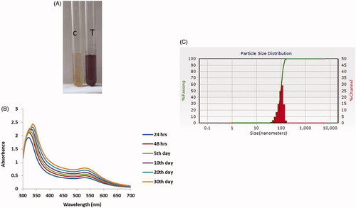
A rising deliberation of Magnolia officinalis extracts that leads to increased intensity of absorption. The UV-visible spectra were documented at time intervals such as of 24, 48, 72 and 92 h from the commencement of reaction with a diverse amount of plant extract. This result corresponds with prior studies in which UV-Vis spectroscopy was used to observe the size and shape of the restricted nanomaterial in the aqueous deferments [Citation31]. The current study illustrates the produced AuNPs exhibit maximum absorbance in the 535 nm range at 96 h. The highest absorbance bands were elevated at different concentrations of AuNPs solution at diverse time positions ().
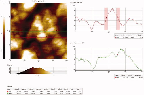
In contrast to other metal nanoparticles, primarily, AuNPs enclose free electrons which put in SPR absorption bands owing to the combined shuddering of electrons of metal nanoparticles in significance with lightwave [Citation32]. Size distributions of amalgamated gold nanoparticles from Magnolia officinalis extract are studied by DLS method. Histogram image specifies the particle size of the AuNPs from Magnolia officinalis is 128 nm (). AFM confirms the size of the developed nanoparticles. In () accurate AFM images afford indulged power during movement of the AuNPs which coincide with previous findings [Citation33]. Moreover, aggregated particles were not observed in the results of particle allocation and size.
Figure 2. Atomic force microscopyanalysis of gold nanoparticles synthesized from Magnolia officinalis.
Furthermore, morphological individuality of AuNPs produced from Magnolia officinalis was supplementary measured and deliberate by HR-TEM analysis (). The high resolution-TEM image illustrates that the morphological arrangement of AuNPs which are observed in, oval, spherical, hexagonal, and triangular with average sizes between 70 and 10 nm. This verdict supports the significantly interconnected with DLS images for AuNPs synthesized from Magnolia officinalis. Energy dispersive studies were scrutinized to evaluate the shape of the particles produced from Magnolia officinalis extract (). Expansion of gold nanostructures was confirmed by TEM figures, which give high-density AuNPs produced by the Magnolia officinalis extract.
Figure 3. (A) High resolution transmission electron microscopy (HR-TEM) and (B) Energy dispersive X-ray (EDX) and analysis of AuNPs synthesized from Magnolia officinalis.
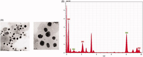
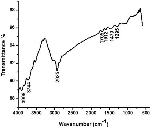
In the present study, a strong signal was acknowledged in the gold region which authenticate the AuNPs formation. In the current study, we acquired the sturdy signals of Au (1.5–2 keV and 2–3.5 keV). In surface plasmon resonance the unique ocular assimilation for metallic crystal was usually found to be 3 KeV [Citation34]. Finally, FTIR is used to distinguish the several functional groups which are there in the synthesized AuNPs from Magnolia officinalis extract by acquiring dissimilar spectrum values. In current study, FTIR showed that peaks at 3908, 3744, 3500, 3255, 3010, 2925, 1705, 1612, 1479 and 1295 cm−1 which identify the aromatic, hydroxyl, carboxyl alkyl and amide groups that are nearby to the surface of AuNPs (. Current finding associates with prior studies in which production of AuNPs from diverse plants extracts which have several functional groups [Citation35].
Figure 4. Fourier transforms infrared (FTIR) spectroscopy analysis of AuNPs synthesized from Magnolia officinalis.
Anticancer potential of AuNPs from Magnolia officinalis lung adenocarcinoma (A549) cells
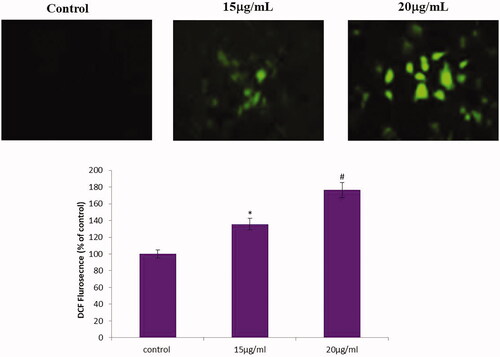
AuNPs were studied for their anticancer actions against A549 cells. Accordingly, anticancer prospective of AuNPs were deliberated by cytotoxicity assay (MTT). In which, elevated AuNPs concentration significantly reserved the A549 cells augmentation for 24 h incubation (. Therefore, we establish that 50 µg/mL concentrations of AuNPs exposed outstanding cell death in A549 cells. Therefore, we have chosen 50 µg/mL concentrations of AuNPs for further apoptotic studies. Amalgamated AuNPs from Dendropanax morbifera leaf extract shows noteworthy anticancer activity in HaCaT cells [Citation36]. In cellular microenvironment, intracellular reactive oxygen species (ROS) in a system’s organization persuades oxidative stress which results in apoptosis [Citation37]. Subsequently, AuNPs arbitrated ROS fabrication was scrutinized by DCFH-DA fluorescent assay. In the current study, we established that AuNPs synthesized from Magnolia officinalis provoke ROS production in A549 cells (. In summary, elevated ROS directs to the fragmentation of nucleus and depolarization of mitochondria, which results in oxidative stress interceded apoptosis [Citation38,Citation39].
Figure 5. Cytotoxic potential of AuNPs from Magnolia officinalis in A549 cells. This experiment was repeated thrice and the bars in the graph represent S.E. (*p < .05)
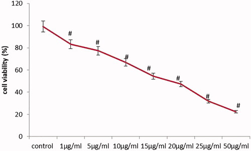
Figure 6. Effect of AuNPs from Magnolia officinalis arbitrated ROS measurements in A549 cells. This experiment was repeated thrice and the bars in the graph represent S.E. (*p < .05, #p < .01).
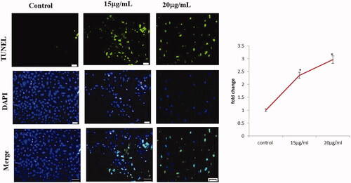
Expansion and succession of tumour and confrontation to several oncologic healing management results in mostly scarce apoptotic stimuli. Apoptosis is a central regulator in tissue maintenance and homeostasis [Citation40]. Accordingly, apoptosis is meticulous and significant alleyway for cancer treatment. AuNPs produced Magnolia officinalis aggravate ROS arbitrated apoptosis was assessed by DAPI/TUNEL dual staining. In the current study, control cells illustrate TUNEL assay stained with green fluorescent which indicates viable cells. Whereas action with AuNPs produced Magnolia officinalis confirmed increased apoptotic cells by scrutinizing DAPI with blue fluorescent cells (. Fragmented nucleus has been considered as imprinted occurrence to authenticate whether the cells are endured apoptosis A549 cells were uncovered with 50 µg/mL of AuNPs for 24 h; final outcome explains the improved number of TUNEL-positive cells. Current findings show that AuNPs influence fragmentation of DNA that results in apoptosis in A549 cells. Preceding findings demonstrate that TUNEL assay is generally engaged to differentiate apoptosis-provoked DNA fragmentation [Citation41].
Figure 7. AuNPs mediated apoptotic morphological studied by TUNEL assay and DAPI/PI staining.
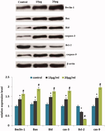
AuNPs intervened apoptosis makers such as Bax, Bcl-2, Bid, β-actin, Beclin-1, caspase-3 were scrutinized by western blotting. shows treatment with AuNPs produced from Magnolia officinalis diminished the expression of Bcl-2, Bid and elevated expression of Bax, Beclin-1 and caspase-3 in A549 cells. This data in a straight line relates DAPI and TUNEL assay. Finally, with these findings, AuNPs produced from Magnolia officinalis effectively convinced apoptosis by adjusting the apoptotic pathway. Preceding studies hold up our current findings in which gold nanoparticles convince apoptosis in human breast cancer cells (MCF-7) [Citation42].
Figure 8. Western blotting images for Bax, Bcl-2, Bid, β-actin, Beclin-1, caspase-3 protein expression in AuNPs treated A549 cells.
Conclusions
Finally, we exemplify an easy, quick, and reproducible technique for the eco-friendly production of AuNPs devoid of classy reducing agents. The efficacy of AuNPs produced from Magnolia officinalis is well established. The physio-chemical uniqueness of production of AuNPs from Magnolia officinalis is recognized by UV-absorbance, DLS, FTIR, EDX, and HR-TEM. AuNPs effectively persuade cytotoxicity and apoptosis by intonating intrinsic apoptotic gene expressions in A549 cells. So, ultimately our study corroborates that production of AuNPs from Magnolia officinalis exhibit anticancer effects.
Conflicts of interest
All the authors declare that there is no potential conflict of interest.
Additional information
Funding
References
- Semenza GL. Targeting HIF-1 for cancer therapy. Nat Rev Cancer. 2003;3:721–732.
- Sepeur S. Nanotechnology: technical basics and applications. Hanover (Germany): Vincentz Network GmbH & Co KG; 2008.
- Maeda H. The enhanced permeability and retention (EPR) effect in tumour vasculature: The key role of tumour-selective macromolecular drug targeting. Adv Enzyme Regul. 2001;41:189–207.
- Sharma HS, Ali SF, Hussain SM, et al. Influence of engineered nanoparticles from metals on the blood-brain barrier permeability, cerebral blood flow, brain edema and neurotoxicity. An experimental study in the rat and mice using biochemical and morphological approaches. J Nanosci Nanotechnol. 2009;9:5055–5072.
- Raveendran P, Fu J, Wallen SL. Completely “green” synthesis and stabilization of metal nanoparticles. J Am Chem Soc. 2003;125:13940–13941.
- Daniel MC, Astruc D. Gold nanoparticles: assembly, supramolecular chemistry, quantum-size-related properties, and applications toward biology, catalysis, and nanotechnology. Chem Rev. 2004;104:293–346.
- Tripathi RM, Shrivastav A, Shrivastav BR. Biogenic gold nanoparticles: as a potential candidate for brain tumour directed drug delivery. Artif Cell Nanomed B. 2014;43:1–7.
- Pattnaik P. Surface plasmon resonance: applications in understanding receptor-ligand interaction. Appl Biochem Biotechnol. 2005;126:79–92.
- Andreeva D. Low temperature water gas shift over gold catalysts. Gold Bull. 2002;35:82–88.
- Bepler G. Lung cancer: provoking new concepts, generating novel ideas, and rekindling enthusiasm. Canc Contr. 2003;1:275–276.
- Kumar R, Lu SK, Minchom A, et al. A phase 1b trial of the combination of an all-oral regimen of capecitabine and erlotinib in advanced non-small cell lung cancer in Caucasian patients. Cancer Chemother Pharmacol. 2016;77:375–383.
- Vineis P, Wild CP. Global cancer patterns: causes and prevention. Lancet. 2014;383:549–557.
- Sheervalilou R, Ansarin K, Fekri Aval S, et al. An update on sputum MicroRNAs in lung cancer diagnosis. Diagn Cytopathol. 2016;44:442–449.
- Torre LA, Bray F, Siegel RL, et al. Global Cancer Statistics, 2012. CA Cancer J Clin. 2015;65:87–108.
- Youlden DR, Cramb SM, Baade PD. The international epidemiology of lung cancer: geographical distribution and secular trends. J Thorac Oncol. 2008;3:819–831.
- Mehta RG, Murillo G, Naithani R, et al. Cancer chemoprevention by natural products: how far have we come? Pharm Res. 2010;27:950–961.
- Seo JU, Kim MH, Kim HM, et al. Anticancer potential of magnolol for lung cancer treatment. Arch Pharm Res. 2011;34:625–633.
- Tse A-W, Wan C-K, Zhu G-Y, et al. Magnolol suppresses NF-kappaB activation and NF-kappaB regulated gene expression through inhibition of IkappaB kinase activation. Mol Immunol. 2007;44:2647–2658.
- Ho KY, Tsai CC, Chen CP, et al. Antimicrobial activity of honokiol and magnolol isolated from Magnolia officinalis. Phytother Res. 2001;15:139–141.
- Fong WF, Tse AK, Poon KH, et al. Magnolol and honokiol enhance HL-60 human leukemia cell differentiation induced by 1, 25-dihydroxyvitamin D3 and retinoic acid. Int J Biochem Cell Biol. 2005;37:427–441.
- Hwang ES, Park KK. Magnolol suppresses metastasis via inhibition of invasion, migration, and matrix metalloproteinase-2/-9 activities in PC-3 human prostate carcinoma cells. Biosci Biotechnol Biochem. 2010;74:961–967.
- McKeown BT, Hurta RA. Magnolol affects expression of IGF-1 and associated binding proteins in human prostate cancer cells in vitro. Anticancer Res. 2014;34:6333–6338.
- Kang YJ, Park HJ, Chung HJ, et al. Wnt/beta-catenin signaling mediates the antitumour activity of magnolol in colorectal cancer cells. Mol Pharmacol. 2012;82:168–177.
- Li ML, Zhang F, Wang XA, et al. Magnolol inhibits growth of gallbladder cancer cells through the p53 pathway. Cancer Sci. 2015;106:1341–1350.
- Shen J, Ma H, Zhang T, et al. Magnolol inhibits the growth of non-small cell lung cancer via inhibiting microtubule polymerization. Cell Physiol Biochem. 2017;42:1789–1801.
- Balupillai A, Gunaseelan S, Govindasamy K, et al. Linalool prevents oxidative stress activated protein kinases in single UVB-exposed human skin cells. PLOS One. 2017;12:e0176699.
- Mittal AK, Chisti Y, Banerjee UC. Synthesis of metallic nanoparticles using plant extracts. Biotechnol Adv. 2013;31:346–356.
- Rai M, Yadav A, Gade A. Current [Corrected] trends in phytosynthesis of metal nanoparticles. Crit Rev Biotechnol. 2008;28:277–284.
- Kumar V, Yadav SK. Plant-mediated synthesis of silver and gold nanoparticles and their applications. J Chem Technol Biotechnol. 2008;84:151–157.
- Aljabali A, Akkam Y, Al Zoubi M, et al. Synthesis of gold nanoparticles using leaf extract of Ziziphus zizyphus and their antimicrobial activity. Nanomaterials 2018;8:174.
- Zhang XF, Liu ZG, Shen W, et al. Silver nanoparticles: synthesis, characterization, properties, applications, and therapeutic approaches. Int J Mol Sci. 2016;17:1534.
- Eustis S, El-Sayed MA. Why gold nanoparticles are more precious than pretty gold: noble metal surface plasmon resonance and its enhancement of the radiative and nonradiative properties of nanocrystals of different shapes. Chem Soc Rev. 2006;35:209–217.
- Kaviya S, Santhanalakshmi J, Viswanathan B, et al. Biosynthesis of silver nanoparticles using citrus sinensis peel extract and its antibacterial activity. Spectrochimica Acta Part A: Mol Biomol Spectroscop. 2011;79:594–598.
- Darwich S, Mougin K, Rao A, et al. Manipulation of gold colloidal nanoparticles with atomic force microscopy in dynamic mode: influence of particle–substrate chemistry and morphology, and of operating conditions. Beilstein J Nanotechnol. 2011;2:85–98.
- Palaniselvam K, Yusoff MM, Maniam GP, et al. Biosynthesis of metallic nanoparticles using plant derivatives and their new avenues in pharmacological applications – an updated report a review. Saudi Pharmac J. 2016;24:473–484.
- Wang C, Mathiyalagan R, Ju Kim Y, et al. Rapid green synthesis of silver and gold nanoparticles using Dendropanax morbifera leaf extract and their anticancer activities. Int J Nanomed. 2016;11:3691–3701.
- Donika I, Zhelev Z, Aoki I, et al. Overproduction of reactive oxygen species - obligatory or not for induction of apoptosis by anticancer drugs. Chin J Cancer Res. 2016;28:383–396.
- Guo C, Sun L, Chen X, et al. Oxidative stress, mitochondrial damage and neurodegenerative diseases. Neural Regen Res. 2013;8:2003–2014.
- Zorov DB, Juhaszova M, Sollott SJ. Mitochondrial reactive oxygen species (ROS) and ROS-induced ROS release. Physiol Rev. 2014;94:909–950.
- Nagata S. Apoptosis and clearance of apoptotic cells. Annu Rev Immunol. 2018;36:489–517.
- Groscurth P, Kressel M. Distinction of apoptotic and necrotic cell death by in situ labelling of fragmented DNA. Cell Tissue Res. 1994;278:549–556.
- Shyur LF, Chen CH, Lo CP, et al. Induction of apoptosis in MCF-7 human breast cancer cells by phytochemicals from Anoectochilus formosanus. J Biomed Sci. 2004;11:928.