 ?Mathematical formulae have been encoded as MathML and are displayed in this HTML version using MathJax in order to improve their display. Uncheck the box to turn MathJax off. This feature requires Javascript. Click on a formula to zoom.
Abstract
While there are many comorbidities associated with obesity, one of the more poorly understood is knee osteoarthritis through obesity. The purpose of this study was to compare the kinematics and kinetics of gait and cumulative knee adductor load, which represents the sum of repetitive exposures to medial knee loading during daily activity, between young obese adults with young, healthy-weight adults. Eight obese and eight healthy-weight young adults participated. Data from a three-dimensional motion capture system and a synchronized floor-mounted force plate were collected during gait trials. Participants wore accelerometers to determine step counts for seven consecutive days. Dependent t-tests were used to identify differences in gait kinematics, kinetics and cumulative knee adductor load between groups. Compared to the healthy-weight participants, obese young adults demonstrated a slower walking speed, greater stance duration, less knee flexion at heel contact, greater knee adduction in early stance and less knee abduction at terminal stance (p < 0.05). The obese young adults had a greater external knee extension moment (p < 0.05) and external rotation moment (p < 0.05) in early stance. The obese group had a greater cumulative knee adductor load. These results provide insight into a potential pathway by which obesity predisposes a healthy young adult for knee osteoarthritis.
Public Interest Statement
This research paper analyzed the effect of obesity on knee joint loading in a young adult population. Obesity is an overwhelming public health problem. Rates of obesity are rising in most segments of the population, including young adults. Obesity is associated with a myriad of health problems, including osteoarthritis of the knee. The link between obesity and osteoarthritis is multifactorial, but likely includes increased loading and alterations in the biomechanics of the joints of the lower extremity with increased weight gain. Identifying biomechanical risk factors for osteoarthritis development in young adults prior to the onset of musculoskeletal disorders may help with applying appropriate interventions. The present study found much more variable walking patterns and greater cumulative loading of the knee in obese young adults than healthy-weight young adults. This work and associated biomechanical research is imperative to preserving the optimal musculoskeletal health of young, obese adults throughout their lives.
Competing interests
The authors declare no competing interest.
1. Introduction
Among Canadian adults between 20 and 39 years of age, obesity rates have risen from 7 to 19% in men, and from 4 to 21% in women over the last 30 years (Shields et al., Citation2010). Obesity elevates the risk for comorbidities, including musculoskeletal disorders such as osteoarthritis (OA) (Orpana, Trembley, & Fines, Citation2007). The increasing rates of obesity among young adults raise concern over the potential increase in the incidence of knee OA. Young adults represent an age group that has just completed adolescence, typically post-eighteen years of age, and has yet to experience the noticeable effects of aging that can often begin in the mid-thirties. Young adults are theoretically in the prime of their musculoskeletal conditioning with peak physical strength, endurance, coordination, and balance outcomes (Lynch et al., Citation1999; Petrella, Kim, Tuggle, Hall, & Bamman, Citation2005), but obesity may undermine this musculoskeletal development and predispose young adults to knee OA.
It is believed that excessive axial loads from obesity promote degeneration of knee joint structures (Andriacchi et al., Citation2004; Harding, Hubley-Kozey, Dunbar, Stanish, & Astephen-Wilson, Citation2012). Each additional kilogram of body mass between obese and healthy-weight adults increases the compressive load on the knee joint by approximately 4 kilograms during activity (Messier, Gutekunst, Davis, & DeVita, Citation2005). In addition, mechanics that bias loads toward the medial knee, calculated using the stance phase knee adduction moment (KAM) (Andriacchi, Koo, & Scanlan, Citation2009; Hunter, Sharma, & Skaife, Citation2009; Lim et al., Citation2009), are linked with degeneration of knee structures (Bennell et al., Citation2011; Miyazaki et al., Citation2002). Both the peak and impulse values from the KAM waveform have been used to quantify medial knee joint loading (Thorp et al., Citation2006). The impulse is the area under the time-varying KAM waveform. This impulse gives information of both the magnitude and duration of medial knee loading (Thorp et al., Citation2006).
A number of systemic factors have also been implicated in the role of obesity in the development and progression of OA, including at the knee. Obesity has been associated with OA at non-weight-bearing joints such as the hand (Sharma et al., Citation2001). If there is a relationship between obesity and hand OA as has been suggested in some research, then another factor other than increased axial forces must be at play. This systemic factor within the joint environment may be derived from a number of sources, including hormonal (estrogen levels) or biochemical (serum lipid or uric acid levels) or external (smoking) (Sharma et al., Citation2001). Systemic factors likely affect all joints that are susceptible to OA, including the knee. As both systemic and increased axial forces may influence the effect of obesity on knee OA, this may explain the higher rates of OA at the knee than other joints.
While obesity increases ground reaction forces (GRF) (Browning & Kram, Citation2007; Browning, McGowan, & Kram, Citation2009; DeVita & Hortobágyi, Citation2003), the effect on knee angles and moments is not as clear, especially in the frontal plane. Compared to healthy-weight counterparts, the peak normalized KAM was greater in obese children (Gushue, Houck, & Lerner, Citation2005), lower in obese adolescents (McMillian, Pulver, Collier, & Williams, Citation2010) and not different in adults (Ko, Stenholm, & Ferrucci, Citation2010; Lai, Leung, Li, & Zhang, Citation2008). Another study demonstrated that obese women had greater unnormalized peak KAM than healthy-weight women when gait speed was controlled (Russell & Hamill, Citation2011). This finding was repeated in obese and healthy-weight, middle-aged adults (Segal, Yack, & Khole, Citation2009). Among obese and healthy-weight healthy adults between 20 and 60 years, the peak KAM increased with increasing age in healthy obese adults, but not in healthy-weight adults, despite no evidence of knee OA (Blazek, Asay, Erhart-Hledik, & Andriacchi, Citation2013). There is a need to identify whether an elevated KAM is present in young obese adults with healthy knees to understand whether increased medial loads and altered joint mechanics exist prior to structural or symptomatic changes associated with OA of the knee.
A measure of the cumulative impact of obesity on medial knee loads during daily activity would be ideal in identifying the mechanical role of obesity in the development of knee OA. Recently, a cumulative knee loading measure was validated on a group of healthy adults, as well as older adults with OA of the knee (Maly, Robbins, Stratford, Birmingham, & Callaghan, Citation2013; Robbins, Birmingham, Jones, Callaghan, & Maly, Citation2009). Cumulative knee adductor loading (CKAL) combines daily steps, as measured by an accelerometer, and the load and duration using the KAM angular impulse per step documented in a gait laboratory. This measure discriminated between healthy and OA knees better than the peak KAM (Maly et al., Citation2013).
This purpose of this study was to determine whether differences exist in three-dimensional knee kinematics and kinetics during gait, and CKAL between an obese young adults and an age-, height-, and sex-matched sample of healthy-weight young adults during walking. As obese young adults are known to typically select a slower walking speed, analyses will also consider the effect of different walking speed on kinematics, kinetics, and cumulative load. The results will identify potential risk factors for the development of knee OA among young adults. It is hypothesized that obese young adults will have slower self-selected walking speed, longer stance duration, greater peak knee angles and moments, greater range of motion, greater KAM impulse, and greater CKAL.
2. Methods
2.1. Study design and sample
A convenience sample of 16 young adults between 19 and 28 years of age, with equal numbers of men and women, was recruited from a local community in Southwestern Ontario, Canada for this cross-sectional study. Of the 16 participants, eight were obese (four men, four women). Obese was defined as a Body Mass Index (BMI) greater than 30 kg/m2 and a waist circumference greater than 0.88 and 1.02 m for women and men, respectively (Price, Uauy, Breeze, Bulpitt, & Fletcher, Citation2006). Eight healthy-weight adults were recruited as age-, height-, and sex-matched controls. Healthy-weight was defined as a BMI between 18 and 25 kg/m2 and a waist circumference of less than 0.80 m and 0.94 m for women and men, respectively (Janssen, Katzmarzyk, & Ross, Citation2003; Lean, Han, & Morrison, Citation1995).
Exclusion criteria included a self-reported history of lower extremity injury such as a sprain, strain or fracture, or lower extremity surgery, inability to walk without the use of a gait aid, persistent knee pain requiring the use of medication, or any cardiovascular or neurological illness that may affect their gait or preclude them from physical exercise. In addition, potential participants were excluded if they answered “yes” to any item on the Physical Activity Readiness Questionnaire (PAR-Q). The PAR-Q is a seven-item screening tool to detect individuals at high health risk when increasing their physical activity (Adams, Citation1999). All participants provided written, informed consent and the experimental protocol was approved by the University of Waterloo Office of Research Ethics.
2.2. Data collection
After being screened for inclusion and exclusion criteria, participants completed the Lower Extremity Functional Scale (LEFS) questionnaire to describe the status of their knee, underwent gait analysis in a motion capture laboratory, and wore an accelerometer during their daily activities for the following seven consecutive days.
The LEFS consists of 20 questions used to determine disability and functional status in the lower limb (Stratford, Kennedy, & Hanna, Citation2004). Scores range between 0 and 80, with a higher score indicating better functional status. The LEFS produces reliable and valid data (Stratford et al., Citation2004) and is useful in classifying a hierarchy of physical functioning (Wang, Hart, Stratford, & Mioduski, Citation2009). LEFS scores were used to describe differences between participant groups in knee function and capacity, as well as ensure no participants had a chronic knee problem.
2.2.1. Gait analysis
Body mass, height, and waist, dominant leg thigh and shank circumferences were measured with the participants dressed in a t-shirt, shorts, and bare feet. The dominant leg was identified by asking the participant which leg they would use to kick a ball.
Next, gait speed parameters were determined for three experimental conditions for the obese participants (self-selected natural, slow, and fast gait speed) and four conditions for the matched controls (self-selected natural, slow, fast, and matched to obese self-selected gait speed). Self-selected natural walking speed was determined over a straight 15 m hallway. From the self-selected walking speed, a 15% slower and 15% faster gait speed was determined for the slow and fast conditions, respectively. Healthy-weight participants completed self-selected natural, slow and fast conditions, and were asked to match the self-selected natural speed performed by their matched obese participant during the experiment. For all conditions, a walking speed range of 2.5% above and below the desired target speed was deemed acceptable (Robbins & Maly, Citation2009).
For each condition, unilateral knee kinematics were measured using an eighteen-camera (six bank) three-dimensional motion capture system at 64 Hz (Optotrak Certus, Northern Digital Inc. Waterloo, Ontario, Canada). To track body segments, infrared emitting diodes called “smart” markers were used. Rigid plates, each with six markers, were placed on the lateral aspect of the mid thigh and mid shank using adhesive and elasticized bands. Five markers were secured to one rigid body, which was placed on the posterior sacrum. As well, four individual markers were placed on the lateral aspect of the calcaneus and dorsum, and distal end of the fifth and first metatarsals. Skin movement artifact is a known error associated with using skin surface markers to track human motion, especially in populations with increased soft tissue. Use of markers affixed to rigid plates can decrease the effect of skin movement artifact by limiting erroneous marker motion with respect to each other. GRF were measured using a floor mounted force platform (OR6-7, Advanced Mechanical Technology Inc., Watertown, MA, USA). Force platform data were sampled at 1,024 Hz and synchronized with the motion analysis system.
All trials were performed barefoot. A 5 s static trial was collected in anatomical position. Subsequent joint angles were calculated with reference to this static trial. A trial requiring hip circumduction (i.e. “hula hoop” motion) was used to identify a functional hip joint center (Schwartz & Rozumalski, Citation2005). Following this, a functional knee joint center was determined from a trial of active, open-kinetic chain knee flexion and extension (Schwartz & Rozumalski, Citation2005).
Participants practiced each walking task to orient themselves to the speed requirements. For each trial, participants walked 4 m in a straight line on a level walkway. Walking speed was controlled using infrared sensors placed at the start and finish of the walkway. Verbal feedback was given immediately to ensure the participants walked at an appropriate gait speed. Five successful walking trials at three different gait speeds of self-selected, slow and fast were completed, for a total of 15 trials for obese participants. The healthy participants completed five extra trials at their matched obese participant’s self-selected speed for a total of 20 trials. The order of the walking speeds was block randomized.
2.2.2. Accelerometry
To measure daily step counts, each participant was given a triaxial accelerometer (ActiGraph GT3X, Fort Walton Beach, USA) to wear for seven consecutive days. Set with an epoch of 60 s, the accelerometer summed step counts and stored this information to memory.
The accelerometer was worn over the midline of the anterior thigh of the dominant leg. Participants were instructed to wear the accelerometer during all waking hours, except when bathing or swimming. While five days is adequate for characterizing physical activity, seven days accounts for both weekday and weekend activity (Robbins et al., Citation2009). Participants were contacted via email once during the week to encourage compliance.
2.3. Data processing
2.3.1. Gait analysis
Filtering of kinematic and kinetic data, as well as kinematic and kinetic data processing was completed using Visual3D (Version 4.29.75, C-Motion, Maryland, USA). A residual analysis was performed to determine the cut-off frequency of 3 Hz for the kinematic, and 20 Hz for the kinetic data. Using inverse dynamics, moments were determined at the knee of the dominant leg in each of the experimental conditions. Lower extremity segment masses, location of mass centers, and moments of inertia were established using subject-specific waist, thigh, and shank circumferences and the default body segment parameters employed by Visual3D.
The amplitude of knee angles was calculated in reference to alignment during the standing static calibration trial. Angles were time-normalized to 100% of gait cycle. Analyses were performed over the stance phase (0–60%) only. Positive values represented adduction, flexion, and internal rotation of the shank relative to the thigh for joint angles in the frontal, sagittal, and transverse planes.
The amplitudes of three-dimensional knee moments were presented in absolute values of Nm to exemplify the effect of body mass on joint loading (Browning & Kram, Citation2007). In the time domain, knee moments were time-normalized to 100% of the gait cycle and two-point ensemble averaged and analyzed over the stance phase, from heel contact (0%) to toe-off (60%). However, non-time-normalized knee moments were used to calculate the KAM impulse.
Knee angles and moments were averaged for the five trials in each of the walking conditions. The peak magnitudes of adduction and abduction, flexion and extension, and internal and external rotation angles and moments were extrapolated and analyzed from the time-normalized, averaged waveform. These extrapolated peaks were single values of the gait cycle that represented the maximum and minimum magnitudes of the stance phase waveforms. These values were consistently found at the same time points of the gait cycle in all three dimensions for all participants. The KAM impulse was calculated by integrating the non-normalized KAM waveform over stance using the trapezoidal rule. This impulse was averaged over five walking trials (Robbins et al., Citation2009).
2.3.2. Accelerometry
Step count data were reviewed to confirm that participants wore the device for at least 12 h. This was done by analyzing how many steps the device recorded in each of the 60 s epochs over each day (Robbins et al., Citation2009). Then, step count data were averaged for the seven collection days.
2.3.3. Cumulative Knee Adductor Load (CKAL)
The CKAL for each participant was determined by multiplying the mean daily step count with the mean KAM impulse for each participant’s self-selected natural, fast, and slow walking speed. As with the self-selected speed analysis, these walking speeds were used as replicates within each participant for the statistical analysis. CKAL was calculated using Equation (1) (Robbins et al., Citation2009).
where CKAL = Cumulative knee adductor load for one day in kiloNewton-meters*seconds (kNm s); M(t) = External KAM in Nm at time (t); a = time (t) at heel strike; b = time (t) at toe-off.
2.4. Statistical analysis
Analyses were conducted in SPSS v 17. Means and standard deviations were computed for anthropometric, spatiotemporal, kinematic, kinetic, and CKAL measures. A two-way repeated measures analysis of variance (ANOVA) was calculated to determine if walking speed (self-selected, fast, slow) or group (obese, control) explained differences in peak kinematics, peak kinetics, and the KAM impulse. There was no effect of walking speed. A power analysis performed on this test demonstrated that at least 80 subjects would be required to detect significance between walking speeds. Therefore, walking speed was removed as a factor in the analysis. This turned the speeds of self-selected natural, fast, and slow into replications for the group factor, and increased the power of the self-selected speed condition analysis. This analysis is subsequently referred to as the combined self-selected speed analysis.
Dependent t-tests were used to determine where differences existed between groups at the combined self-selected speed condition. In addition, peak and range of knee angle and moments in the obese and control groups were compared at the matched speed condition using dependent t-tests. Finally, the same strategy was used to identify differences between groups in CKAL, the KAM impulse and steps taken per day. For all comparisons, significance was set to ∞ = 0.05.
3. Results
3.1. Sample
Two obese participants displayed unusual KAM waveforms. These participants demonstrated temporal and magnitude alterations in their adduction waveform that were dissimilar to the typical double-peak adduction waveform. Their adduction waveform showed some similarities to those demonstrated in lateral knee OA patients (Lynn, Reid, & Costigan, Citation2007). Thus, the decision was made to remove their data, and that of their healthy-weight matches from the data-set. These participants are discussed in Section 3.1.1 of the results. A deeper discussion of this choice is made in Section 4.1. All analyses presented outside of Section 3.1.1 are performed on the reduced subject data-set of n = 12.
displays anthropometric, self-selected walking speed means, and LEFS scores for both groups (n = 12). The significant group differences were anthropometric measures that distinguish obesity, specifically body mass, waist circumference, and BMI. The LEFS scores (out of 80) displayed in show the obese group to be of a slightly lower mean functional status. As the difference between the two participant groups is less than 7 points (), both groups had a similar clinical status (Wang et al., Citation2009).
Table 1. Anthropometrics, Lower Extremity Functional Scores (LEFS), and self-selected natural walking speed means and standard deviations (SD) for the obese and healthy participant groups
3.1.1. Removed obese subjects
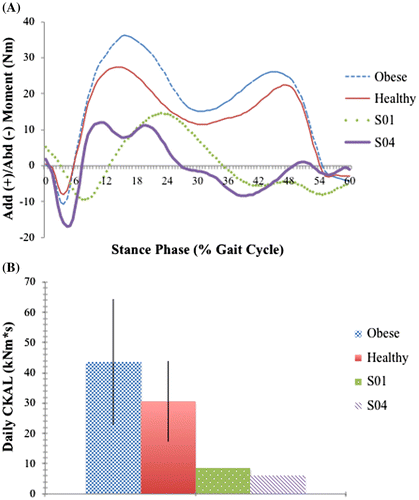
Two obese participants—S01 and S04—were found to have unusual frontal waveforms that did not conform to what is typically observed. The atypical shape and magnitude of the frontal moments throughout stance phase from S01 and S04 warranted individual investigation (A). Both participants had peak frontal moments and adduction impulses that were much smaller than all other participants. The resulting CKAL for both participants were exceptionally low (B). These unusual observations were only seen in the frontal plane—sagittal and transverse plane data for S01 and S04 were as expected. Neither participant was deemed a statistical outlier—both fell within two standard deviations of the obese group mean frontal moment peak. However, the S01 and S04 frontal moments were uniquely different from all other participants.
Figure 1. The knee adduction moment and CKAL of the two removed obese subjects, referred to as S01 and S04 compared to the mean obese and healthy-weight group knee adduction moments and CKAL.
Notes: The adduction waveforms of both S01 and S04 do not conform to typical knee adduction moments seen in individuals without musculoskeletal pathology. The CKAL for S04 and S01 is calculated from the self-selected natural walking speed, and is expressed in kNm s.

3.2. Combined self-selected walking speed
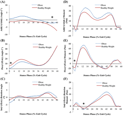
summarizes the comparison between groups for knee angles and moments at the combined self-selected walking speed. The average group adduction/abduction, flexion/extension and internal/external rotation angle, and moment waveforms are shown in . Significant group differences were found in the peak adduction and flexion knee angles. The peak extensor and peak external rotation moments that occurred at heel contact and early stance, respectively, were different between groups. The peak KAM was not different between groups at a combined self-selected walking speed (p = 0.12) ().
Table 2. Results for the knee angles and moments for obese and healthy-weight participants at the self-selected speed condition
Figure 2. Average (A) adduction/abduction, (B) flexion/extension, (C) internal/external rotation angles, (D) adduction/abduction, (E) flexion/extension, (F) internal/external rotation moment waveforms for both participant groups at the combined self-selected speed condition.
Notes: In the self-selected speed condition, mean curves were determined using the three speeds of fast, self-selected, and slow. The healthy-weight group is represented by the solid line and the obese group is represented by the dotted line. A positive value on the vertical axis represents an adduction, flexion or internal rotation moment. Significant mean differences between groups are indicated by an asterisk where they occurred in the gait cycle.

3.3. Matched walking speed
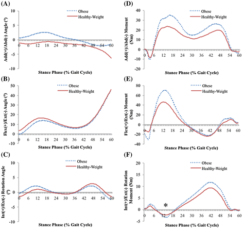
The results of the mean matched speed kinematic and kinetic analysis are displayed in . The average group adduction/abduction, flexion/extension and internal/external rotation angle, and moment waveforms are shown in . Unlike in the combined self-selected speed analysis, no significant differences were found between groups at the matched speed in either the peak knee angles or moments.
Table 3. Results for the knee angles and moments for obese and healthy-weight participants at the matched speed condition
Figure 3. Average (A) adduction/abduction, (B) flexion/extension, (C) internal/external rotation angles, (D) adduction/abduction, (E) flexion/extension, (F) internal/external rotation moment waveforms for both participant groups at the matched speed condition.
Notes: The matched speed mean curves were determined using the self-selected speed for the obese group and the matched speed for the healthy-weight group. The healthy-weight group is represented by the solid line and the obese group is represented by the dotted line. A positive value on the vertical axis represents an adduction, flexion or internal rotation moment. Significant mean differences between groups are indicated by an asterisk where they occurred in the gait cycle.

3.4. CKAL
Results from the CKAL and its component measures are in . The healthy-weight group had a similar mean number of steps per day as the obese group. Stance duration (p = 0.003), the KAM impulse (p = 0.049), and CKAL (p = 0.025) were different between groups, such that those in the obese group experienced greater exposure to medial knee loading than healthy-weight counterparts.
Table 4. Group differences in stance duration (s), steps per day, moment impulse (Nm s), and cumulative knee adductor load (kNm s)
4. Discussion
The purpose of this study was to identify whether differences existed in three-dimensional knee kinematics and kinetics during gait, and CKAL, between obese young adults and an age-, height-, and sex-matched sample of healthy-weight adults during walking. Though asymptomatic, young adults with obesity demonstrated several walking mechanics that were different than age-, height-, and sex-matched controls. Those with obesity selected a slower gait speed. The peak waveform analysis of joint angles and moments also indicated that those with obesity demonstrated a greater knee adduction angle throughout stance, less peak knee flexion at heel strike, as well as greater peak knee extension and external rotation moments during heel strike and early stance, respectively. When the healthy-weight group was required to walk at a gait speed matched with the obese group these statistical differences in gait mechanics were eliminated. However, shape and magnitude trends in all knee angles and moments remained consistent between both participant groups when walking speeds were matched. The matched speed analysis highlighted the effect of a small sample size in the present study. The sample of obese young adults in the present study demonstrated greater total exposure to medial knee loading, reflected by CKAL, compared to healthy counterparts.
While several kinematic and kinetic gait abnormalities were noted between the obese and healthy weight groups at combined self-selected gait speed, there were no significant differences found between groups at the matched speed. Comparisons between the combined self-selected speed waveforms in and the matched speed waveforms in show that there is a similar magnitude of difference between participant groups in both walking speed conditions. A power analysis approximated that 15 subjects would be required in each participant group to obtain statistical significance in peaks such as the knee adduction and extension moment. Low statistical power for the matched speed analysis partially explains the lack of significant kinematic and kinetic differences. However, it is also possible that the differences noted between the groups at combined self-selected walking speeds are due to differences in natural walking speed. Previous research has shown limited significant changes in knee kinematics with changing walking speed, except mild reductions in knee flexion with decreasing walking speed (Lelas, Merriman, Riley, & Kerrigan, Citation2003; Winter, Citation1991), as was seen in the present study. The minimal change in magnitude of the knee angles at a matched speed suggest that differences in combined self-selected walking speed are not the strongest explanation for the observed group differences in the combined self-selected speed kinematic analysis.
At a combined self-selected speed, obese participants made heel contact with less knee flexion than the healthy-weight participants, a difference between groups that extended into midstance (B). Several hypotheses could explain why this gait abnormality was observed. Maintaining a straighter leg at heel contact may be a strategy to decrease the moment arm about and knee joint loading in the sagittal plane. Alternatively, the extended knee angle could be a consequence of knee extensor weakness or compromised knee extensor activity relative to body mass. Previous work on sagittal plane kinematics in the obese is inconsistent. Findings from the current study are in agreement with results in DeVita and Hortobágyi (Citation2003) and McMillian et al. (Citation2010) where differences in sagittal plane angles were found between obese and healthy-weight populations. In the current study, obese participants had less knee flexion than healthy-mass counterparts. At the combined self-selected speed, the obese group had a statistically significant greater knee extensor moment at heel contact (E) than the healthy-weight group by more than 17 Nm. Considered with the mentioned temporal-spatial gait adaptations, the straight leg posture in early stance may have reduced the peak flexion moment in early to midstance. This kinematic difference in knee extension in early stance may be one reason for the lack of differences between participant groups in the peak knee flexion moment. Browning and Kram (Citation2007) and Spyropoulos, Pisciotta, Pavlou, Cairns, and Simon (Citation1991) demonstrated that sagittal knee joint angles in the obese were not different from a healthy-weight group. While Spyropoulos only analyzed knee kinematics, Browning & Kram found that the sagittal knee moment was greater in an obese group. This is particularly interesting, as Browning and Kram are the only others to present the absolute magnitude of knee moments, as was done in the present analysis. Differences between studies in the age group analyzed, the magnitude of obesity as measured by BMI, and methodological choices—particularly the use of a treadmill-mounted force plate to measure kinetic by Browning & Kram—may account for differences between other studies and the present one.
Aside from the greater peak knee extensor moment in the obese young adults, the only other between group difference in peak knee moments was a greater external rotation moment at early stance in the obese group. Very little work has been done examining the axial rotational moments about the knee. Alterations to the range of the rotational knee moment following anterior cruciate ligament injury have been implicated in the development of OA, by placing increased loads on specific regions of articular cartilage (Andriacchi & Dyrby, Citation2005), and thus may be worth deeper examination in obese populations who are at risk for OA development. Harding et al. (Citation2012) reported rotational moments with reduced range in healthy obese adults, suggesting limited mobility in the transverse plane. This finding of reduced range of rotational moments is not in agreement with the present results. However, the present cohort was younger than those studied by Harding et al. (Citation2012). In obese knee OA populations, those with moderate OA have a greater internal rotation moment in late stance, while those with severe OA had a reduced internal rotation moment (Astephen, Deluzio, Caldwell, Dunbar, & Hubley-Kozey, Citation2008). The rotational moments exhibited by the present obese young adults could be explored to determine whether these moments increase the risk for development of knee OA.
There were no significant differences between the participant groups in the peak KAM. The magnitude of the peak KAM was highly variable between individual obese participants in the present study. This may suggest inconsistent strategies for controlling the frontal moment in obese young adults. Alternatively, the high variability may be a consequence of treating the obese group as homogeneous. It may have been more appropriate to have analyzed the obese participants by the distribution of body mass. Obese adults with the majority of their excess weight centrally located demonstrated higher KAMs than those with their excess weight located in the lower body (Segal et al., Citation2009). These differences were negated once the KAM was normalized to body mass (Segal et al., Citation2009). The variability in peak KAM in the present study may be a reflection of the high variability in BMI within the obese group. Categorization of obese participant groups based on mass distribution should be recommended when analyzing biomechanical changes due to body mass. A power analysis indicated that double the number of participants in each group, for a total of 12 in each, would have been sufficient to detect differences between participant groups in the peak KAM. This suggests that with a larger study sample size, differences could be detected between obese and healthy-weight young adults in the peak KAM.
Despite issues with a small sample size, differences were detected between groups in the KAM impulse. The important factor that distinguished between groups in the KAM impulse in the present study was stance duration. Despite the variability in the obese group peak adduction moment and the low sample size, a significantly larger KAM impulse in the obese group was identified, and distinguished medial compartment loading differences between groups. The slower natural walking speed of the obese group significantly increased their stance duration, which significantly increased the KAM impulse despite BMI variability within the participant group. Longer stance duration resulted in greater overall medial compartment loading in obese young adults compared to their healthy counterparts, a factor the peak KAM is unable to detect. This finding is strengthened by the results of the CKAL measure between groups, which also suggest that the present cohort of obese young adults are incurring greater cumulative stress in the medial compartment of their knee joint. A greater adduction moment from a one-stride gait analysis is a strong risk factor for development and progression of knee OA (Astephen et al., Citation2008; Baliunas et al., Citation2002; Issa & Sharma, Citation2006). Since the CKAL uses the impulse of the KAM as one of its primary components, then it is possible that a similar risk level for knee OA can be inferred. The altered kinematics, together with the greater CKAL in young obese adults could indicate a pathological loading environment at the knee that may increase risk for knee OA. However, a longitudinal study would be required to establish whether a greater CKAL would lead to knee OA.
4.1. Decision to remove obese participants
Due to uncertainty in the origin of the unusual frontal moments for two obese participants (S01 and S04), they, along with their healthy matches, were removed from the data-set. Though the removal of these participants reduced an already small research cohort and may have limited the generalizability of the study results to obese young adult population, the authors believe the choice was warranted. A very thorough investigation of the data for these two participants was undertaken and there was no evidence that there were any errors during data collection or processing that contributed to the aberrant results. Data processing was standardized to ensure all participant data underwent the same sequence of processing events. All of these procedures were performed to ensure testing reliability and validity. Therefore, the frontal moment changes in S01 and S04 may be a display of compensatory gait mechanisms as a consequence of excess body weight.
It could be argued that removing the data of two obese subjects from the kinematic and kinetic group analysis restricted the applicable populations of obese and healthy-weight participants. The full data-set may be more representative of the possible biomechanical features and knee joint measures in young adults who are obese. A future study with a much larger sample size would be needed to demonstrate whether the results of the case studies are anomalies or representative of a significant portion of the obese population. In the present study they have been treated as anomalies. Therefore, the frontal moments from S01 and S04 need to be analyzed under a separate lens than the other participants to formulate a biomechanical foundation for their unusual frontal moments.
Two specific biomechanical factors may be responsible for the frontal moment anomalies—knee malalignment and a toe-out gait pattern. Knee alignment was measured using a marker-based method (Hunt, Birmingham, Jenkyn, Giffin, & Jones, Citation2008). Both participants had valgus knee alignment values that were well beyond neutral and different from the mean of the obese participant group (). Knee alignment can have a very large effect of the loading of the knee joint (Sharma et al., Citation2001). This valgus knee alignment is likely a strong contributor to the uniquely small knee adduction moments in S01 and S04. A greater valgus alignment decreases the knee adduction moment. This is the result of the load-bearing axis shifting away from the medial compartment of the knee in a valgus knee alignment (Andrews, Noyes, Hewett, & Andriacchi, Citation1996; Brouwer et al., Citation2007; Hurwitz, Ryals, Case, Block, & Andriacchi, Citation2002).
Table 5. The marker-based knee alignment of the two obese subjects removed from the data analysis during their self-selected natural walking speed
Another factor that may play a role in the frontal knee moments of S01 and S04 is the toe-out angle. While not quantitatively measured in the present study, toe-out gait may have factored into the results of the knee kinematics and kinetics. A greater toe-out angle has been shown to significantly decrease the late stance peak of the frontal knee moment (Guo, Axe, & Manal, Citation2007; Jenkyn, Hunt, Jones, Giffin, & Birmingham, Citation2008; Rutherford, Hubley-Kozey, Deluzio, Stanish, & Dunbar, Citation2008). By increasing toe-out angle, the ankle inversion moment decreases, which decreases the knee adduction moment (Guo et al., Citation2007; Jenkyn et al., Citation2008). This reduces the load on the medial compartment of the knee joint, much like a valgus knee alignment (Rutherford et al., Citation2008).
If the data from S01 and S04 are representative of true joint loading due to changes to the biomechanics of the lower extremity, the CKAL measure as it is presently interpreted may not be a useful tool in the case of these two participants. One of the two inputs of the CKAL—the frontal moment impulse—would skew the individual CKAL of S01 and S04 and possibly misrepresent the loading environment of their knees. The CKAL values from these two participants suggest minimal daily cumulative joint loading (B). This cumulative load result is problematic. As the primary measure of joint loading in the CKAL is the knee adduction moment, it only truly represents the ratio of loading in the medial compartment. Less medial compartment loading, by way of a decreased adduction moment, equates to a smaller CKAL, but not to less total knee joint loading. It may only represent a changed ratio of medial to lateral loading.
4.2. Limitations
The most significant limitation in this study was the small sample size. The study sample size was originally small. The removal of the two obese participants and their healthy-matches reduced an already small research cohort. It is difficult to ascertain if such a small sample size could be representative of the obese young adult population as a whole. The small sample size in this study resulted in the high variability of the estimated gait measures of interest. This reduced the power associated with statistical tests, particularly the peak KAM and matched speed analyses. With a larger sample size, statistical differences may have been detected between the obese and healthy-weight young adults. However, it is also possible that a larger sample size would have confirmed the present results of this study. The uncertainty regarding the standard errors of estimates of this study limits the generalizability of the study results. The presented results should be considered initial findings in an understudied population. Future research involving a larger sample size will provide better evidence for the initial results demonstrated in the present study.
Furthermore, the obese participant group was less homogenous in their body mass than the healthy-weight group, which may have introduced confounding factors to the analysis. Using skin markers to determine position and motion of the joint becomes problematic, as errors are likely to occur as a result of skin movement. Greater subcutaneous tissue between the skin and bone also made palpating bony anatomical landmarks more difficult with the obese participants. Errors due to skin artifact may have been magnified in the obese participant group due to their increased body mass. Methods to circumvent skin movement are limited when using motion capture. However, the use of markers secured to rigid plates, as done in this study, can aid in reducing erroneous marker positions due to skin movement. In addition, the walking tasks completed in this study were of low speed and impact. This would constrain the amount of skin movement as compared to a more ballistic movement or task. Finally, in determining steps per day through an accelerometer, there were possible limitations in compliance as well as the sensitivity of the device. In particular, some physical activities are not captured such as standing.
5. Conclusion
Recent statistics show that obesity rates in the youngest categories of the Canadian population are rising at an unprecedented rate, making the understanding of how obesity interacts with OA development paramount (Shields et al., Citation2010). Obese young adults were found to have walking characteristics that were different from their healthy-weight peers. These included slower walking speed, greater stance duration, less knee flexion at heel contact, more adduction during stance, and greater knee extension and external rotation moments in early stance. When walking at a matched speed to the control group, similar trends in kinematics and kinetics were demonstrated between obese and healthy-weight young adults. Although the peak KAM was not different between groups, the obese young adults had a greater magnitude of KAM impulse and therefore a resulting larger CKAL. While these results suggest that obese young adults may incur greater medial compartment loading at the knee, it remains to be seen whether these biomechanical differences result in a greater risk for knee OA.
Additional information
Funding
Notes on contributors
Kathleen F.E. MacLean
Kathleen F.E. MacLean is a PhD candidate in Kinesiology at the University of Waterloo.
Jack P. Callaghan
Jack P. Callaghan is a professor in Kinesiology at the University of Waterloo, and is the Canadian Research Chair in Spine Biomechanics and Injury Prevention.
Monica R. Maly
Monica R. Maly is an associate professor in Rehabilitation Science at McMaster University and holds a Canadian Institutes of Health (CIHR) Research New Investigator Award.
Combining specializations in biomechanics, gait and rehabilitation, the authors have collaborated on measures related to quantifying cumulative loading of the knee and its impact on the development of musculoskeletal pathologies of the knee. Of particular interest is how abnormal biomechanics and overloading of the knee joint is associated with the genesis and progression of osteoarthritis of the knee, which is presently a significant public health concern that may worsen with increasing obesity rates and sedentary lifestyles.
References
- Adams, R. (1999). Revised physical activity readiness questionnaire. Canadian Family Physician, 992, 1004–1005.
- Andrews, M., Noyes, F. R., Hewett, T. E., & Andriacchi, T. P. (1996). Lower limb alignment and foot angle and related to stance phase knee adduction in normal subjects: A critical analysis of the reliability of gait analysis data. Journal of Orthopedic Research, 14, 289–295.
- Andriacchi, T. P., & Dyrby, C. O. (2005). Interactions between kinematics and loading during walking for the normal and ACL deficient knee. Journal of Biomechanics, 38, 293–298.10.1016/j.jbiomech.2004.02.010
- Andriacchi, T. P., Koo, S., & Scanlan, S. F. (2009). Gait mechanics influence healthy cartilage morphology and osteoarthritis of the knee. The Journal of Bone & Joint Surgery, 91, 95–101.
- Andriacchi, T. P., Mundermann, A., Smith, R. L., Alexander, E. J., Dyrby, C. O., & Koo, S. (2004). A framework for the in vivo pathomechanics of osteoarthritis at the knee. Annals of Biomedical Engineering, 32, 448–457.
- Astephen, J. L., Deluzio, K. J., Caldwell, G. E., Dunbar, M. J., & Hubley-Kozey, C. L. (2008). Gait and neuromuscular pattern changes are associated with differences in knee osteoarthritis severity levels. Journal of Biomechanics, 41, 868–876.10.1016/j.jbiomech.2007.10.016
- Baliunas, A. J., Hurwitz, D. E., Ryals, A. B., Karrar, A., Case, J. P., Block, J. A., & Andriacchi, T. P. (2002). Increased knee joint loads during walking are present in subjects with knee osteoarthritis. Osteoarthritis and Cartilage, 10, 573–579.10.1053/joca.2002.0797
- Bennell, K., Bowles, K., Wang, Y., Cicuttini, F., Davies-Tuck, M., & Hinman, R. (2011). Higher dynamic medial knee load predicts greater cartilage loss over 12 months in medial knee osteoarthritis. Annals of the Rheumatic Diseases, 70, 1770–1774.10.1136/ard.2010.147082
- Blazek, K., Asay, J. L., Erhart-Hledik, J., & Andriacchi, T. (2013). Adduction moment increases with age in healthy obese individuals. Journal of Orthopaedic Research, 31, 1414–1422.10.1002/jor.v31.9
- Brouwer, G. M., van Tol, A. W., Bergink, A. P., Belo, J. N., Bernsen, R. M. D., Reijman, M., … Bierma-Zeinstra, S. M. (2007). Association between valgus and varus alignment and the development and progression of radiographic osteoarthritis of the knee. Arthritis & Rheumatism, 56, 1204–1211.
- Browning, R. C., & Kram, R. (2007). Effects of obesity on the biomechanics of walking at different speeds. Medicine & Science in Sports & Exercise, 39, 1632–1641.
- Browning, R. C., McGowan, C. P., & Kram, R. (2009). Obesity does not increase external mechanical work per kilogram body mass during walking. Journal of Biomechanics, 42, 2273–2278.10.1016/j.jbiomech.2009.06.046
- DeVita, P., & Hortobágyi, T. (2003). Obesity is not associated with increased knee joint torque and power during level walking. Journal of Biomechanics, 36, 1355–1362.10.1016/S0021-9290(03)00119-2
- Guo, M., Axe, M. J., & Manal, K. (2007). The influence of foot progression angle on the knee adduction moment during walking and stair climbing in pain free individuals with knee osteoarthritis. Gait & Posture, 26, 436–441.
- Gushue, D. L., Houck, J., & Lerner, A. L. (2005). Effects of childhood obesity on three-dimensional knee joint biomechanics during walking. Journal of Pediatric Orthopaedics, 25, 763–768.10.1097/01.bpo.0000176163.17098.f4
- Harding, G. T., Hubley-Kozey, C. L., Dunbar, M. J., Stanish, W. D., & Astephen-Wilson, J. L. (2012). Body mass index affects knee joint mechanics during gait differently with and without moderate knee osteoarthritis. Osteoarthritis and Cartilage, 20, 1234–1242.10.1016/j.joca.2012.08.004
- Hunt, M. A., Birmingham, T. B., Jenkyn, T. R., Giffin, J. R., & Jones, I. C. (2008). Measures of frontal plane lower limb alignment obtained from static radiographs and dynamic gait analysis. Gait & Posture, 27, 635–640.
- Hunter, D. J., Sharma, L., & Skaife, T. (2009). Alignment and osteoarthritis of the knee. The Journal of Bone & Joint Surgery, 91, 85–89.
- Hurwitz, D. E., Ryals, A. B., Case, J. P., Block, J. A., & Andriacchi, T. P. (2002). The knee adduction moment during gait in subjects with knee osteoarthritis is more closely correlated with static alignment than radiographic disease severity, toe out angle and pain. Journal of Orthopaedic Research, 20, 101–107.
- Issa, S. N., & Sharma, L. (2006). Epidemiology of osteoarthritis: An update. Current Rheumatology Reports, 8, 7–15.
- Janssen, I., Katzmarzyk, P. T., & Ross, R. (2003). Waist circumference and not body mass index explains obesity-related health risk. The American Journal of Clinical Nutrition, 79, 379–384.
- Jenkyn, T. R., Hunt, M. A., Jones, I. C., Giffin, J. R., & Birmingham, T. B. (2008). Toe-out gait in patients with knee osteoarthritis partially transforms external knee adduction moment into flexion moment during early stance phase of gait: A tri-planar kinetic mechanism. Journal of Biomechanics, 41, 276–283.
- Ko, S., Stenholm, S., & Ferrucci, L. (2010). Characteristic gait patterns in older adults with obesity – Results from the Baltimore longitudinal study of aging. Journal of Biomechanics, 43, 1104–1110.10.1016/j.jbiomech.2009.12.004
- Lai, P. P. K., Leung, A. K. L., Li, A. N. M., & Zhang, M. (2008). Three-dimensional gait analysis of obese adults. Clinical Biomechanics, 23, S2–S6.
- Lean, M. E., Han, T. S., & Morrison, C. E. (1995). Waist circumference as a measure for indicating need for weight management. BMJ, 311, 158–161.10.1136/bmj.311.6998.158
- Lelas, J. L., Merriman, G. J., Riley, P. O., & Kerrigan, D. C. (2003). Predicting peak kinematic and kinetic parameters from gait speed. Gait & Posture, 17, 106–112.
- Lim, B. W., Kemp, G., Metcalf, B., Wrigley, T. V., Bennell, K. L., Crossley, K. M., & Hinman, R. S. (2009). The association of quadriceps strength with the knee adduction moment in medial knee osteoarthritis. Arthritis & Rheumatism, 61, 451–458.
- Lynch, N. A., Metter, E. J., Lindle, R. S., Fozard, J. L., Tobin, J. D., Roy, T. A., … Hurley, B. F. (1999). Muscle quality. I. Age-associated differences between arm and leg muscle groups. Journal of Applied Physiology, 86, 188–194.
- Lynn, S. K., Reid, S. M., & Costigan, P. A. (2007). The influence of gait pattern on signs of knee osteoarthritis in older adults over a 5–11 year follow-up period: A case study analysis. The Knee, 14, 22–28.10.1016/j.knee.2006.09.002
- Maly, M. R., Robbins, S. M., Stratford, P. W., Birmingham, T. B., & Callaghan, J. P. (2013). Cumulative knee adductor load distinguishes between healthy and osteoarthritic knees—A proof of principle study. Gait & Posture, 37, 397–401.
- McMillian, A. G., Pulver, A. M. E., Collier, D. N., & Williams, D. S. B. (2010). Sagittal and frontal plane joint mechanics throughout the stance phase of walking in adolescents who are obese. Gait & Posture, 32, 263–268.
- Messier, S. P., Gutekunst, D. J., Davis, C., & DeVita, P. (2005). Weight loss reduces knee-joint loads in overweight and obese older adults with knee osteoarthritis. Arthritis & Rheumatism, 57, 2026–2032.
- Miyazaki, T., Wada, M., Kawahara, H., Sato, M., Baba, H., & Shimada, S. (2002). Dynamic load at baseline can predict radiographic disease progression in medial compartment knee osteoarthritis. Annals of the Rheumatic Diseases, 61, 617–622.10.1136/ard.61.7.617
- Orpana, H. M., Trembley, M. S., & Fines, P. (2007). Trends in weight change among Canadian adults. Statistics Canada: Health Reports, 18, 9–16.
- Petrella, J. K., Kim, J. S., Tuggle, S. C., Hall, S. R., & Bamman, M. M. (2005). Age differences in knee extension power, contractile velocity and fatigability. Journal of Applied Physiology, 98, 211–220.
- Price, G. M., Uauy, R., Breeze, E., Bulpitt, C. J., & Fletcher, A. E. (2006). Weight, shape, and mortality risk in older persons: elevated waist-hip ratio, not high body mass index, is associated with a greater risk of death. The American Journal of Clinical Nutrition, 84, 449–460.
- Robbins, S. M., Birmingham, T. B., Jones, G. R., Callaghan, J. P., & Maly, M. R. (2009). Developing an estimate of daily cumulative loading for the knee: Examining test-retest reliability. Gait & Posture, 30, 497–501.
- Robbins, S. M., & Maly, M. R. (2009). The effect of gait speed on the knee adduction moment depends on waveform summary measures. Gait and Posture, 30, 543–546.10.1016/j.gaitpost.2009.08.236
- Russell, E. M., & Hamill, J. (2011). Lateral wedges decrease biomechanical risk factors for knee osteoarthritis in obese women. Journal of Biomechanics, 44, 2286–2291.10.1016/j.jbiomech.2011.05.033
- Rutherford, D. J., Hubley-Kozey, C. L., Deluzio, J., Stanish, W. D., & Dunbar, M. (2008). Foot progression angle and the knee adduction moment: a cross-sectional investigation in knee osteoarthritis. Osteoarthritis & Cartilage, 16, 883–889.
- Schwartz, M. H., & Rozumalski, A. (2005). A new method for estimating joint parameters from motion data. Journal of Biomechanics, 38, 107–116.10.1016/j.jbiomech.2004.03.009
- Segal, N. A., Yack, J., & Khole, P. (2009). Weight, rather than obesity distribution, explains peak external knee adduction moment during level gait. American Journal of Physical Medicine & Rehabilitation, 88, 180–246.
- Sharma, L., Song, J., Felson, D. T., Cahue, S., Shamiyeh, E., & Dunlop, D. D. (2001). The role of knee alignment in disease progression and functional decline in knee osteoarthritis. The Journal of the American Medical Association, 286, 188–195.10.1001/jama.286.2.188
- Shields, M., Tremblay, M. S., Laviolette, M., Craig, C. L., Jenssen, I., & Gorber, S. C. (2010). Fitness of Canadian adults: Results from the 2007–2009 Canadian Health measures survey. Statistics Canada: Health Reports, 21, 1–16. Catalogue No. 82-003-XPE
- Spyropoulos, P., Pisciotta, J. C., Pavlou, K. N., Cairns, M. A., & Simon, S. R. (1991). Biomechanics gait analysis in obese men. Archives of Physical Medicine and Rehabilitation, 72, 1065–1070.
- Stratford, P. W., Kennedy, D. M., & Hanna, S. E. (2004). Condition-specific Western Ontario McMaster osteoarthritis index was not superior to region-specific lower extremity functional Scale at detecting change. Journal of Clinical Epidemiology, 57, 1025–1032.10.1016/j.jclinepi.2004.03.008
- Thorp, L. E., Sumner, D. R., Block, J. A., Moisio, K. C., Shott, S., & Wimmer, M. A. (2006). Knee joint loading differs in individuals with mild compared with moderate medial knee osteoarthritis. Arthritis & Rheumatism, 54, 3842–3849.
- Wang, Y. C., Hart, D. L., Stratford, P. W., & Mioduski, J. E. (2009). Clinical interpretation of a lower-extremity functional scale-derived computerized adaptive test. Physical Therapy, 89, 957–968.10.2522/ptj.20080359
- Winter, D. A. (1991). The biomechanics and motor control of human gait: Normal, elderly and pathological (2nd ed.). Waterloo, ON: University of Waterloo Press.
