Abstract
Background: Patients with cirrhosis are perceived to be at increased risk for bleeding given the presence of coagulopathy leading to unnecessary head imaging studies. Our aim is to determine the prevalence of intracranial hemorrhage (ICH) in patients with cirrhosis using gold standard autopsy data. Methods: An autopsy-based case control study was performed in a tertiary care hospital with a specialized Liver Unit. Autopsies without both head and abdominal examinations were excluded. Results: Between 1986 and 2003, a total of 679 autopsies were performed. 37 autopsies were excluded and 642 autopsies were available for final review. A total of 21 (4.3%) patients in the cirrhosis group had ICH compared to 12 (7.9%) patients in the control group (p = 0.115). Since Alzheimer’s patients may be at higher risk for ICH given the presence of cerebral amyloid angiopathy, a separate analysis was performed after excluding these patients. The results were similar (4.3% vs. 9.3%, p = 0.061). The prevalence of old strokes (1.2% vs. 13.9%, p < 0.001), acute strokes (0% vs. 4%, p < 0.001), and Alzheimer’s disease (1% vs. 29.1%, p < 0.001) were found to be higher in the control group compared to the cirrhosis group. Conclusion: Despite the presence of coagulopathy, patients with cirrhosis are at no increased risk for intracranial bleeding compared to controls without cirrhosis.
Public Interest Statement
The medical evaluation of patients with cirrhosis contributes significant cost to health care systems worldwide. A common complication of cirrhosis is a form of confusion called hepatic encephalopathy (HE). Common causes of HE are infection, gastrointestinal bleeding, electrolyte abnormalities, and constipation. Despite the easy bleeding seen in patients with cirrhosis, an uncommon cause of HE is intracranial hemorrhage (ICH). But computed tomography scans (better known as CT scans) are obtained routinely on these patients leading to increased health care costs. In our study, we have used information obtained from autopsies to show that the prevalence of ICH is not increased in patients with cirrhosis compared to those without cirrhosis. Our results suggest that CT scans are ordered unnecessarily in patients with cirrhosis and that the more common triggers of HE should be given priority during the medical evaluation. By decreasing unnecessary testing, health care costs may be reduced.
Competing Interests
The authors declare no competing interest.
1. Introduction
The care of patients with cirrhosis in the United States is a costly endeavor (Kim, Gross, Poterucha, Locke, & Dickson, Citation2001; Otgonsuren et al., Citation2015). Approximately one-third of hospital admissions for patients with cirrhosis are because of altered mental status (Rahimi, Elliott, & Rockey, Citation2013). Further, coagulopathy is a common hematologic derangement seen in patients with cirrhosis and this patient population is anecdotally regarded to be at higher risk for intracranial hemorrhage (ICH) than the general population. Therefore, the use of computed tomography (CT) of the head is ubiquitous in the evaluation of patients with cirrhosis who present with altered mental status. Overall, an estimated 1 in 14 patients presenting to the emergency department receives a CT scan of the head (Malatt et al., Citation2014) and this proportion is likely higher when patients with cirrhosis are involved.
A recent retrospective cohort study utilizing review of International Classification of Diseases, 9th Revision codes has suggested that routine non-contrast CT of the head is low yield in detecting ICH when high- and low-risk indications were compared (Donovan et al., Citation2015). The aim of our study was to compare the prevalence of ICH and survey the various types of neuropathology between patients with cirrhosis and non-cirrhotic controls using autopsy, the gold standard.
2. Methods
Our autopsy-based case control study took place at a single center tertiary care hospital in Los Angeles County. The site was unique in that was a referral center with specialized services for both liver disease and Alzheimer’s disease. All autopsies performed between 1986 and 2003 were reviewed and data were extracted. The study was approved by the Institutional Review Board at Ranch Los Amigos (Downey, California). Only autopsies with included examinations of both the head and abdomen were included in our analysis. Any autopsies missing either of these examinations were excluded. Patients with confirmed cirrhosis based on autopsy results were placed in the cirrhosis group, while those that did not have cirrhosis on autopsy were placed in the control group.
Admission labs including white blood cell count, hemoglobin, platelets, international normalized ratio (INR), total bilirubin, aspartate aminotransferase (AST), alanine aminotransferase (ALT), alkaline phosphatase, and creatinine levels were recorded for all patients. The sodium level was recorded for patients in the cirrhosis group only and no patients were excluded on the basis of serum sodium level. No information regarding amount of lactulose given to each patient was recorded.
Three senior pathologists performed all of the autopsies in our study. The chronicity of stroke (old versus acute) was determined by characteristic neuropathologic findings on autopsy. Similarly, the diagnosis of cirrhosis was confirmed by gross and microscopic evaluations of the liver at the time of autopsy.
3. Statistical analysis
Continuous variables were summarized using means, standard deviations, medians, and quartiles. Categorical variables were summarized using frequencies and percentages. Continuous variables were compared between cirrhosis and control using Wilcoxon rank-sum tests, while categorical variables were compared using Pearson’s chi-squared test and Fisher’s exact test as appropriate. A p-value < 0.05 was considered statistically significant. All analyses were performed using R v. 3.1.2 (www.r-project.org).
4. Results
Between 1986 and 2003, 679 autopsies were available for review. In the cirrhosis group, 519 autopsies were performed. Of these, 28 autopsies were excluded because they did not include examinations of both the head and abdomen. Thus, 491 autopsies were available for review in the cirrhosis group. In the control group, 160 autopsies were performed. Of these, nine autopsies were excluded because they did not include examinations of both the head and abdomen. Thus, 151 autopsies were available for review in the control group. The vast majority of patients in the cirrhosis group died of complications related to cirrhosis including variceal bleeding, sepsis from spontaneous bacterial peritonitis or other bacterial infections, and hepatorenal syndrome. On the other hand , most of the patients in the control group died of cardiopulmonary disease, Alzheimer’s disease, and infection.
The baseline characteristics of the cirrhosis and control groups are listed in . Generally, the cirrhosis group was composed of younger (mean age ± standard deviation 47 ± 10.9), Hispanic males, while the control group was older (mean age ± standard deviation 62.2 ± 18.2) and more diverse racially. Total bilirubin, alkaline phosphatase, AST, ALT, creatinine, and INR were all higher in the cirrhosis group reflecting advanced liver disease and presence of coagulopathy. Serum sodium levels were not recorded for the control group. In the cirrhosis group, the mean sodium level was 130.0 mEq/L (standard deviation ± 7.2 mEq/L). The mean change in serum sodium in the cirrhosis group was −0.84 mEq/L (standard deviation ± 9.1 mEq/L).
Table 1. Baseline characteristics
Alcohol was the primary cause of chronic liver disease in 74% of subjects in the cirrhosis group. Other etiologies included: Chronic hepatitis C virus (HCV) (7%), HCV plus alcohol (6%), and chronic hepatitis B (7%). Cirrhosis from other causes including non-alcoholic steatohepatitis, primary sclerosing cholangitis, primary biliary cirrhosis, autoimmune hepatitis, hemochromatosis, and Wilson’s disease comprised the remaining 6%.
The different types of neuropathology found on autopsy are shown in . Overall, there was no difference in prevalence of ICH between the cirrhosis and control groups (4.3% vs. 7.9%, p = 0.115). Since Alzheimer’s patients may be at higher risk for ICH given the presence of cerebral amyloid angiopathy, a separate analysis was performed after excluding these patients. Results were similar (4.3% vs. 9.3%, p = 0.061). Our sample size of 486 cirrhosis cases and 107 controls, after exclusion of patients with Alzheimer’s, provides 80% power to detect a difference in incidence rates of ICH of 0.076, assuming a reference incidence of 0.043, a Pearson chi-squared test, and an alpha of 0.05. We were thus underpowered to detect differences smaller than this.
Table 2. Survey of neuropathology
The prevalence of old strokes (1.2% vs. 13.9%, p < 0.001), acute strokes (0% vs. 4%, p < 0.001), and Alzheimer’s disease (1% vs. 29.1%, p < 0.001) were found to be higher in the control group compared to the cirrhosis group. To compare the prevalence of any life-threatening neurologic finding in the cirrhosis and control groups, we used the outcome of ICH or acute stroke. We found that 21 cirrhosis patients (4.3%) had either ICH or acute stroke, while 16 control patients (15.0%) had either ICH or acute stroke, a difference, which was statistically significant (p < 0.001).
There were seven cases of central pontine myelinolysis (CPM) in the cirrhosis and none in the control group, but this difference was not statistically significant. Overall, 91% of the cirrhosis group had no significant brain pathology compared to 58.9% in the control group.
5. Discussion
The results of our study indicate that despite the presence of coagulopathy, patients with cirrhosis are at no increased risk for ICH compared to controls using autopsy, the gold standard test. One potential weakness of our control sample is the high prevalence of Alzheimer’s disease (29%). This high prevalence was because the study took place in a specialized referral center for Alzheimer’s disease. Alzheimer’s disease may increase one’s risk of ICH due to cerebral amyloid angiopathy. However, even after removing Alzheimer’s patients from our analysis, the results remained unchanged.
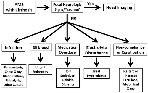
Thus, our data suggest that the routine use of head imaging in patients with cirrhosis would be low yield for the diagnosis of ICH. In our cohort of patients, ICH was uncommon (4.3% vs. 7.9% in the control group, p = 0.115). On the other hand, hepatic encephalopathy (HE) in patients with cirrhosis is quite common affecting up to 30% of patients with decompensated liver disease (Romero-Gomez, Boza, Garci'a-Valdecasas, Garci'a, & Aguilar-Reina, Citation2001). Thus evaluating for common precipitants of HE such as infection, inciting medications, gastrointestinal bleeding, electrolyte abnormalities, and constipation should be the initial step prior to cross-sectional imaging of the head in the absence of trauma or focal neurologic signs (). The higher prevalence of old stroke, new stroke, and Alzheimer’s disease in the older control group would not be unexpected especially since the study site was an Alzheimer’s referral center. When our analysis was expanded to compare the outcome of ICH or acute stroke between the groups, we found that the control group had significantly more ICH or acute strokes compared to the cirrhosis group (15.0% vs. 4.3%, p < 0.001) further suggesting that head imaging would be low yield in finding life-threatening neurologic processes in patient with cirrhosis.
Figure 1. Proposed clinical algorithm for patients with cirrhosis presenting with altered mental status (AMS).

While not statistically significant, there were seven cases of CPM in the cirrhosis group and none in the control group. Of note, in four of these cases, the patients were asymptomatic and neurologic examinations were unremarkable. The other three patients were in coma. Of these three patients in coma, only one received active correction of serum sodium for hyponatremia, which was done at a rate of less than 8 meq/L/day. Thus, this patient was symptomatic despite following what is generally regarded as a safe rate of correction. Asymptomatic CPM is a relatively uncommon entity in the general population. A large autopsy study involving a Veteran’s Affairs population found the prevalence of asymptomatic CPM to be 0.5% (Newell & Kleinschmidt-DeMasters, Citation1996). CPM is associated with multiple conditions including alcoholism, which could explain why the prevalence of CPM in our study was higher (1.4%) (Sullivan & Pfefferbaum, Citation2001).
The strength of our study is the use of autopsy data to confirm the diagnoses of cirrhosis and ICH in the cirrhosis and control groups. To our knowledge, no published data regarding the neuropathology as determined by autopsy of patients with cirrhosis exist. Limitations of our study reflect the retrospective design. Clinical information regarding patient symptoms, physical exam findings, and medications administered were not available for analysis. Thus no firm conclusions may be drawn from our data, which can definitively guide the care of patients with cirrhosis presenting with altered mental status.
Our study also suggests that laboratory parameters such as INR and platelet level do not reliably predict bleeding. The cirrhosis group had statistically higher INR and lower platelet levels (), but ICH prevalence was no different than controls. In another study, thromboelastogaphy guided blood product administration was compared to blood product administration guided by traditional parameters (INR and platelets). The thromboelastography group used fewer blood products and there was no difference in complications from bleeding (De Pietri et al., Citation2016). Further, the larger implication is that by overreacting to the INR and platelet levels in patients with cirrhosis, medicals costs increase and patient safety may be compromised (Rahimi & O’Leary, Citation2016).
In conclusion, despite the presence of coagulopathy, patients with cirrhosis are at no increased risk for ICH compared to non-cirrhotic controls using autopsy, the gold standard test. Therefore, routine head imaging of patients with cirrhosis would be low yield in detecting ICH and unnecessary head imaging studies in these patients can be avoided.
Additional information
Funding
Notes on contributors
Eric W. Chak
Eric W. Chak MD, MPH is an assistant professor of Internal Medicine at the University of California, Davis Medical Center in Sacramento, California. He holds dual board certification in Internal Medicine and Gastroenterology. Chak’s primary research activities involve the use of information technology to increase screening for chronic hepatitis B and next-generation sequencing techniques to determine the biologic factors that affect hepatitis B-related liver cancer survival among Asian Americans. The current research that we report relates to Chak’s clinical interest in the care of patients with cirrhosis and the proper use of health care resources in the management of these complicated patients.
References
- De Pietri, L., Bianchini, M., Montalti, R., De Maria, N., Di Maira, T., Begliomini, B., ... Villa, E. (2016). Thrombelastography-guided blood product use before invasive procedures in cirrhosis with severe coagulopathy: A randomized, controlled trial. Hepatology, 63, 566–573.10.1002/hep.v63.2
- Donovan, L. M., Kress, W. L., Strnad, L. C., Sarwar, A., Patwardhan, V., Piatkowski, G., ... Afdhal, N. H. (2015). Low likelihood of intracranial hemorrhage in patients with cirrhosis and altered mental status. Clinical Gastroenterology and Hepatology, 13, 165–169.10.1016/j.cgh.2014.05.022
- Kim, W. R., Gross, Jr., J. B., Poterucha, J. J., Locke, 3rd, G. R., & Dickson, E. R. (2001). Outcome of hospital care of liver disease associated with hepatitis C in the United States. Hepatology, 33, 201–206.10.1053/jhep.2001.20798
- Malatt, C., Zawaideh, M., Chao, C., Hesselink, J. R., Lee, R. R., & Chen, J. Y. (2014). Head computed tomography in the emergency department: A collection of easily missed findings that are life-threatening or life-changing. The Journal of Emergency Medicine, 47, 646–659.10.1016/j.jemermed.2014.06.042
- Newell, K. L., & Kleinschmidt-DeMasters, B. K. (1996). Central pontine myelinolysis at autopsy; a twelve year retrospective analysis. Journal of the Neurological Sciences, 142, 134–139.10.1016/0022-510X(96)00175-X
- Otgonsuren, M., Henry, L., Hunt, S., Venkatesan, C., Mishra, A., & Younossi, Z. M. (2015). Resource utilization and survival among medicare patients with advanced liver disease. Digestive Diseases and Sciences, 60, 320–332.10.1007/s10620-014-3318-9
- Rahimi, R. S., Elliott, A. C., & Rockey, D. C. (2013). Altered mental status in cirrhosis: Etiologies and outcomes. Journal of Investigative Medicine, 61, 695–700.
- Rahimi, R. S., & O’Leary, J. G. (2016). Transfusing common sense instead of blood products into coagulation testing in patients with cirrhosis: Overtreatment ≠ safety. Hepatology, 63, 368–370.10.1002/hep.v63.2
- Romero-Gomez, M., Boza, F., Garci'a-Valdecasas, M. S., Garci'a, E., & Aguilar-Reina, J. (2001). Subclinical hepatic encephalopathy predicts the development of overt hepatic encephalopathy. The American Journal of Gastroenterology, 96, 2718–2723.10.1111/ajg.2001.96.issue-9
- Sullivan, E. V., & Pfefferbaum, A. (2001). Magnetic resonance relaxometry reveals central pontine abnormalities in clinically asymptomatic alcoholic men. Alcoholism: Clinical and Experimental Research, 25, 1206–1212.10.1111/acer.2001.25.issue-8
