Abstract
Chilaiditi syndrome is an occasional radiographic anomaly characterized by the interposition of the bowels between the liver and the diaphragm. The presence of Chilaiditi syndrome can cause serious problem on percutaneous endoscopic gastrostomy (PEG) procedure, because the anomalous position of the transverse colon can obstruct the procedure and might lead to occurrence of complications. Our report describes a PEG-induced gastro-colo-cutaneous fistula in a patient with Chilaiditi syndrome which resulted in minimally invasive laparoscopic gastrostomy. As most patients who need a PEG placement have impaired consciousness, the majority of PEG-related complications are overlooked or discovered long after the procedure. However, we recognized the complication immediately after the PEG procedure and successfully treated by laparoscopic surgery.
Public Interest Statement
Our report is intended to alert that PEG procedure can cause complication of gastro-colo-cutaneous fistura in patients of Chilaiditi syndrome. Furthermore, we report and recommend low-invasive salvage surgery under laparoscopy if the complication was detected early.
Competing Interests
The authors declare no competing interest.
1. Introduction
In situations where normal or nasogastric feeding is insufficient to maintain a patient’s health, percutaneous endoscopic gastrostomy (PEG) has been recommended to provide sufficient enteral nutrition. Due to the anatomical and positional relationship of the stomach and the colon, the placement of a PEG tube, particularly when it is intended to support long-term usage, predisposes the patient to several complications, such as gastro-colic and colo-cutaneous fistulas (Schrag et al., Citation2007).
In 1910, a Greek radiologist, D. Chilaiditi, first reported an incidental radiographic finding, which was characterized by the presence of hepato-diaphragmatic interposition of the colon or the small bowel. As a result of this anatomical anomaly, the transverse colon is often located between the stomach and the abdominal wall in these patients (Kamiyoshihara, Ibe, & Takeyoshi, Citation2009). This interposition of the bowels between the liver and the diaphragm occasionally causes bowel obstruction or volvulus (Antonacci et al., Citation2011; de Acosta et al., Citation2011; Plorde & Edmond, Citation1996). In this case report, we describe the development of a gastro-colo-cutaneous fistula during the implantation of a PEG tube due to injury to the transverse colon in a patient with a Chilaiditi syndrome. The fistula was successfully treated by laparoscopic surgery.
2. Case presentation
An 84-year-old female was referred to our hospital for a PEG placement to improve her nutritional condition. She had undergone endoscopic retrograde cholangiopancreatography to remove stones in her common bile duct (CBD) at a previous hospital. While receiving several treatments and interventions for CBD stones, her general health status had decreased gradually but significantly. At first, she received enteral nutrition via a nasogastric tube, which dramatically improved her condition. We discussed with her relatives the indication for a PEG placement and they consented to the procedure.
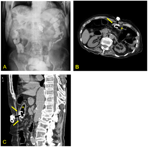
The first attempt at PEG implantation was aborted as the endoscopist could not identify an appropriate location in the stomach where the light of the endoscope could be seen through her abdomen. As part of this patient’s colon was interposed between her liver and diaphragm, a Chilaiditi sign (Figure (A)), the endoscopist prescribed a laxative and glycerin enema to reduce the volume of the colon. The procedure was re-attempted the following day with difficulty identifying an appropriate location for implantation of the PEG still encountered. An abdominal computed tomography (CT) scan was performed immediately after the procedure using the contrast medium gastrografin injected via the PEG tube to identify the possibility of colonic injury. A pinching of the transverse colon between the stomach and the abdominal wall was identified, indicative of a gastro-colo-cutaneous fistula (Figure (B) and (C)). Although the patient did not show signs of peritonitis or colon obstruction, we decided to perform a salvage surgery as we anticipated future stenosis of the colon. Moreover, the surgery would minimize the risk of colonic injury from any future PEG tube replacement. A laparoscopic partial colectomy and gastrostomy were conducted the next day.
Figure 1. (A) Abdominal radiograph showing segmental interposition of the colon between the diaphragm and the liver. The PEG tube appears to be implanted in the middle of the transverse colon. (B, C) Abdominal computed tomography scan (transverse and sagittal plane) showing pinching of the colon between the stomach and abdominal wall.
* Indicates the stomach.

3. Surgical procedures
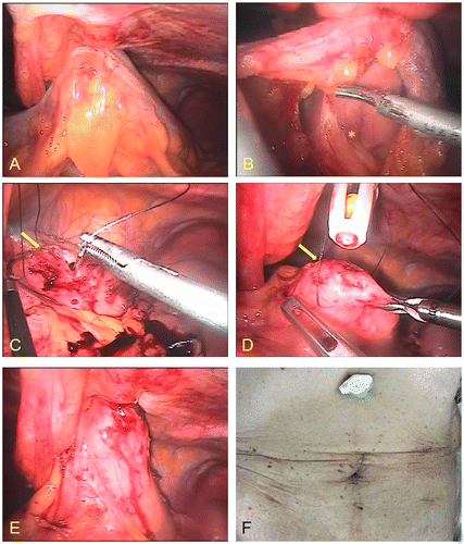
The surgery was performed with the patient in a supine position. An umbilical incision was made to insert the main trocar, and a pneumoperitoneum was initiated. No signs of peritonitis were identified. However, when the transverse colon was lifted and fixed to the abdominal wall, a slight subserosal hemorrhage was revealed (Figure (A)). Three other trocars were inserted on both sides of the abdomen.
Figure 2. Intra-operative photographs (A–E). (A) The colon is lifted and fixed to the abdominal wall; a slight subserosal hemorrhage is visible. (B) The stomach is visible during removal of the transverse colon from the mesocolon; * indicates the stomach. (C) Suturing of the abdominal wall and the stomach via the same hole in the skin through which the PEG tube was inserted; arrows indicate the hole of the stomach. (D) A new tube was introduced into the stomach. (E) Image shows the anterior wall of the stomach fixed to the abdominal wall. (F) Image of the incisions a few days after surgery.

The pinched section of the transverse colon was removed from the mesocolon using a harmonic scalpel (Ethicon Endo-Surgery, Blue Ash, Ohio, USA). Tissue was resected until the omental bursa was reached. At this point, the stomach, which was also lifted and fixed to the abdominal wall, could be observed behind the colon (Figure (B)). The larger omentum was removed from the colon and the colon was then severed using linear staplers (Endo GIA™, 60 mm Medium/Thick) (Covidien, Minneapolis, Minnesota, USA). After removing the PEG tube, the resected colon, which was perforated, was placed in a plastic bag to prevent contamination of the abdominal cavity. Next, a pair of 3-0 nylon sutures was inserted into the abdominal cavity from both sides of the hole in the skin through which the PEG tube was inserted. Suture needles were used to lift and fix the anterior wall of the stomach to the abdominal wall (Figure (C)). Prior to ligation using the nylon sutures, a new PEG tube (Kangaroo™, 20 Fr × 2.5 cm) (Covidien) was introduced through the same hole in the skin (Figure (D)). After expanding the bumper of the PEG tube in the stomach, the stomach was fixed to the abdominal wall with the nylon sutures (Figure (E)). Subsequently, contrast radiography was performed to ensure absence of gastrografin extravasation.
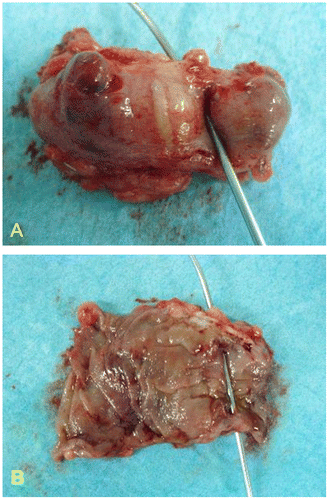
The umbilical incision was then extended to create a colonic anastomosis. After inserting a wound retractor (Alexis Wound Protectors/Retractors, M size) (Applied Medical, Rancho Santa Margarita, California, USA) to prevent surgical site infection (SSI), both edges of the colon were pulled out and a functional end-to-end anastomosis was created using linear staplers. After restarting the pneumoperitoneum, the abdominal cavity was washed with 3,000 ml of normal saline, and a JVAC drain (Ethicon Endo-Surgery) was then placed in the left subphrenic area. An adhesion prevention sheet (Seprafilm™) (Kaken Pharmaceutical, Tokyo, JAPAN) was placed behind the umbilical wound before closing the incisions. The resected colon was perforated at both the anterior and posterior wall indicating penetration of the PEG tube, although no histological abnormalities were observed (Figure (A) and (B)).
Figure 3. Resected specimen confirming penetration of the colon by the PEG tube: (A) serosal view, (B) mucosal view.

Enteral nutrition via the PEG was initiated on post-operative day 1, with no occurrence of significant major or minor complication, including surgical site infection or PEG-related skin troubles (Figure (F)).
4. Discussion
Previous studies have reported on PEG-related complications identified months and even years after complications during a PEG placement (Kim et al., Citation2014; Pitsinis & Roberts, Citation2003; Yamazaki, Sakai, Hatakeyama, & Hoshiyama, Citation1999). Schrag et al. (Citation2007) advocated adherence to the following three principles for PEG placement to avoid complications: endoscopic gastric distension, endoscopically visible focal finger invagination and transillumination. In our case, the transillumination of the stomach wall was not strong enough to prove that there were no other organs between the stomach and abdominal wall; therefore, the procedure could not be performed. Strict compliance with these three conditions may be essential to avoid complications related to PEG placement.
The incidence of the Chilaiditi sign has been estimated at 0.025 to 0.28%, and is four times more likely to occur in males than in females (Alva, Shetty-Alva, & Longo, Citation2008). Persons with Chilaiditi sign typically do not present with any symptoms. However, the Chilaiditi syndrome is known to sometimes cause abdominal and chest symptoms due to bowel obstruction or volvulus of the bowels (Saber & Michael, Citation2005). We did not find any study that demonstrated a clear relationship between this syndrome and PEG-related complications. An interesting report by Fukita et al. (Citation2014) demonstrated a unique method where colonoscopy was simultaneously used to mobilize the transverse colon in order to avoid colonic injury in a similar patient. If this anomaly is identified prior to PEG placement, this technique can be beneficial.
Bertolini, Christa, and Michael (Citation2014) reported a novel technique for closing colonic fistulas caused by buried bumper syndrome (BBS), defined as a continuous mechanical stimulation and sustained severe compression between the internal and external PEG bumpers, using colonoscopy. This technique, known as the over-the-scope-clip system (OTSC), however, has been reported to be difficult to perform or contraindicated as an early salvage method in the early stages of fistula development.
However, we successfully performed the minimally invasive salvage surgery without complications, immediately after PEG placement, after confirmation of the presence of an accidental fistula on the contrast CT. Our management strategy may be debatable with respect to the necessity of a salvage surgery because no significant signs of bowel obstruction or peritonitis were observed the day following PEG implantation. However, we performed the salvage procedure based on anticipated future complications, such as stenosis of the colon due to inflammation and fibrosis, and also the risk of colon injury due to any future PEG tube replacement. As we performed the salvage procedure prior to occurrence of any adhesion or inflammation, the procedure was safe. As long as the patient’s condition is adequate for general anesthesia and pneumoperitoneum, we advocate proceeding with a laparoscopic salvage surgery if a fistula complication is identified in the early phase after PEG implantation.
Ethical statement
The publication of this manuscript is approved by the ethical committee of our hospital and under the informed consent of the patient’s relatives.
Funding
The authors received no direct funding for this research.
Additional information
Notes on contributors
Hokahiro Katayama
In our hospital we engage in general surgery. While the number of patients are small, we eagerly try to apply low invasive surgery.
References
- Alva, S., Shetty-Alva, N., & Longo, W. E. (2008). Image of the month. Chilaiditi sign or syndrome. Archives of Surgery , 143, 93–94.10.1001/archsurg.2007.12-a
- Antonacci, N., Di Saverio, S., Biscardi, A., Giorgini, E., Villani, S., & Tugnoli, G. (2011). Dyspnea and large bowel obstruction: A misleading Chilaiditi syndrome. The American Journal of Surgery, 202, e45–e47.10.1016/j.amjsurg.2010.10.003
- Bertolini, R., Christa, M., & Michael, C. S. (2014). First report of colonoscopic closure of a gastrocolocutaneous PEG migration with over-the-scope-clip-system. World Journal of Gastroenterology, 20, 11439–11442.10.3748/wjg.v20.i32.11439
- de Acosta, A., David, A. M., Aberle, C. M., Khair, G., Streicher, A., Bartholomew, J., & Kacey, D. (2011). Chilaiditi syndrome complicated by a closed-loop small bowel obstruction. Gastroenterology & Hepatology, 8, 274.
- Fukita, Y., Katakura, Y., Adachi, S., Yasuda, I., Asaki, T., Toyomizu, M., & Ishibashi, H. (2014). Colonoscopy-assisted percutaneous endoscopic gastrostomy to avoid a gastrocolocutaneous fistula of the transverse colon. Endoscopy, 46, E60.
- Kamiyoshihara, M., Ibe, T., & Takeyoshi, I. (2009). Chilaiditi's sign mimicking a traumatic diaphragmatic hernia. The Annals of Thoracic Surgery, 87, 959–961.10.1016/j.athoracsur.2008.07.033
- Kim, H. S., Bang, C. S., Kim, Y. S., Kwon, O. K., Park, M. S., Eom, J. H., … Kim, D. J. (2014). Two cases of gastrocolocutaneous fistula with a long asymptomatic period after percutaneous endoscopic gastrostomy. Intestinal Research, 12, 251–255.10.5217/ir.2014.12.3.251
- Pitsinis, V., & Roberts, P. (2003). Gastrocolic fistula as a complication of percutaneous endoscopic gastrostomy. European Journal of Clinical Nutrition, 57, 876–878.10.1038/sj.ejcn.1601687
- Plorde, J. J., & Edmond, J. R. (1996). Transverse colon volvulus and associated Chilaiditi’s syndrome: case report and literature review. American Journal of Gastroenterology, 91, 12.
- Saber, A. A., & Michael, J. B. (2005). Chilaiditi’s syndrome: What should every surgeon know? The American Surgeon, 1, 261–263.
- Schrag, S. P., Sharma, R., Jaik, N. P., Seamon, M. J., Lukaszczyk, J. J., Martin, N. D., … Stawicki, S. P. (2007). Complications related to percutaneous endoscopic gastrostomy (PEG) tubes. A comprehensive clinical review. Journal of Gastrointestinal and Liver Diseases, 16, 407–418.
- Yamazaki, T., Sakai, Y., Hatakeyama, K., & Hoshiyama, Y. (1999). Colocutaneous fistula after percutaneous endoscopic gastrostomy in a remnant stomach. Surgical Endoscopy, 13, 280–282.10.1007/s004649900964
