Abstract
In this paper, we comment on a 4 year-old boy with a complex and large suprasellar arachnoid cyst extending to the adjacent temporal lobe. Its successful decompression using a unilateral endoscopic approach (confirmed on post-operative imaging) and excellent clinical recovery, highlights this as a useful technique to manage such cases.
Public Interest Statement
This case report demonstrates an unusual presentation of a relatively uncommon neurosurgical problem. As can be seen in the imaging of the patient’s head, there is a large mass, representing fluid, that has extended significantly. Indeed, on close inspection, you can see it distorting some of the surrounding structures, including where the optic nerves cross. The case demonstrates successful management endoscopically (essentially “keyhole” neurosurgery) with restoration of the flow of the fluid and reduced distortion of surrounding structures. There are multiple different approaches to this type of problem and this once again supports existing literature that the endoscopic management is effective and well tolerated.
Competing interests
The authors declare no competing interest.
1. Introduction
Arachnoid cysts have been shown to account for approximately 1% of all intracranial space occupying lesions and are commonly identified in children, with a prevalence of 2.6% in patients aged 18 years or less. Suprasellar arachnoid cysts account for between 2–16% of these cases (Gui, Wang, Zong, Zhang, & Li, Citation2011; Oberbauer, Haase, & Purcher, Citation1992). These cysts are found incidentally or with multiple symptoms including hydrocephalus and visual disturbance. They are considered predominantly as a congenital abnormality, associated with hyperplastic arachnoid cells and increased amounts of collagen within the walls of the cyst. Management has remained varied over time and surgical options include microsurgical excision/fenestration, cysto-peritoneal or ventriculo-peritoneal shunt insertion and endoscopic fenestration, with the current vogue favouring the endoscopic techniques (Gui et al., Citation2011). The latter is usually performed via a single burr hole although Fujio et al. have recently reported a case in which a bilateral approach via two burr holes was used in order to achieve a larger fenestration and thus greater decompression of the cyst (Fujio et al., Citation2016). In this paper, we report on a child that was found to have a large suprasellar arachnoid cyst with extension into the adjacent temporal lobe. The scan appearances were unusual for typical arachnoid cysts and we managed this case successfully using an endoscopic technique.
2. Case presentation
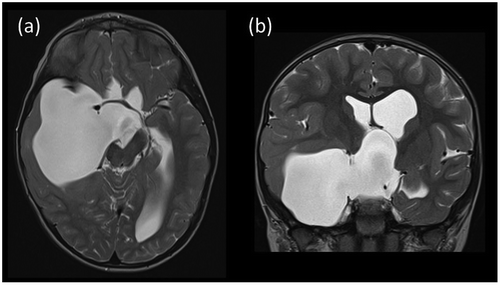
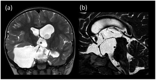
A 4 year-old boy was referred to our neurosurgical unit following a four-month history of “vacant episodes” and “occasional vomiting”. The child had no history of headache and his GCS was 15 on examination. He also complained of transient visual loss and episodes of blurred vision in the left eye at the time of referral, although examination of the eyes was normal with no evidence of papilloedema on fundoscopy. EEG was performed at the referring hospital and this showed high amplitude irregular slow waves with spike complexes in the right anterio mid-temporal region and subsequent MRI revealed a large suprasellar cyst extending into the right temporal lobe, shown in Figure . The child was commenced on leviteracetam and transferred to our neurosurgical unit where he underwent endoscopic marsupialisation of the cyst via a right frontal approach, leading to good decompression of the cyst, shown on his initial post-operative scan in Figure . The endoscopic technique was carried out via a right frontal burr hole at Kocher’s point, thus providing the best trajectory toward the frontal horn of the lateral ventricle, through which the cyst could be decompressed. Once a fenestration was made in the lateral wall of the suprasellar component, aiming for as large a window as possible, the endoscope was advanced towards the temporal aspect of the cyst wall and further fenestrations were made to restore CSF flow. Care was taken to avoid important structures encountered during this process, such as the basilar artery and perforators, the optic chiasm, the oculomotor nerve and the internal carotid artery. The aim was to perform numerous fenestrations, ideally acting in avascular areas, to allow for maximum decompression of the cyst.
Figure 1. Pre-operative MRI. (a) Axial and (b) coronal T2-weighted images showing the large right sided arachnoid cyst with suprasellar and right middle cranial fossa components.

Figure 2. Post-operative MRI. (a) Coronal T2-weighted image showing partial decompression of the arachnoid cyst. Note that the third ventricle (asterisk) has now partially re-expanded with descent of the previously elevated third ventricular floor (arrow). (b) Midline sagittal constructive interference in the steady-state (CISS) image showing the fenestration in the floor of the third ventricle with small CSF flow voids indicating patency.

3. Discussion
We report successful treatment of a complex suprasellar cyst with temporal extension using a unilateral endoscopic approach. Post-operative imaging confirmed adequate decompression and the presence of flow voids through each compartment of the cyst. Clinically the patient recovered remarkably well with no deterioration in vision and no further absence seizures. He continues to have follow-up with regular visual tests and endocrinology review to monitor for precocious puberty that can be associated with suprasellar cysts and most likely their compression on the pituitary stalk.
Suprasellar arachnoid cysts are a particularly complex pathology, often taking many years to present and are frequently found incidentally causing no symptoms whatsoever. The exact cause of arachnoid cysts is unknown but most are thought to be congenital in nature. We speculate that aberrant flow of CSF through cisterns, in this case possibly the suprasellar and basal cisterns, due to arachnoid adhesions/aberrant formation maybe a reason for the development of these cysts. However, there are some incidences of arachnoid cysts forming in association with haemorrhage, neoplasm and leptomeningitis (Gui et al., Citation2011). Because of their complex nature and commonly asymptomatic mode of presentation, the decision to treat is not taken lightly. It can be difficult to ascertain the extent of symptomatology in children, as found in this case. EEG findings as well as evidence of distortion effect on the optic chiasm on MRI imaging suggested that the cyst was the likely cause of these symptoms and so the decision to treat surgically was undertaken. As previously stated, endoscopic fenestration is the favoured choice as the literature shows excellent outcomes with a relatively low complication compared to those of shunting the cyst, microsurgery via craniotomy and percutaneous ventricle-cystostomy; reported 92% cure or improvement via endoscopic fenestration versus 81% for other surgical methods (Gui et al., Citation2011). Neuro-endoscopic fenestration has also been shown to be the safer of the surgical options avoiding risks such as subdural effusion and intracranial haematoma which can occur with microsurgery via craniotomy and there is no introduction of a foreign body, such as in a shunt procedure, which can block or become infected, leading to potentially life threatening CNS infection. Neuro-endoscopic fenestration does, however, carry risks of damage to CNS structures, infection, bleeding and failure of the procedure to relieve symptoms and therefore should not be carried out without careful consideration of risk versus benefits.
For this child, the most recent post-operative MRI scan showed that there was a marked reduction in the size of the large suprasellar and the right middle cranial fossa arachnoid cyst, with a corresponding reduction in distortion of the optic chiasm and pituitary stalk and reduced elevation of the floor of third ventricle. The MRI imaging showed a small perforation in the floor of the third ventricle/roof of the suprasellar cyst with some subtle flow artefact in this region suggesting continued patency of the endoscopic fenestration. There was no significant change in the ventricular dimensions at this stage.
To conclude, this report demonstrates a successful decompression of a highly complex suprasellar arachnoid cyst using the neuro-endoscopic technique, which we continue to advocate as the treatment of choice for these cases.
Funding
The authors received no direct funding for this research.
Additional information
Notes on contributors
O. MacCormac
Oscar MacCormac is currently a neurosurgery registrar at St Mary’s Hospital in Paddington, London, UK. He undertook his first year as a junior doctor at Queen’s Medical Centre, Nottingham, UK. Here he met Mr Natalwala and Prof Vloeberghs. Following working with these colleagues, this case was encountered and it was deemed pertinent to the literature to (1) highlight the effectiveness of endoscopic management and also the unusual size and presentation of the arachnoid cyst. Prof Vloeberghs operates using this method on similar cases with excellent results.
References
- Fujio, S., Bunyamin, J., Hirano, H., Oyoshi, T., Sadamura, Y., Bohara, M., & Arita, K. (2016). A novel bilateral endoscopic approach for suprasellar arachnoid cysts: A case report. Pediatric Neurosurgery, 51(1), 30–34.
- Gui, S. B., Wang, X. S., Zong, X. Y., Zhang, Y. Z., & Li, C. Z. (2011, May 18). Supracellar cysts: Clinical presentation, surgical indications and optimal surgical treatments. BMC Neurology, 11, 52.10.1186/1471-2377-11-52
- Oberbauer, R. W., Haase, J., & Purcher, R. (1992). Arachnoid cysts in children: A European co-operative study. Childs Nervous System, 8(5), 281–286.10.1007/BF00300797
