Abstract
Recent reports suggest the use of mouthpieces may be beneficial at improving aerobic and anaerobic exercise performance. However, the mechanisms of these reported improvements have yet to be elucidated. The purpose of this study was to explore the possible mechanisms of improved performance using the ArmourBite® mouthpiece. Using a within subject randomized treatment design, 15 experienced resistance trained males (19–26 years of age) performed 6 sets of 10 repetitions of free weight back squats at 80% of 1RM with and without a mouthpiece. Blood samples were collected before exercise, after 3 sets (Mid), immediately post (Post), 30 min post (Post-30), 60 min post (Post-60) and 120 min post (Post-120) exercise. Samples were analyzed for lactate and ELISA was used to determine cortisol. Mouthpiece use resulted in more repetitions completed without assistance (54.36 ± 0.61 vs. 53.27 ± 0.79, p = 0.046) and fewer assisted repetitions (6.73 ± 0.79 vs. 5.64 ± 0.61 repetitions, p = 0.046) compared to the control group. Lactate concentrations were lower in the treatment vs. control group at the Post (11.54 ± 2.23 vs. 13.07 ± 2.96 mmol/L, p = 0.023) Post- 30 (4.45 ± 1.94 vs. 5.41 ± 1.90 mmol/L, p = 0.021), and Post-60 (2.07 ± 0.94 vs. 2.55 ± 0.96 mmol/L, p = 0.048) sampling periods. Mouthpiece use lowered cortisol levels at Mid and Post-30 (19.39 ± 6.90 vs. 27.84 ± 14.56 μg/dL, p = 0.02 (22.91 ± 8.47 vs. 31.81 ± 10.79 μg/dL, p = 0.04). Cortisol AUC values showed significant differences within the AUC Pre-Post control and treatment (55.16 ± 23.84 vs. 41.95 ± 2.65 μg/dL, p = 0.02) groups. These data suggest that mouthpiece use may increase performance and decrease stress when used during intense resistance exercise.
Public Interest Statement
Mouthguards/mouthpieces have traditionally been used for protection during collision sports. However, our research finds that mouthpieces may also provide a performance benefit to the user during strenuous exercise. This article finds that individuals who used a lower custom-fitted mouthpiece experienced improvements during intensive resistance exercise. More specifically, with mouthpiece use, their blood lactate levels were 13% lower immediately after exercise and they had a 39% reduction in cortisol levels 30 minutes post exercise. What does this mean? For those who take part in strenuous anaerobic exercise, this improvement in both lactate and cortisol could benefit recovery mechanisms which would then improve subsequent training sessions. Thus, it is evident from this study that wearing a mouthpiece promoted positive physiological changes. More exploration into why this occurred is needed in order to understand the effect of mouthpiece use during exercise.
Competing Interests
The authors declare no competing interest.
1. Background
Mouthguard use during sport has been utilized as a method to prevent oral-facial injury, with a review of dental trauma literature citing participation in sport as being the greatest cause of dental injury (Glendor, Citation2009; Hughston, Citation1980). However, the use of such appliances has also been cited to improve athletic performance. Early research in this area focused on the use of MORA devices (mandibular orthopedic repositioning appliance) stated to improve performance along with the protection of teeth (Fonder, Citation1976). The review of this research occurring in the late seventies and early eighties, was varied and provided no solid evidence that these devices improved performance based on the methodology used in the studies. Smith, Stenger, and Grunwaldt found improvements in strength with MORA devices in professional football players, with Grunwaldt finding an 8–11% improvement in Cybex muscle testing in these athletes with the oral appliance (Smith, Citation1982; Stenger, Citation1977). Yet in testing college athletes, researchers were unable to detect differences in strength with the use of an oral appliance (Welch, Edington, & Ritter, Citation1986; Yates, Koen, Semenick, & Kuftinec, Citation1984). However; problems existed for each of these studies, ranging from small sample sizes to varied fitness levels of athletes, to lack of uniformity of devices used between studies. Thus, research in the area of mouthpiece use during exercise, and measurement of these parameters, remained stagnant until the early 2000s when interest in this topic renewed partly due to the subjective feedback provided by athletes wearing mouthpieces designed by Shock Doctor, Bite Tech and Makkar Athletic; mouthguard companies that marketed the effectiveness of mouthguard use during exercise for performance enhancement.
Recently, a review of the anaerobic parameters during exercise showed that when using a mouthpiece, improvements include increased torque, bench throw power and force, vertical jump, and the Wingate anaerobic test (Arent, McKenna, & Golem, Citation2010; Buscà, Morales, Solana-Tramunt, Miró, & García, Citation2016; Dunn-Lewis et al., Citation2012; Durante-Pereira et al., Citation2008; Ebben, Leigh, & Geiser, Citation2008; Morales, Buscà, Solana-Tramunt, & Miró, Citation2015). Dunn-Lewis and colleagues cited significant increases in bench throw power and force, increased rate of power production in the vertical jump with the Pure Balance mouthguard vs. no mouthguard and an over the counter mouthguard, citing improvements in both men and women in the area of upper body power exercises (Dunn-Lewis et al., Citation2012). Busca and colleagues and Durante-Pereira and colleagues in testing countermovement vertical jumps cited significant improvements in mean power and height in the mouthpiece vs. no mouthpiece condition (Buscà et al., Citation2016; Durante-Pereira et al., Citation2008). Other studies found significant improvements, 4% peak power and 1% mean power, during the Wingate test using an oral appliance (Arent et al., Citation2010; Morales et al., Citation2015). Morales and colleagues also measured lactate during anaerobic testing and cited a significant 8% decrease in lactate measures with mouthpiece use (Morales et al., Citation2015). Although this research appears to support the use of a mouthpiece during anaerobic exercise, more research in the area of objective measures is needed to quantify a mouthpiece effect. To do this, Garner and colleagues have assessed lactate and cortisol during both aerobic and anaerobic exercise, finding significant improvements in both lactate and cortisol with mouthpiece use (Garner, Dudgeon, & McDivitt, Citation2011; Garner & McDivitt, Citation2009b, Citation2015). Specifically as it relates to anaerobic protocols, they found a 51% reduction in cortisol levels 10 min post intensive bout of resistance training. This is supported in the animal literature in which stressed rats had a significantly lowered stress response while biting on a stick vs. no stick (Hori et al., Citation2004; Sasaguri et al., Citation2005). Yet, there is a paucity of data in the area of mouthpiece use and effect on lactate and cortisol during resistance exercise. Thus, the purposes of this study were to determine the effect of mouthpiece use on free weight back squat performance as well as the effect on lactate and cortisol measures mid exercise, immediately post, and 30, 60 and 120 min post exercise.
2. Methods
2.1. Experimental approach to the problem
The study was conducted using a within-subject randomized treatment design. One week following an initial visit, participants reported to the lab and were randomly assigned to either a control (no mouthpiece) or treatment (mouthpiece) group. Subjects remained blind to the treatment until their first scheduled testing day and received the other treatment during their second testing day, scheduled for one week later at the same time of day controlling for diurnal variations. Subjects were instructed to refrain from intense physical activity or exercise for 24 h prior to testing as well as food and drink except for water 2 h prior to testing (Stachowicz & Lebiedzińska, Citation2016). The influence of overall diet on cortisol secretion is inconclusive, however; the military environment of our subject pool resulted in a very homogenous diet. After arriving for testing, researchers placed an IV catheter into a superficial forearm vein for each testing sequence. Following IV insertion, subjects followed a standardized 20-min no activity period before the first blood sample to control for the stress of IV placement. Subjects in the treatment group were individually fitted by the researcher with the boil and bite UnderArmour ArmourBite® (UnderArmour, Baltimore, Maryland) mandibular mouthpiece molded to mandibular characteristics before IV insertion and were instructed to wear the mouthpiece throughout the entire testing period. Six blood samples were taken during each testing protocol to analyze blood lactate and serum cortisol: Pre-testing, after set 3 (mid), immediately post exercise (post), Post-30 min exercise, Post-60 min exercise, and Post-120 min exercise. Following a standardized warm-up, subjects began the standardized testing procedure, which consisted of 6 sets of 10 repetitions at 80% of the subject’s respective 1 RM with 2-min rest between sets. Assisted repetitions were used to help subjects complete all repetitions within a set.
2.2. Subjects
Fifteen experienced resistance trained male participants (19–26 years of age, height: 178 ± 6.3 cm, weight: 87 ± 13.6 kg, and body fat: 16 ± 7%) with no prior performance mouthpiece use volunteered to investigate the performance benefit and response of serum cortisol and blood lactate with and without the use of ArmourBite® mouthpiece during and following heavy resistance exercise. Subjects were recruited through posters, announcements, word of mouth, and referrals that briefly explained the study. The study was explained to the participants upon their initial contact with the researchers and again during their first visit. The initial meeting included: written consent approved by the institution’s Internal Review Board, a completed Health Questionnaire and Physical Activity Readiness Questionnaire, one repetition maximum (1 RM) test for the free weight back squat, and anthropometric measurements (body composition by BodPod™, height, and weight).
2.3. Procedures
All subjects performed the same 1RM free weight back squat protocol: After a standardized warm up, subjects performed 2 warm-up sets with 10 repetitions at 50% and 4 repetitions at 70% of the estimated 1 RM. Subjects then made their first attempt with their estimated 1RM. Researchers sought to achieve subjects’ 1RM within 4 attempts. If subjects succeeded on their attempts the load was increased. If subjects failed on their attempt, researchers reduced the load by 15–20 lb and a re-attempt was made. Once subjects failed twice, the subjects’ 1RM was recorded with the last successful attempt. Subjects were scheduled to begin testing at least 7 days following their initial visit. Testing sessions were scheduled based on subject’s availability. Testing session time remained consistent for each trial. No mouthpiece was utilized during the initial testing period to ascertain estimated 1 RM.
Before any testing began, all subjects were fitted with the UnderArmour ArmourBite® (UnderArmour, Baltimore, Maryland) mandibular mouthpiece following standardized fitting instructions from the manufacturer. The mouthpiece was immersed in boiling water for 1 min then fitted to the subject’s lower teeth by placing the bite pads on the teeth and then having the subjects bite down. The fit of the mouthpiece was assessed by research team members to ensure that the mouthpiece was properly placed so that the upper teeth and the lower teeth were separated by the bite pads. If the mouthpiece did not fit properly, the mouthpiece was re-boiled, and the process repeated until proper placement was achieved. In addition, during all testing which involved the mouthpiece, subjects were instructed to bite down on the mouthpiece while completing the exercise session.
The testing protocol consisted of a standardized warm-up period of 10-min light resistance cycling (Monark Ergomedic 828 E, Varberg, Sweden), 5-min of jumping rope, 1 set of 10 repetitions at 50% of the subject’s 1 RM free weight back squat, and 1 set of 4 repetitions at 70% of the subjects 1 RM. Subjects were then given a chance to stretch ad libitum before initiation of the exercise protocol known to elicit increases in serum cortisol, which consisted of 6 sets of 10 repetitions at 80% of the subject’s respective 1 RM with a 2-min rest following each set (Kraemer & Ratamess, Citation2005; Ratamess et al., Citation2005). If a subject could not complete 10 repetitions on a given set, a certified strength and conditioning specialist provided the subject the necessary assistance required to complete each subsequent repetition for that set. All assistance was administered following the National Strength and Conditioning Associated standardized spotting protocol (Sands et al., Citation2012). If 10 repetitions were not completed on a set, the load was adjusted so subjects could complete 10 repetitions for each subsequent set. Subjects completed an identical protocol for both testing days.
2.4. Sample collection and analysis
Blood samples were collected through an 18-gauge, 1.75 inch indwelling IV catheter (Exel Safelet Cath, Exelint International Corporation, Los Angeles, California) placed into a superficial forearm vein. Researchers attached a 5 cm extension tube and a three-way stopcock with swivel male Luer-lok to the catheter. After placement of the catheter, researchers used 4 mL of heparinized saline (Heparin Lock Flush Solution 100 units/mL, BD, Franklin Lakes, New Jersey) in the extension tube to maintain patency of the line. A 2 mL heparinized saline flush was also performed at the conclusion of every blood draw except for the final. The catheter, extension tube, and stopcock were secured with paper tape (3 M, St. Paul, Minnesota) and Coban wrap (3 M, St. Paul, Minnesota). Following IV insertion, a standardized 20-min no activity period before the first blood sample was utilized to control for the stress of IV placement.
Researchers collected approximately 24 mL of blood per sample in order to quantify lactate and cortisol. Using a 5 mL syringe with a Luer-lok tip, researchers drew approximately 4 mL of blood that was discarded as waste to clear the IV line of any heparinized blood. Using two 10 mL syringes with a Luer-lok tip, researchers collected 20 mL of blood. Blood samples were used to monitor hemoglobin, hematocrit and lactate, and for collection of blood serum and plasma for later examination of stress hormones. Serum tubes were placed in centrifuge (Horizon Premier, Fisher Healthcare, Philipsburg, Pennsylvania) and spun at 3,000 RPM for 15 min. Samples were then collected with transfer pipets (Samco Scientific Corporation, San Fernando, California) and placed in storage containers (Eppendorf North American, Hauppauge, New York) and immediately stored at −35°C (Thermo Scientific, USA) for immunoassay at a later date.
Hematocrit (Hct) and Hemoglobin (Hb) were measured to account for changes in plasma volume. Samples for Hct, Hb and lactate were taken from the remaining 6 ml of blood from the samples drawn. Blood lactate concentrations were measured using Lactate Plus distributed by Nova BioMedical (Waltham, MA). Three blood lactate concentrations were recorded and averaged together at each sampling period. Hct samples were determined in triplicate by placing blood in hemato-clad heparinized 75 mm Hct tubes (Drummond Scientific Company, Broomall, Pennsylvania), packed with clay at one end, and wiped with a Kim Wipe (KimTech, Roswell, Georgia) before spinning. Hematocrit samples were centrifuged for 5 min at 10,000 rpm with a ZIPocrit (LW Scientific, Lawrenceville, Georgia) centrifuge. Hct values were determined using a metric ruler by the same research assistant. For Hb, blood samples were placed in HemoPoint H2 Microcuvettes and then analyzed in duplicate by a HemoPoint H2 (Stanbio, Boerne, Texas). If the two Hb values were more than 10% different from each other, then a third measurement was performed. Duplicate and triplicate samples values were averaged for each variable at each time point.
Enzyme-linked immunoassay analyses (ELISA) were used for detection of serum cortisol (Cortisol ELISA, ALPCO Diagnostic, Salem, New Hampshire). Samples were thawed only once and were assayed in duplicate following manufactures instructions. ELISA plates were analyzed using a Molecular Devices microplate reader (ELx808, BioTek Instruments, Winooski, Vermount). Intra assay coefficients of variation (CV) for cortisol were 6%, and inter assay CV for cortisol were 16% μg/dL. Minimum sensitivity of serum cortisol was 0.4 μg/dL.
2.5. Statistical analyses
Data evaluations were performed using Microsoft Excel with StatPlus supplementing analyses when needed. One way repeated measures analysis of variance (ANOVA) tests were used to determine any differences within the control and treatment groups. Dependent measures T-tests were used to compare between groups to determine if mouthpiece use had significant effects. A Bonferroni adjustment was made to protect against the chance of committing a type I error due to the low power associated with the small sample size. Mean area under the curve (AUC) analyses were performed using the trapezoidal method for three different time periods for both groups: All six sample periods (AUC All-6), Pre-testing through immediately Post (AUC Pre-Post), and immediately Post through Post-120 min (AUC Post- Post-120). Statistical significance was set at p < 0.05. All data are presented as mean ± standard deviation.
3. Results
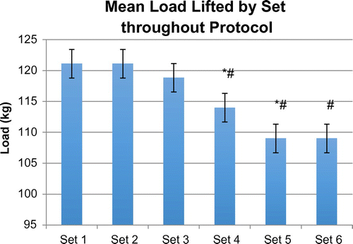
Based on previous research it was hypothesized that the use of the ArmourBite® mouthpiece would decrease serum cortisol levels and lactate levels during heavy resistance exercise. No significant changes in plasma volume were found between groups during the trial period. Due to the testing protocol, loads (weight lifted) were identical between groups. Loads decreased significantly between set 3 and set 4 (119 ± 21 vs. 114 ± 20 kg, p = 0.01) and from set 4 to set 5 (114 ± 20 vs. 109 ± 19 kg, p = 0.028). Loads did not decrease significantly with sets 1 through 3. However; set 4, set 5, and set 6 loads decreased significantly compared to set 1 (121 ± 24 vs. 114 ± 20, 109 ± 19, and 109 ± 19 kg; Set 4, p = 0.016; Set 5, p = 0.013; Set 6, p = 0.013). See Figure for details of loads lifted. These results were expected, as the exercise protocol is known to cause muscular fatigue.
Figure 1. Mean load throughout the exercise protocol for all subjects and treatments. Identical load procedure was performed for both groups.

Total mean non-assisted repetitions (i.e., completed under the subject’s own power) were different between control and treatment groups with the treatment group completing more non-assisted repetitions before failure (53.27 ± 0.79 vs. 54.36 ± 0.61 repetitions, p = 0.046). Consequently, total mean assisted repetitions were also different between control and treatment groups with the control group requiring significantly more assisted repetitions (6.73 ± 0.79 vs. 5.64 ± 0.61 repetitions, p = 0.046).
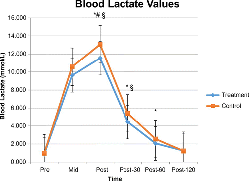
Blood lactate concentrations were significantly lower in the treatment group vs. control group at the Post (11.54 ± 2.23 vs. 13.07 ± 2.96 mmol/L, p = 0.02), Post- 30 (4.45 ± 1.94 vs. 5.41 ± 1.90 mmol/L, p = 0.02), and Post-60 (2.07 ± 0.94 vs. 2.55 ± 0.96 mmol/L, p = 0.048) sampling periods as outlined in Figure . As expected both groups showed significant increases in blood lactate compared to rest at the Mid and Post time points; however, by Post-30 lactate concentrations no longer differed significantly from baseline levels in the treatment group while control group levels remained significantly elevated.
Figure 2. Mean lactate concentrations for treatment and control groups throughout testing period.

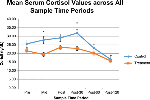
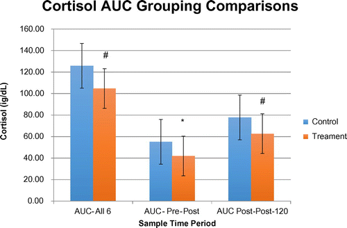
Serum cortisol levels were lower at the Mid and Post-30 time points in the treatment group vs. control group (19.39 ± 6.90 vs. 27.84 ± 14.56 μg/dL, p = 0.02; 22.91 ± 8.47 vs. 31.81 ± 10.79 μg/dL, p = 0.04) while the remaining time points trended lower, but not significant, for the treatment group compared to the control group as seen in Figure . Surprisingly, cortisol values within both groups never experienced significant increases from pre-exercise values during the Mid and Post sampling period (Treatment: 21.55 ± 9.82, vs. Mid 19.39 ± 6.90 and 23.56 ± 4.47 μg/dL; Control: 25.58 ± 10.88 vs. 27.84 ± 14.56 and 29.06 ± 12.63 μg/dL); however, cortisol values of the treatment group returned to baseline levels by Post-30 while the control group peaked at the Post-30 sampling period (22.91 ± 8.47 vs. 31.81 ± 10.79 μg/dL). AUC analysis for cortisol values showed significant differences within the AUC Pre-Post for control and treatment (55.16 ± 23.84 vs. 41.95 ± 12.65 μg/dL, p = 0.02) groups. While the AUC All-6 and AUC Post - Post-120 showed lower trend values between the control and treatment groups but no significant differences (125.81 ± 31.99 vs. 104.67 ± 33.16 μg/dL, p = 0.053; 77.74 ± 24.03 vs. 62.72 ± 25.39 id/dL, p = 0.11). See Figure for differences in cortisol AUC values between groups.
4. Discussion
Previous investigations of mouthpiece use during exercise have shown reductions in salivary cortisol levels (Garner, Dudgeon, Scheett, McDivitt, Citation2011; Garner & McDivitt, Citation2009b, Citation2015), lactate concentration (Garner & McDivitt, Citation2009a, Citation2009b, Citation2015), and changes in respiratory kinetics (Garner, Citation2015; Garner, Dudgeon, Scheett, & McDivitt, Citation2011). These results suggest the possible involvement of the two major stress axes: the hypothalamic-pituitary-adrenal (HPA) axis (CitationGarner et al., 2011) and subsequently possible involvement of the sympathetic-adrenal-medullary (SAM) axis (Garner, Dudgeon, McDivitt, Citation2011) due to mutual activation in the stress response (Chrousos, Citation2007; Fatouros et al., Citation2010; Tsigos & Chrousos, Citation2002; Vanltallie, Citation2002). This suggests that mouthpiece use can acutely impact neuroendocrine physiology, thereby impact substrate availability and utilization, and ultimately play a role in performance outcomes. The purpose of this investigation was, firstly, to investigate the effects of performance mouthpiece use on the serum cortisol response to resistance exercise. Secondly, the purpose of this study was to investigate the effect of performance mouthpiece use on blood lactate levels before, during, and after intense resistance exercise. Finally, this study aimed at determining if performance mouthpiece use could improve resistance exercise performance. Based on current research, it is hypothesized that serum cortisol and blood lactate concentrations will decrease as a result of performance mouthpiece use during and following a resistance exercise session and performance will be enhanced.
Total mean non-assisted repetitions completed proved to be different between groups with the treatment group completing significantly (p < 0.05) more repetitions before failure. Conversely, total mean assisted repetitions were significantly (p < 0.05) higher with the control group. Further, mean non-assisted repetitions trended higher with the treatment group during every set except sets 1 and 2. Mean weight lifted followed a similar trend with the treatment group exhibiting a trend of more weight lifted during the latter sets. The higher trend of non-assisted repetitions led to a significantly higher (p < 0.05) total mean weight lifted for the treatment group compared to the control group. In this application it appears that performance mouthpiece use during heavy resistance exercise has led to the completion of more repetitions before failure, thus resulting in more total weight lifted.
Lactate data from this study could provide some insight into explaining the difference in total weight lifted and non-assisted repetitions. This study showed lactate, which is released in proportion to the perceived “stress” of the activity, to be consistently lower in the treatment group compared to the control group immediately post exercise through 60 min post exercise. This blunted lactate response in the treatment group coincides with the trend for fewer assisted repetitions in the same group. Increases in blood lactate caused by the accumulation of hydrogen ions with high intensity exercise results in metabolic acidosis leading to fatigue, thereby hindering athletic performance (Amis et al., Citation2000). While this study did not provide a mechanism for the lactate reduction, these results have shown that mouthpiece use decreases blood lactate accumulation thereby possibly delaying the onset of fatigue resulting in more physical work performed.
Animal studies have been successful at linking biting and chewing mechanisms with a reduction in the stress response during stress-induced activities. A 2004 study by Hori and colleagues (Hori et al., Citation2004) investigated the possible suppression of the stress-induced expression of corticotrophin releasing hormone (CRH) in the paraventricular nucleus (PVN) of the rat hypothalamus through nonfunctional biting. Researchers restrained the bodies of rats, known to activate the HPA axis, for either 30 or 60 min to examine the expression of CRH in the cells of the PVN during biting. Rats biting on a wooden stick during the restraint period had a significant (p < 0.05) reduction in the expression of CRH than those not biting, regardless of the restraint time period. The secretion of CRH from the PVN results in the release of adrenocorticotropic hormone (ACTH) from the anterior pituitary (Chrousos, Citation2007; Tsigos & Chrousos, Citation2002). ACTH secretion stimulates the release of cortisol from the adrenal cortex (Chrousos, Citation2007; Tsigos & Chrousos, Citation2002). Researchers noted the interaction of the hypothalamus with the cerebral cortex and the limbic system, thereby, integrating the autonomic and endocrine functions. Researchers speculated that it is because of this interaction that suppression of central noradrenergic transmission might be the mechanism for the suppression of CRH by biting.
Thus, examining the clenching response may provide the key to understanding outcomes noted in mouthguard/mouthpiece use during exercise. Clenching has been shown to affect cerebral activity in activation of the cortical areas in the brain and in increasing blood flow (Hasegawa et al., Citation2007; Hori et al., Citation2004; Sasaguri et al., Citation2005; Shibusawa et al., Citation2009; Tahara et al., Citation2007). Specifically, researchers have cited that restrained and stressed rats, when biting on a stick, had reductions in corticotrophin releasing factor and c-Fos in the hypothalamus which may be modulated by suppression of extracellular signal-regulated protein kinase 1/2 (pERK 1/2) in the paraventricular nucleus (Hori et al., Citation2004; Sasaguri et al., Citation2005; Kaneko et al., Citation2004). Human studies have also supported this link with lowered cortisol levels during biting down on a mouthpiece or chewing during stress (Garner, Dudgeon, Scheett et al., Citation2011; Tahara et al., Citation2007). Garner and colleagues (Garner, Dudgeon, Scheett et al., Citation2011) examined salivary cortisol levels with mouthpiece use following a 1-h intense resistance training session in 28 trained, college-aged males. Researchers found a significant 51% difference (p = 0.02) in salivary cortisol levels 10-min post exercise between mouthpiece and no treatment groups. Perhaps the most interesting finding was that cortisol divergence did not appear between treatment and control groups until post-exercise, which researchers believe could be due to the stress response remaining unaffected by mouthpiece use during the exercise protocol (Garner, Dudgeon, Scheett et al., Citation2011).
This link between the hypothalamus and the involvement in the jaw musculature via clenching may be explained by neuronal projections from the lateral hypothalamus connecting to the trigeminal motor nucleus in the rat model (Mascaro et al., Citation2009). In addition, it was observed that the trigeminal motor nucleus was innervated by corticotrophin releasing factor immunoreactive fibers within the amygdala (Mascaro et al., Citation2009). Research also suggests that activation within the dorsolateral prefrontal cortex (DLPFC, an area in the cerebral cortex) is most likely dependent on continuous teeth contact as occurs during clenching, and that intensity of the clenching most likely influences that magnitude of the cerebral activity within the sensorimotor cortex (area in cerebral cortex responsible for motor function) (Iida et al., Citation2012; Shibusawa et al., Citation2009). Finally, Qin et al. (Citation2009) noted that the function of the DLPFC most likely is affected by the HPA axis by decreasing levels of the catecholamines (Qin et al., Citation2009). These findings are of significance as it relates to mouthpiece use during exercise as they provide potential explanation for the cited decreases in cortisol and lactate while clenching on a mouthpiece during exercise (Garner, Dudgeon, Scheett, et al., Citation2011; Garner & McDivitt, Citation2009a, Citation2009b, Citation2015).
Our findings indicate a differing cortisol response to high-intensity exercise with the use of a mouthpiece. Peak cortisol levels were lower with mouthpiece use and the overall cortisol production was blunted with mouthpiece use as evidenced the AUC data. This study was the first to investigate serum cortisol levels with mouthpiece use during high-intensity resistance exercise; however, previous research has also cited salivary cortisol augmentation with mouthpiece use during exercise (Garner, Dudgeon, Scheett et al., Citation2011; Garner & McDivitt, Citation2009b). While cortisol’s importance during exercise (i.e. gluconeogenesis) is an important step to provide energy during prolonged or intense exercise, its catabolic nature post-exercise could hinder or delay recovery. Our findings suggest a possible performance benefit, particularly post-exercise, as we have shown a quicker return to baseline with both salivary cortisol (Garner, Dudgeon, Scheett et al., Citation2011) and less overall cortisol production (present study).
Early research linking human performance and mouthpiece use was plagued by poor research methodology and called for a greater contribution and collaboration between the scientific community and clinical researchers. Research now favors proper design and analysis, with researchers beginning to investigate the mechanisms behind oral appliance use and human performance. The design of the performance mouthpiece in this study used a wedge component designed to reposition the mandible. This design results in a separation of the teeth with a more favorable position of the mandible (Garner, Dudgeon, Scheett et al., Citation2011) causing a decreased amount of stress placed upon the mandible, thereby decreasing the stress response through possible actions on the motor areas of the brain (Iida et al., Citation2010; Tahara et al., Citation2007). Researchers have recently been able to map brain activity during clenching and chewing (Hasegawa et al., Citation2007; Momose et al., Citation1997; Shibusawa et al., Citation2009; Tahara et al., Citation2007) and have found that clenching and chewing not only resulted in activation of the autonomic nervous system areas of the brain, but also resulted in stimulation of the hypothalamus (Tahara et al., Citation2007).
5. Practical application
Due to the importance of stress hormones to provide increased blood flow, oxygen, and substrates to working muscles and the desire to suspend the stress response upon cessation of exercise to inhibit catabolic mechanisms, finding a way to decrease the stress response during the post exercise period would be valuable. Further, delaying the accumulation of lactate concentrations by prolonging the onset of muscle acidosis caused by the accumulation of hydrogen ions would thereby improve athletic performance (Garner & McDivitt, Citation2015; Gelb et al., Citation1996). This particular study demonstrated decreases in cortisol production during and after exercise with a general trend of lower cortisol values throughout the exercise protocol with performance mouthpiece use. Further, lactate was found to be significantly lower during the post exercise time period. Much of the research in the area of mouthguard/mouthpiece use during exercise finds minimal to no acute benefits, with much of the data being conflicting. However; this research along with the earlier research related to cortisol, suggests the potential impact of mouthpiece use on recovery and subsequent training sessions. However, research related to performance mouthpiece use is still emerging, thus it is imperative researchers continue to investigate possible mechanisms of action. Most of the research associated with mouthpiece/mouthguard use during exercise does not seek to elucidate the mechanisms of why a mouthpiece/mouthguard may be beneficial (Allen et al., Citation2014). Yet our group feels it is critical to clarify the potential physiological mechanisms in order to better understand the acute effects within the specific population studied and exercise protocol chosen. Bridging the gap between exercise physiology and dental research will provide valuable knowledge for the practitioner, exercise scientist, and the athlete and avoid much of the guesswork utilized in past research.
Funding
This work was supported by The Citadel Foundation; the Bite Tech, Inc.
Acknowledgements
The authors would like to thank The Citadel Foundation and Bite Tech, Inc. for partial funding of this research.
Additional information
Notes on contributors
W.D. Dudgeon
W.D. Dudgeon received a PhD in exercise science with a focus on applied physiology at the University of South Carolina in 2006. He is currently a tenured, associate professor at the College of Charleston and the head of department of health and human performance. His research interests include mouthpiece use during exercise and sport nutrition.
The Human Performance Laboratory at The Citadel has spent the last 13 years assessing the effect of mouthpiece use on human performance parameters during exercise. Parameters assessed have included oropharyngeal width and diameter, cortisol (blood and salivary), lactate levels, respiratory changes and human work output. The research in our laboratory has suggested a physiological effect during both aerobic and anaerobic performance. Theories which support these changes may include increased cerebral blood flow that occurs during clenching on a mouthpiece and improved placement of the genioglossus which reduces the respiratory rate during aerobic exercise. The latest research presented in this paper demonstrates blood marker changes in both lactate and cortisol after a bout of intense resistance exercise. Future research needs must assess the causes for these physiological changed with mouthpiece use during exercise
References
- Allen, C. R., Dabbs, N. C., Zachary, C. S., & Garner, J. C. (2014). The acute effect of a commercial bite-aligning mouthpiece on strength and power in recreationally trained men. Journal of Strength and Conditioning Research, 28(2), 499–503.10.1519/JSC.0b013e3182a95250
- Amis, T., Di Somma, E., Bacha, F., & Wheatley, J. (2000). Influence of intra-oral maxillary sports mouthguards on the airflow dynamics or oral breathing. MSSE, 32, 284–290.
- Arent, S. M., McKenna, J., & Golem, D. L. (2010). Effects of neuromuscular dentistry-desgined mouthguard on muscular endurance and anaerobic power. Comparative Exercise Physiology, 7, 73–79.
- Buscà, B., Morales, J., Solana-Tramunt, M., Miró, A., & García, M. (2016). Effects of jaw clenching while wearing a customized bite-aligning mouthpiece on strength in healthy young men. Journal of Strength and Conditioning Research, 30(4), 1102–1110.10.1519/JSC.0000000000001192
- Chrousos, G. P. (2007). Organization and integration of the endorine system. Sleep Medicine Clinics, 2(2), 125–145.10.1016/j.jsmc.2007.04.004
- Dunn-Lewis, C., Luk, H. Y., Comstock, B. A., Szivak, T. K., Hooper, D. R., Kupchak, B. R., … Denegar, C. R. (2012). The effects of a customized over-the-counter mouth guard on neuromuscular force and power production in trained men and women. Journal of Strength and Conditioning Research, 26(4), 1085–1093.10.1519/JSC.0b013e31824b4d5b
- Durante-Pereira, D. M. V., del Rey-Santamaria, M., Javierre-Garces, C., Barbany‐Cairó, J., Paredes‐Garcia, J., Valmaseda‐Castellón, E., … Gay‐Escoda, C. (2008). Wearability and physiological effects of custom-fitted vs self-adapted mouthguards. Dental Traumatology, 24(4), 439–442.10.1111/edt.2008.24.issue-4
- Ebben, W. P., Leigh, D. H., & Geiser, C. F. (2008). The effect of remote voluntary contractions on knee extensor torque. Medicine & Science in Sports & Exercise, 40(10), 1805–1809.10.1249/MSS.0b013e31817dc4ad
- Fatouros, I., Chatzinikolaou, A., Paltoglou, G., Petridou, A., Avloniti, A., Jamurtas, A., … Margeli, A. (2010). Acute resistance exercise results in catecholaminergic rather than hypothalamic-pituitary-adrenal axis stimulation during exercise in young men. Stress, 13, 468.
- Fonder, A. C. (1976). The profound effect of the 1973 Nobel Prize on dentistry. Basal Facts, 1, 21–29.
- Garner, D. P. (2015). Effects of various mouthpieces on respiratory physiology during steady state exercise in college-aged subjects. General Dentistry, 63, 30–34.
- Garner, D. P., Dudgeon, W. D., & McDivitt, E. (2011). The effects of mouthpiece use on cortisol levels during an intense bout of resistance exercise. Journal of Strength and Conditioning Research, 25(10), 2866–2871.10.1519/JSC.0b013e31820ae849
- Garner, D. P., Dudgeon, W. D., Scheett, T. P., & McDivitt, E. J. (2011). The effects of mouthpiece use on gas exchange parameters during steady-state exercise in college-aged men and women. The Journal of the American Dental Association, 142(9), 1041–1047.10.14219/jada.archive.2011.0325
- Garner, D. P., & McDivitt, E. (2009a). Effects of mouthpiece use on airway openings and lactate levels in healthy college males. Compendium of Continuing Education in Dentistry, 30(2), 9–13.
- Garner, D. P., & McDivitt, E. J. (2009b). The effects of mouthpiece use on salivary cortisol and lactate levels during exercise. Medicine & Science in Sports & Exercise, 41, S448.
- Garner, D. P., & McDivitt, E. J. (2015). Effects of mouthpiece use on lactate and cortisol levels during and after 30 min of treadmill running. Effects of mouthpiece use on lactate and cortisol levels during and after 30 min of treadmill running, 3. doi:10.11131/2015/101148
- Gelb, H., Mehta, N. R., & Forgione, A. G. (1996). The relationship between jaw posture and muscular strength in sport dentistry: A reappraisal. CRANIO®, 14(4), 320–325.10.1080/08869634.1996.11745984
- Glendor, U. (2009). Aetiology and risk factors related to traumatic dental injuries - a review of the literature. Dental Traumatology, 25(1), 19–31.10.1111/edt.2009.25.issue-1
- Hasegawa, Y., Ono, T., Hori, K., & Nokubi, T. (2007). Influence of human jaw movement on cerebral blood flow. Journal of Dental Research, 86(1), 64–68.10.1177/154405910708600110
- Hori, N., Yuyama, N., & Tamura, K. (2004). Biting suppresses stress-induced expression of corticotropin-releasing Factor (CRF) in the rat hypothalamus. Journal of Dental Research, 83(2), 124–128.10.1177/154405910408300208
- Hughston, J. C. (1980). Prevention of dental injuries in sports. The American Journal of Sports Medicine, 8(2), 61–62.10.1177/036354658000800201
- Iida, T., Kato, M., Komiyama, O., Suzuki, H., Asano, T., Kuroki, T., … Kawara, M. (2010). Comparison of cerebral activity during teeth clenching and fist clenching: A functional magnetic resonance imaging study. European Journal of Oral Sciences, 118(6), 635–641.10.1111/eos.2010.118.issue-6
- Iida, T., Sakayanagi, M., Svensson, P., Komiyama, O., Hirayama, T., Kaneda, T., … Kawara, M. (2012). Influence of periodontal afferent inputs for human cerebral blood oxygenation during jaw movements. Experimental Brain Research, 216(3), 375–384.10.1007/s00221-011-2941-3
- Kaneko, M., Hori, N., Yuyama, N., Sasaguri, K., Slavicek, R., & Sato, S. (2004). Biting supresses Fos expression in various regions of the rat brain: Further evidence that the masticatory organ functions to manage stress. Stomatologic, 101, 151–156.
- Kraemer, W. J., & Ratamess, N. A. (2005). Hormonal responses and adaptations to resistance exercise and training. Sports Medicine, 35(4), 339–361.10.2165/00007256-200535040-00004
- Mascaro, M. B., Prosdócimi, F. C., Bittencourt, J. C., & Elias, C. F. (2009). Forebrain projections to brainstem nuclei involved in the control of mandibular movements in rats. European Journal of Oral Sciences, 117(6), 676–684.10.1111/j.1600-0722.2009.00686.x
- Momose, T., Nishikawa, J., Watanabe, T., Sasaki, Y., Senda, M., Kubota, K., … Minakuchi, S. (1997). Effect of mastication on regional cerebral blood flow in humans examined by positron-emission tomography with 15O-labelled water and magnetic resonance imaging. Archives of Oral Biology, 42(1), 57–61.10.1016/S0003-9969(96)00081-7
- Morales, J., Buscà, B., Solana-Tramunt, M., & Miró, A. (2015). Acute effects of jaw clenching using a customized mouthguard on anaerobic ability and ventilatory flows. Human Movement Science, 44, 270–276.10.1016/j.humov.2015.09.008
- Qin, S., Hermans, E. J., van Marle, H. J. F., Luo, J., & Fernández, G. (2009). Acute psyhological stress reduces working memory-related activing in the dorsolateral prefrontal cortex. Biological Psychiatry, 66(1), 25–32.10.1016/j.biopsych.2009.03.006
- Ratamess, N. A., Kraemer, W. J., Volek, J. S., Maresh, C. M., VanHeest, J. L., Sharman, M. J., …, Deshenes, M. R. (2005). Androgen receptor content following heavy resistance exercise in men. The Journal of Steroid Biochemistry and Molecular Biology, 93(1), 35–42.10.1016/j.jsbmb.2004.10.019
- Sands, W., Wurth, J., & Hewit, J. (2012). The National Strength and Conditioning Association’s Basics of Strength and Conditioning Manual. Chapter 3: Technique Fundamentals and Spotting. Retrieved from https://www.nsca.com/uploadedFiles/NSCA/Resources/PDF/Publications/basics_of_strength_and_conditioning_manual.pdf
- Sasaguri, K., Kikuchi, M., Hori, N., Yuyama, N., Onozuka, M., & Sato, S. (2005). Suppression of stress immobilization-induced phosphorylation of ERK 1/2 by biting in the rat hypothalamic paraventricular nucleus. Neuroscience Letters, 383(1–2), 160–164.10.1016/j.neulet.2005.04.011
- Shibusawa, M., Takeda, T., Nakajima, K., Ishigami, K., & Sakatani, K. (2009). Functional near infrared spectroscopy study on primary motor and sensory cortex response to clenching. Neuroscience Letters, 449(2), 98–102.10.1016/j.neulet.2008.10.073
- Smith, S. D. (1982). Adjusting mouthguards kinesiologically in professional football players. New York State Dental Journal, 48, 298–301.
- Stachowicz, M., & Lebiedzińska, A. (2016). The effect of diet components on the level of cortisol. European Food Research and Technology, 242(12), 2001–2009.10.1007/s00217-016-2772-3
- Stenger, J. M. (1977). Physiologic dentistry with Notre Dame athletes. Basal Facts, 2, 8–18.
- Tahara, Y., Sakurai, K., & Ando, T. (2007). Influence of chewing and clenching on salivary cortisol levels as an indicator of stress. Journal of Prosthodontics, 16(2), 129–135.10.1111/jopr.2007.16.issue-2
- Tsigos, C., & Chrousos, G. P. (2002). Hypothalamic-pituitary-adrenal axis, neuroendocrine factors and stress. Journal of Psychosomatic Research, 53(4), 865–871.10.1016/S0022-3999(02)00429-4
- Vanltallie, T. (2002). Stress: A risk factor for serious illness. Metabolism, 51, 6.
- Welch, M. J., Edington, D. M., & Ritter, R. (1986). Muscular strength and temporomandibular joint repositioning. Journal of Orthopaedic & Sports Physical Therapy, 7(5), 236–239.10.2519/jospt.1986.7.5.236
- Yates, J. W., Koen, T. J., Semenick, D. M., & Kuftinec, M. M. (1984). Effect of a mandibular orthopedic repositioning appliance on muscular strength. The Journal of the American Dental Association, 108(3), 331–333.10.14219/jada.archive.1984.0026


