
ABSTRACT
Eukaryotic parasites represent a serious human health threat requiring health professionals with parasitology skills to counteract this threat. However, recent surveys highlight an erosion of teaching of parasitology in medical and veterinary schools, despite reports of increasing instances of food and waterborne parasitic infections. To address this we developed a web-based resource, DMU e-Parasitology®, to facilitate the teaching and learning of parasitology, comprising four sections: theoretical; virtual laboratory; virtual microscopy; virtual clinical case studies. Testing the package was performed using a questionnaire given to ninety-five Pharmacy students in 2017/18 to assess effectiveness of the package as a teaching and learning tool. 89.5% of students reported appropriate acquisition of knowledge of the pathology, prevention and treatment of some parasitic diseases. 82.1% also welcomed the clinical specialism of the package as it helped them to acquire basic diagnostic skills, through learning infective features/morphology of the parasites.
1. Introduction
Recent cross-border parasite outbreaks in the European Union (EU) involving different human pathogens such as Cyclospora cayetanensis (Nichols et al., Citation2015) and future threats related to globalisation, climate and vector ecology changes (Momčilović et al., Citation2019), for example, parasitic infections in non-endemic regions such as Schistosoma haematobium in Corsica (France) (Boissier et al., Citation2016) or Plasmodium falciparum in Italy (Day, Citation2017), in conjunction with the appearance of emerging and re-emerging human parasites in Europe, e.g. Entamoeba histolytica (Escolà-Vergé et al., Citation2017), have highlighted the relevance of the study of parasitology to train future professionals from the health sector to tackle these human threats. However, Bruschi (Citation2009) has reported a significant decrease of the time dedicated to the study of this discipline and a reduction of the number of parasitology departments in European Higher Education Institutes (HEIs). The erosion of the teaching of medical parasitology has also been observed in HEIs from other countries such as China (Peng et al., Citation2012) or the United States (Acholonu, Citation2003), despite the increasing number of food and waterborne parasitic infections that is being reported in these countries.
Additionally, this downward trend of the teaching of parasitology has also been observed in veterinary schools in Europe (Madeira de Carvalho & Alho, Citation2018). Thus, Liu et al. (Citation2018) have highlighted challenges in relation to the control and prevention of important parasitic pathogens affecting animals, which can impact humans, for which specific education is needed. The phenomenon of a reduction of teaching parasitology should be appropriately addressed in the European Higher Education Area (EHEA) to be able to respond to both current and future parasitic threats and minimise morbidity and mortality indices produced by human-related parasites, in both humans and (companion and wild) animals. Providing human health science students with the most relevant knowledge of this scientific subject to protect humans and animals represents a real challenge, as students cannot acquire a comprehensive knowledge of parasitology that covers the exponential increase of parasitology knowledge that scientific progress is generating. Therefore, medical parasitology curricula should be appropriately adapted to ensure that students are provided with the most up-to-date body of core knowledge and acquire the necessary skills and tools to resolve clinical problems, receive professional competencies and acquire the appropriate tools for promoting life-long learning, so students are able to resolve clinical problems and analyse and acquire newly-generated information (Fox, Blake, & Jacobs, Citation2018; Joachim et al., Citation2018).
1.1. Current status of teaching parasitology in England
The current status of teaching parasitology, specifically of medical parasitology, in England and the United Kingdom (UK) it is unknown. Only a few reviews have documented the status of the teaching of this subject recently, but were related to veterinary parasitology (Fox et al., Citation2018). An initial cursory screening using Google® with the search terms ‘parasitology’, ‘microbiology and parasitology’, ‘parasites’, 'parasitic infection' and ‘protozoan’ in conjunction with ‘teaching’, ‘module’ and ‘degree’, we found that the time dedicated to the teaching of parasitology in England is minimal or non-existent in taught health sciences degrees and seems to be highly dependent on the degree programme. Our teaching innovation group has recently reported the current status of teaching medical parasitology in an English HEI, specifically at De Montfort University (Leicester, UK). We have found that parasitology is only taught in three undergraduate programmes and in differing detail in a few modules (Peña-Fernández et al., Citation2018b): BSc Biomedical Science [in two modules, Basic Microbiology (1st year) and Medical Microbiology (3rd year)]; MPharm Pharmacy [in three modules: Fundamental Cell Biology and Physiology (1st year); Inflammation, Cancer and Infection (3rd year) and Travel Medicine (Elective Module; 4th year)]; and finally in the BSc Pharmaceutical and Cosmetics Science [Basic Microbiology (1st year) and Applied Microbiology (Elective Module; 3rd year)].
1.2. Contemporary strategies to enhance status of teaching parasitology
The literature has suggested various approaches to address the erosion of teaching parasitology such as: the necessity of increasing the number of hours related to teaching medical parasitology; re-naming departments and laboratories so they explicitly refer to this scientific field and do not imply a broader coverage as might be implied by use of the broader speciality term ‘microbiology’ (this can jeopardise the relevance of learning parasitology in human health to focus more in other pathogens such as bacteria and viruses); re-classifying parasites in different groups such as micro- and macroparasites (Meléndez, Citation2003). Additionally, and as for other medical sciences, more funds to support research on parasitology will be needed (Fox et al., Citation2018), as they will impact on the enhancement of the teaching status of parasitology.
The use of technical advances such as web-based and virtual environments in medical sciences has shown them to be effective in increasing student engagement and provide flexible learning in terms of time and space (Reid, Thomson, & McGlade, Citation2016). E-learning resources are increasingly relevant resources in higher education to tackle the challenges that human health course in universities are currently facing such as larger student cohorts together with the limited number of academic staff, the delivery of cutting-edge topics and the progressive increase of medical knowledge that requires constant adaptation (Strube, Raue, & Janecek, Citation2018). Additionally, the World Health Organisation (WHO) and the United Nations (UN) have suggested e-Learning as a potential innovative, flexible, interactive and adaptive resource that can be used to address the increasing shortage of health professionals (Al-Shorbaji et al., Citation2015), which it is envisaged to increase to 18 million by 2030 (Law et al., Citation2018). However, modern technology requires a certain degree of knowledge in the user and presents some difficulties including accessibility problems, support and frequent updates.
2. Objectives
This article will cast light on novel methods and tools that three European universities are creating to enhance the teaching of parasitology and address most of the challenges described above for future health-care professionals and HEIs to face and control parasitic infections. An overall evaluation of the effectiveness of the tools created at DMU will be provided with suggestions to easily incorporate them in any human health undergraduate and/or postgraduate programmes that want to use new ways to deliver the teaching of parasitology, which in turn could improve the current status of teaching parasitology in Europe.
3. DMU e-Parasitology
To facilitate the introduction of teaching parasitology and/or enhancing current training delivered in Europe, De Montfort University (DMU, UK) is leading an international teaching project to develop a robust on-line package for teaching and learning parasitology named DMU Parasitology®, whose creation started in 2016. This novel on-line resource is aimed to provide instant access to a range of up-to-date engaging learning materials, self-assessments and clinical case studies, videos, photographs of specimens, designs, etc. on parasitology from virtually any place and at any time, as long as the user has a web-enabled device and access to the Internet. DMU Parasitology® also will facilitate academics to teach students in different ways to enhance and facilitate the acquisition of knowledge, and will provide tools and resources for implementing ‘blended learning’, i.e. combining online digital media with traditional classroom methods (Liu et al., Citation2016; van Doorn, Nijsse, & Ploeger, Citation2018). Finally, this package is aimed at helping academics and teaching regulators enhance the teaching status of parasitology, especially in HEIs with low staff:student ratios and/or resources.
DMU Parasitology® is being created, developed and designed by a group parasitologists and academics from DMU and the Spanish Universities of San Pablo CEU (USP-CEU, Madrid) and Miguel Hernández de Elche (UMH, Alicante), in conjunction with practising Biomedical Scientists from the UK National Health Service (NHS) and technicians from cell and parasite culture units at the participating universities. These biomedical scientists are registered by the HCPC (UK Health and Care Professions Council), which is the UK regulatory body that certifies their professions. Additionally, graduated and final year students of Graphic Design and Illustration at DMU are helping with the creation and design of highly engaging artworks, illustrations and graphic designs, e.g. life cycles, morphological structures and features and eggs. Although a common perception may be that ‘artists’ could have difficulties in communicating science, our team has seen that they could enhance and facilitate the communication of science when working with scientists. Artists can create an impact on society and therefore students, through using different resources such as visually impacting images or artwork. Artworks, illustrations and graphic designs are critical to build e-Learning environments as they will facilitate understanding and enhance knowledge transfer/retention, facilitate student engagement, visual model-based reasoning and simplify the communication of complex concepts.
The development of DMU Parasitology® will be completed and made publicly available on the DMU website http://parasitology.dmu.ac.uk winter 2018. We are building four differentiated sections, or modules, following the recommendations for developing teaching parasitology (Acholonu, Citation2003; Kofer, Hofer, & Hartmann, Citation2017), which in turn will aid academics to use this resource to create multidisciplinary and translational parasitology training. We are also following guidelines to build on-line and/or e-Learning environments (Yavner et al., Citation2015). The four modules are equipped with different units and resources, which are interconnected to facilitate navigation and searching of information by users. Units are being created following the principles of curriculum development for parasitology training and considering adult and behavioural theory (Sisson, Hill-Briggs, & Levine, Citation2010), so the modules will cover not only foundation/core knowledge but also cutting edge parasitology discoveries, which will make the final DMU Parasitology® package appropriate for both students (undergraduate and postgraduate) and professional users. The scaffolding and structure of the web-page, units and resources is being built using Articulate 360 software (Articulate Global, Inc.), to facilitate the creation of a highly interactive package, using high-quality photographs of specimens, videos, designs and artworks to enhance student engagement.
In addition to these four sections, the novel package has different ‘tabs’ accessible from the home page, which provide detailed information about the team, publications and contact details. The team is creating different videos with navigation instructions and demos, which will be available here: http://parasitology.dmu.ac.uk/about.htm.
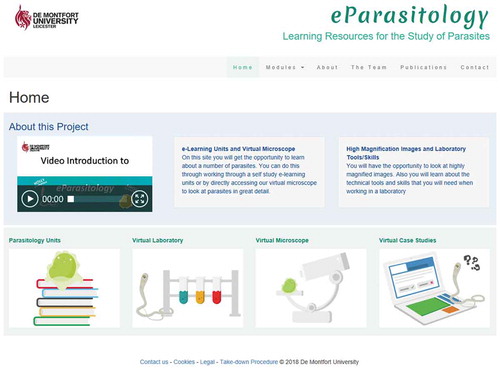
The four sections, which can be seen in , are briefly described below.
Figure 1. Overview of the main page of the DMU e-Parasitology package (Image courtesy of DMU, 2018). Available at: http://parasitology.dmu.ac.uk/index.htm
3.1. Theoretical module
The theoretical module, named as Parasitology Units, is composed of different units with up-to-date information to study major and rare (emerging and re-emerging) parasitic human diseases, focusing on diagnoses or identification, management, treatment, control and prevention of these serious and chronic disabling parasitic diseases. Units were closely developed by DMU web designers to enhance learning and create the most effective package possible, as staff with such expertise can creatively integrate content with the power and flexibility of the Web rather than copy lecture or previous materials on the webpage (Cook & Dupras, Citation2004).
To facilitate the study and identification of the different parasitic diseases, this module is equipped with different subsections that cover the three main classes of human parasites: protozoa, helminths and ectoparasites. For its part, the three common taxa considered for helminths, i.e. trematodes, cestodes and nematodes, have also been subsequently subdivided using subheadings. An additional main heading to cover the study of fungal parasites has been also made available. Under this heading of ‘fungi’, we have addressed the study of the emerging group of pathogens, microsporidia (Corsaro et al., Citation2016), as their study is gaining in relevance due to their impact on human morbidity and public health (Ramanan & Pritt, Citation2014), despite discussion about whether this group of parasites should be re-considered as protozoans (Kirk, Citation2018).
To develop a robust theoretical module, the first developed unit, which covers the study of Toxocara spp., was tested with volunteer 3rd year students enrolled in the bilingual (Spanish and English) Pharmacy and Biotechnology degree at USP-CEU in 2016/17. In the second term of this course and after studying a complete module regarding human parasitology, these students were provided with a link to the Toxocara unit and were asked to provide comprehensive feedback and suggestions to improve the unit. The Toxocara unit was used as a model unit to build this section after addressing the students’ feedback, as briefly described in Peña-Fernández et al. (Citation2017a).
Therefore, all the theoretical units follow the same scaffolding and the same headings, which are aligned with the strategy suggested to provide a solid foundation of parasitology relevant for future health professionals (Strube et al., Citation2018): a) infectious agent, morphology, life-cycle and transmission, b) clinical and pathologic features, c) risk factors and epidemiology, d) diagnosis, e) treatment and f) control and prevention. To facilitate navigation, these headings were converted in an index, presented in a panel/menu to the left hand side of the unit homepage (as can be seen in ). Units are also equipped with different buttons and hyperlinks to navigate across the unit or to jump to another section.
Figure 2. Overview of two formative assessments of the unit Toxocara in the DMU e-Parasitology (Image courtesy of DMU, 2018). Available at: http://parasitology.dmu.ac.uk/learn/modules/toxocara/story.html
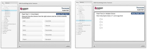
Finally, formative assessments in form of short quizzes, games or exercises are embedded in each unit at selected points to facilitate assimilation and engagement (), which can be attempted unlimited times and provide instant feedback in conjunction with encouraging messages to enhance students’ effort and engagement. Self-assessments present different degrees of difficulty, so the user will face gradually more challenging questions throughout the unit requiring him/her to reflect on the information provided and to put the material into a less familiar context, rather than using simple knowledge recall, a strategy that we are following to enhance critical thinking, clinical reasoning and reflection, important skills for future health-care professionals as they are required to gain new knowledge throughout their careers. These skills are also needed for diagnoses and management of patients. Completion of the different self-assessment tasks available in each unit is voluntarily, so the user can decide whether to attempt or not as progression through the unit is not limited by their completion. Self-assessments are necessary to build robust e-Learning packages, as they encourage active learning (self-assessment, reflection and feedback), which will allow the user to evaluate their acquisition of knowledge and skills as well as enhance their engagement through the unit (Cook & Dupras, Citation2004).
3.2. Virtual laboratory module
This section will be equipped with a range of different units related to traditional and novel biomedical laboratory techniques and equipment for detecting, identifying and investigating any human pathogens such as bacteria and viruses, but with a particular focus on parasites.
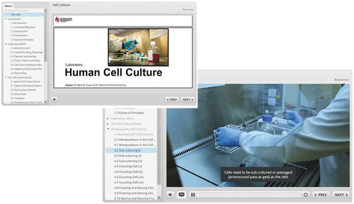
We are following a similar methodology to that used with the theoretical module but using other technological resources including videos to enhance understanding and engagement. The virtual laboratory module is also divided into the following subsections that present a myriad of units distributed under different headings, as follows: microscopes (e.g. electron microscope); molecular biology (e.g. polymerase chain reaction and gel electrophoresis); biological safety cabinets and cell/parasite culture (which includes comprehensive information about how to work with a biological safety cabinet in a biosafety level II and III laboratories); biochemical and immunological techniques (e.g. magnetic immunoseparation); histology (e.g. microtome and histological staining techniques such as haematoxylin and eosin); parasitological staining techniques, such as Kinyoun stain, and immunological techniques, such as IFAT (Indirect Fluorescent Antibody Technique). Finally, a series of units about sampling preparation for parasitological analysis will be also provided, which will include coprological analysis. The virtual laboratory units are highly interactive and are being built using the same scaffolding as for the theoretical ones. In contrast, they have will not have a fixed index as the theoretical modules have, as they will depend on the content to be addressed; however, all the virtual laboratory units will be provided with an introduction, a description of the equipment/technique in conjunction with explanatory videos and conclusions, and equipped with self-assessments. Thus, a characteristic of this section is that units provide explanatory or short videos of academics and/or technicians working hands-on in real conditions with the laboratory equipment and/or performing a specific technique, so the user will receive a complete and ‘real’ experience mimicking working in a laboratory. The videos are very short (no longer than 3 min to avoid disengagement) and can be paused or moved forward or backward (); moreover, they are strategically embedded throughout each unit and supported by a text and photographs/images to reinforce the information shown in the videos. The videos come with audio and subtitles in English to make DMU Parasitology® more inclusive to a variety of users (). A short description of the first virtual laboratory unit developed for cell culture has been discussed in Peña-Fernández, Hurtado, Del Águila, and Evans (Citation2017b); culture of cells is relevant for future parasitologists as many of the practices employed for the culture of human and mammalian cells are applicable to the culture of parasites. Moreover, the culture of mammalian cells would be required for the culture of obligate intracellular parasites such as Plasmodium spp. (Ahmed, Citation2014).
Figure 3. Overview of the unit Human Cell Culture in the virtual laboratory in the DMU e-Parasitology (Images courtesy of DMU; 2018). Available at: http://parasitology.dmu.ac.uk/learn/laboratory.htm
The virtual laboratory environment could help technicians and students across the world to learn how to work in a biomedical laboratory as well as to perform techniques to identify and diagnose human parasites. This resource is supported by two relevant DMU Parasitology® tools: the virtual microscope and virtual library, which are explained below. Moreover, the virtual laboratory module can be used to enhance the practical learning of undergraduate students before attending the laboratory sessions as well as to academics and health-care professionals to develop CPD training. Finally, this module may impact on the teaching of laboratory techniques and skills in developing countries due to their limited resources.
3.3. Virtual microscope, which includes the virtual library
Despite recent advances in molecular biology and new laboratory techniques/equipment, microscopy identification of parasites and/or their infective forms remains the cornerstone of parasitic disease diagnosis (Momčilović et al., Citation2019), especially in developing countries. Thus, knowing the most significant morphological characteristics of eggs (for helminths) and cysts/trophozoites (for protozoans), and how to examine their sizes, shapes and numbers in different clinical samples to establish the parasite(s) involved, the degree of infection (or parasitaemia) and select the most appropriate treatment, remains as a key skill for future parasitologists and/or health-care professionals, particularly for major human diseases such as malaria (Bailey et al., Citation2013).
However, microscopic identification of parasites to species level requires comprehensive training, skills, knowledge and experience (Ahmed et al., Citation2016). As a result, and to facilitate future training of diagnosis, parasitologists are facing different challenges including shortage of skilled personnel to train others, lack of appropriate specimens to provide training and high expectations in the identification of parasites that health authorities demand of diagnostic parasitology (Yang et al., Citation2001), we are building a module with a real slide collection of parasites and their specimens in a range of different clinical samples and cultures. To build this section, real clinical slides are being appropriately scanned, photographed (at x40 magnification objective) and digitised to produce high magnification digital images of the entire glass slide. By using the gadget Zoomofy® (Zoomify, Inc.), each digital slide is equipped with the functionality of a microscope to provide a similar experience to working with a light, fluorescence or inverted microscope. Students can zoom in and out and move around throughout the complete digital slide to facilitate identification and learning of the different morphological characteristics and parasite features for diagnostic purposes (Peña-Fernández et al., Citation2018b).
Ordinary micrographs or photographs are limited to a preselected area and magnification, but the virtual slides created will enable students to study any part of the digitised slide at different magnifications; this strategy will facilitate acquisition of knowledge of the morphological characteristics of the parasites for diagnosis as well as learning how to use a microscope. Thus, different studies have pointed out the potential benefits of using digital slides and virtual microscopes versus the normal light microscope, which include remote access, development of interactive activities in the classroom with web-enabled devices, economic advantages in a world with larger numbers of students and lower number of resources and teaching staff, etc. (Saco et al., Citation2016). Moreover, Ahmed et al. (Citation2016) have reported similar sensitivity and specificity rates for identification of parasites for both digitised slides and real slide preparations, which will support the use of web microscopes for teaching and learning parasitology.
In a similar way as with the other modules, the virtual microscope is also subdivided in different sections and subsections to cover the three major groups of human-pathogenic parasites, i.e. protozoa, helminths and arthropods; as well as other parasites closely related to fungi and common artefacts (non-parasite material) which are commonly found in real conditions, so the user of this package will be able to gain relevant skills to avoid potential false-positives in future situations in real work. We consider that the DMU e-Parasitology’s® virtual library could be used as a high quality and reliable image bank of specimens digitised slides, which could be specifically useful for parasitology diagnostic technicians in developing countries and endemic areas (Linder et al., Citation2008), as well as for postgraduate students that will undertake parasitology research.
3.4. Virtual clinical case studies module
The fourth module is equipped with a series of highly engaging and interactive virtual case studies in medical and clinical parasitology with different levels of difficulty (easy, medium and difficult) for students and users with different backgrounds in parasitology and for promoting active learning and increasing engagement. The virtual case studies are being created following a previously successful novel teaching strategy implemented by our team to facilitate reflection and critical thinking in which we introduced mini-case studies within lectures delivered to final year DMU BSc. Biomedical Science students in 2016/17 (Peña-Fernández, Peña, Potiwat, Coope, & Magnet, Citation2018b). Those mini-case studies were inspired on those designed by the Laboratory Identification of Parasitic Diseases (DPDx) of the Centers for Disease Control and Prevention (CDC, USA, CDC (Centres for Disease Control and Prevention), Citation2018)
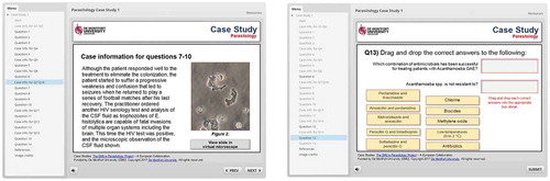
Briefly, the virtual case studies provide the user with a medical history of a patient and different clinical slides to diagnose the parasite(s) involved by observing their morphological characteristics and identifiable structures using the virtual microscope, as it is shown in . Then, the user will navigate through different interactive questions and mini quizzes to reach diagnoses, management, treatment and tailor prevention techniques for the clinical case study proposed. Questions and mini quizzes are formulated in different styles throughout the case study to promote application of knowledge, development of judgement and clinical thinking and reflection (Cook & Dupras, Citation2004); and present different degrees of difficulty so the user is challenged and their interest is maintained. Some questions require the user to make decisions, such as requesting more information, order a different test, tailor interventions to protect human health, etc. The virtual clinical case units could also facilitate the acquisition of problem-solving skills, which are required for clinical reasoning. A brief description of the first virtual case study unit has been provided in Peña-Fernández et al. (Citation2018b).
Figure 4. Overview of the first virtual case study created in the DMU e-Parasitology (Image courtesy of DMU, 2018). Available at: http://parasitology.dmu.ac.uk/learn/case_studies/cs1/story_html5.html
3.5. Peer review and validation
Technology-enhanced learning has a myriad of applications as highlighted above that can be used to tackle most of the challenges that HEIs are currently facing including large numbers of students enrolled in human health programmes and shortages of resources, staff and specimens. However, the international academic community have some concerns with the quality of the teaching and learning materials provided in on-line resources and whether they have been peer-reviewed and appropriately developed from professionals in the area (Fox et al., Citation2018). To develop a scientifically rigorous DMU Parasitology®, a group of international parasitologists has been recently recruited to peer review the package with a focus on the theoretical units developed. Moreover, this group of professionals will also provide feedback to improve the package and units where appropriate, once we have addressed the students’ feedback from the different participating universities, which is being currently collected, as part of the validation process. This paper provides, discusses and analyses the comprehensive feedback collected in one of the participating universities, which is provided below.
3.6. Methods
The DMU Parasitology® package has been tested with a focus group of students appropriately selected from one of the participating universities with a knowledge in parasitology to validate the package and meet the objectives described. The focus group was formed by 109 fourth year Pharmacy students enrolled in the Clinical Analysis & Diagnostics II module [6 ECTS; 4th year module (UMH, Citation2018)] in 2017/18 at UMH. These students previously completed an entire module on medical parasitology, specifically during their second year of studies.
Briefly, we have created and delivered a highly specific and interactive workshop following previous successful experience in the creation of novel training (Peña-Fernández, González-Muñoz, & Peña, Citation2015), in which students were asked to complete one of the virtual case studies and needed to navigate throughout the entire package and use all the tools/sections (specifically the virtual microscope, library and the theoretical section) to resolve the clinical case scenario proposed. The workshop was timetabled in small groups in a computer room and students worked in pairs to encourage team work. Results were discussed with the students even though they were prompted with instant feedback when completing the virtual case study, in order to clarify erroneous knowledge or misinformation and identifying potential issues/challenges found by students during their completion to enhance the case study. Additionally, DMU Parasitology® was used to enhance the lectures and teaching delivered in this module so students received a holistic experience of the novel website before testing the package in this session.
At the end of this session, a validated feedback-questionnaire was distributed to collect students’ feedback and opinion on DMU Parasitology®. The questionnaire presented three sets of questions, to facilitate validation of the product: a) assessment of the structure and format; b) assessment of the use; c) comments and suggestions. A Likert scale was provided for each specific question except for the last set that contained open-questions (free-response), so participants were able to comment on their overall experience and suggest future improvements. Students were informed that the anonymous data provided could be used in a study, so written approval from participants was obtained. Feedback-questionnaires have been described as an appropriate tool to measure the degree of satisfaction of the teaching and learning processes (Peña-Fernández et al., Citation2015). Ethical approval was provided by the Research Ethics Committee at DMU (Ref. 1851; 8 December 2016).
4. Results and discussion
A total of 95 students appropriately completed the questionnaire provided – questionnaires that were only partially completed were discarded. The results for the first two sections are depicted in and .
Table 1. Responses (%) to the feedback-questionnaire used to assess the structure and format of the DMU e-Parasitology package as a learning tool
Table 2. Responses (%) to the feedback-questionnaire used to assess the use of the DMU e-Parasitology package as a learning tool
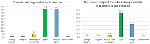
The vast majority of fourth year Pharmacy students at UMH in 2017/18 have indicated that the overall design of DMU Parasitology® is appropriate and engaging [95.8% (these percentages are provided as the sum of agreed and strongly agreed recorded responses); and ], and the website is interactive (91.6%; and ). In a similar manner, students reported that each module individually has been appropriately built to enhance learning and understanding, as follows (): 94.8% of agreement was recorded for the theoretical module, 90.5% for the virtual laboratory and 82.1% for the virtual microscope/library. A more detailed analysis of these responses showed that a very small percentage of students indicated that the scaffolding of the theoretical module was not appropriate (2.1%; ), which could be attributed to the amount of information that these students needed to process in a very short time (as reflected in some of the comments provided in the open-answer section of the questionnaire). Additionally, a high percentage of responses were recorded for the option ‘neither agree/nor disagree’ with respect to the appropriateness of the structure of the virtual microscope (more than 15%; ), which could be attributed to the fact that only a few virtual slides were available at the time that these students used the package (first term, 2017/18 academic course).
Figure 5. Students’ opinion on the structure of DMU e-Parasitology (%) and whether the resource was interactive and engaging
Careful analysis of the feedback provided has highlighted the effectiveness of this novel web-based resource in promoting and enhancing student learning and overall experience on medical parasitology. Thus, UMH students reported high levels of enjoyment (88.4%; ) and satisfaction (90.5%; ) with all aspects of DMU Parasitology®; and a high proportion of students (88.4%; ) have highlighted that the package helped with their studies and would recommend it to peers and for the study of modules/subjects not related to parasitology, because of its broad relevance for medical sciences (89.5%; ). Overall satisfaction would be supported by the fact that most students suggested the creation of similar resources for other disciplines (89.4%; ), a statement for which no student disagreed.
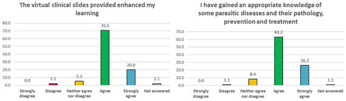
In relation to learning (), 88.4% indicated that the formative assessments and quizzes presented in the units enhanced their learning in conjunction with the virtual clinical slides of specimens (). 89.5% participants highlighted they gained appropriate knowledge of the pathology, prevention and treatment of some parasitic diseases (); and 82.1% indicated that they learnt basic skills to investigate parasitic disease. The percentage of responses indicating disagreement with these statements was minimal, as indicated in .
Figure 6. Students’ opinion (%) on the acquisition of knowledge after using the DMU e-Parasitology package
Regarding the information collected in the open-answer questions, UMH students indicated that the package was interactive and useful for the study of parasitology and related subjects in their programmes. They particularly enjoyed the clinical specialism of the package as it was suitable for the acquisition of basic diagnostic skills through learning the key morphological characteristics of the parasites. They pointed out very minor suggestions for improvement, mostly related to enhance the navigation, reduce the content of some slides, provide more virtual slides of specimens, increase the number of virtual case studies and facilitate the content in another language including Spanish.
The overall analysis of the results collected so far indicates that DMU Parasitology® successfully aided students from an overseas non-UK institution to acquire a certain degree of comprehension of parasitology and some clinical diagnostic skills, which are essential to respond to outbreaks of infection and protect the public as well as face future challenges due to these pathogens. The package seemed successful in facilitating students to acquire essential basic work skills for identification and diagnoses, management, treatment and control of the parasites studied during the specific workshop (Entamoeba histolytica and free-living amoebas). Regarding other parasites that were briefly discussed in the introductory lectures to show the DMU Parasitology® package to these students, which included the study of Toxocara spp. and Plasmodium spp., participants learnt to identify and study these pathogens using a microscope and the ability to suggesting different techniques for identification to species level in order to provide the most appropriate treatment and management to the patients simulated in the case study. Moreover, students were able to acquire different transversal competences including critical thinking and reflection, research skills, communication and team work, problem-solving skills, etc. during the completion of the specific workshop. Therefore, although these results should be considered as preliminary as to date the package has been only tested with a group of undergraduate students from one of the participating universities, it could be pointed out that DMU Parasitology® was successful in promoting active learning and for the acquisition of clinical reasoning, diagnostic skills and medical parasitology learning, to create attentive future health professionals with a comprehensive knowledge of parasitology to respond to considerable future challenges and life-long demands. Finally, the package increased engagement in the study of this subject.
On the other hand, more than 18 academics and professionals from the participating universities have benefited from DMU Parasitology® so far to teach relevant aspects of this discipline that otherwise would be difficult to provide due to time constraints and the number of students. We are also planning to collect feedback from academics that have used this package to aid their work but have not been involved in its development; and we will collect data about external use of DMU Parasitology®. The next step of the project will help us in identifying sections or tools that academics find more useful, to expand on them and collect suggestions for improvement. Of particular interest for us will be to identify institutions that have used the package for enhancing their parasitology teaching and/or created parasitology training through using our package. Please contact the team if you would like to collaborate in this project.
5. Conclusions
DMU Parasitology® could aid human health students gain significant knowledge of parasitology by promoting self-learning and overcoming barriers of time, space, equipment and resources. The scaffolding and structure of the package have been described as appropriate and relevant to study parasitology, and it has shown to be effective in promoting self-learning, increasing students’ engagement and facilitating the acquisition of essential work skills and clinical reasoning to future health professionals in the area of parasitology. The final user will be provided with a holistic experience to study major and rare (emerging and re-emerging) human parasites.
Therefore, this package can assist academics by teaching relevant aspects of medical parasitology which otherwise will be difficult to provide due to time constraints, the number of students, limitation of resources and/or increasing anti-parasitic drug resistances that requires different strategies to be tackled. Finally, DMU Parasitology® could be useful to enhance the current status of teaching parasitology in Europe and other developed countries as well as to ensure high-quality education of future health professionals, as the status of teaching this subject is being eroded despite the increasing number of outbreaks due to human-related parasites.
Disclosure statement
No potential conflict of interest was reported by the authors.
Additional information
Funding
Related Research Data
References
- Acholonu, A.D. (2003). Trends in teaching parasitology: The American situation. Trends Parasitol, 19, 6–9. doi:10.1016/S1471-4922(02)00002-8
- Ahmed, L., Seal, L.H., Ainley, C., De la Salle, B., Brereton, M., Hyde, K., … Gilmore, W.S. (2016). Web-based virtual microscopy of digitized blood slides for malaria diagnosis: An effective tool for skills assessment in different countries and environments. J Med Internet Res, 18(8), e213. doi:10.2196/jmir.6027
- Ahmed, N.H. (2014). Cultivation of parasites. Trop Parasitol, 4(2), 80–89. doi:10.4103/2229-5070.138534
- Al-Shorbaji, N., Atun, R., Car, J., Majeed, A., & Wheeler, E. (2015). eLearning for undergraduate health professional education: A systematic review informing a radical transformation of health workforce development: World Health Organization (WHO). Retrieved from https://whoeducationguidelines.org/content/elearning-report
- Bailey, J.W., Williams, J., Bain, B.J., Parker-Williams, J., & Chiodini, P.L. (2013). General haematology task force of the british committee for standards in haematology. guideline: The laboratory diagnosis of malaria. General haematology task force of the british committee for standards in haematology. Br J Haematol, 163(5), 573–580. doi:10.1111/bjh.2013.163.issue-5
- Boissier, J., Grech-Angelini, S., Webster, B.L., Allienne, J.F., Huyse, T., Mas-Coma, S., … Mitta, G. (2016). Outbreak of urogenital schistosomiasis in Corsica (France): An epidemiological case study. Lancet Infect Dis, 16(8), 971–979. doi:10.1016/S1473-3099(16)00175-4
- Bruschi, F. (2009). How parasitology is taught in medical faculties in Europe? parasitology, lost? Parasitol Res, 105(6), 1759–1762. doi:10.1007/s00436-009-1594-7
- CDC (Centres for Disease Control and Prevention). (2018). Laboratory identification of Parasitic Diseases of Public Health Concern (DPDx). Monthly Case Studies. Retrieved from https://www.cdc.gov/dpdx/monthlyCaseStudies/2017/index.html
- Cook, D.A., & Dupras, D.M. (2004). A practical guide to developing effective web-based learning. J Gen Intern Med, 19(6), 698–707. doi:10.1111/j.1525-1497.2004.30029.x
- Corsaro, D., Michel, R., Walochnik, J., Venditti, D., Müller, K.D., Hauröder, B., & Wylezich, C. (2016). Molecular identification of Nucleophaga terricolae sp. nov. (Rozellomycota), and new insights on the origin of the microsporidia. Parasitol Res, 115(8), 3003–3011. doi:10.1007/s00436-016-5055-9
- Day, M. (2017). Autopsy results confirm 4 year old Italian girl died from malaria. BMJ, 358, j4235. doi:10.1136/bmj.j4235
- Escolà-Vergé, L., Arando, M., Vall, M., Rovira, R., Espasa, M., Sulleiro, E., … Barberá, M.J. (2017). Outbreak of intestinal amoebiasis among men who have sex with men, Barcelona (Spain), October 2016 and January 2017. Euro Surveill, 22(30). doi:10.2807/1560-7917.ES.2017.22.30.30581
- Fox, M., Blake, D., & Jacobs, D. (2018). Veterinary parasitology teaching at London - meeting the ‘Day-One Competency’ needs of new veterinarians. Vet Parasitol, 254, 131–134. doi:10.1016/j.vetpar.2018.01.029
- Joachim, A., Hinney, B., Duscher, G., Preusche, I., & Winter, P. (2018). Teaching parasitology in a modular veterinary curriculum - The Vienna experience. Vet Parasitol, 252, 101–106. doi:10.1016/j.vetpar.2018.02.002
- Kirk, P.M. (2018). Microsporidia: Unicellular spore-forming protozoan parasites (version Nov 2015). In Y. Roskov, L. Abucay, T. Orrell, D. Nicolson, N. Bailly, P.M. Kirk, … L. Penev (Eds.), Species 2000 & ITIS catalogue of life, 20th december 2017 Digital resource at www.catalogueoflife.org/col. Species 2000 Naturalis, Leiden: the Netherlands. ISSN (p. 2405–8858).
- Kofer, J., Hofer, H., & Hartmann, S. (2017). Next-generation parasitologists: Structured training programs meet educational challenges. Trends Parasitol, 33(6), 423–425. doi:10.1016/j.pt.2017.03.008
- Law, G.C., Apfelbacher, C., Posadzki, P.P., Kemp, S., & Tudor Car, L. (2018). Choice of outcomes and measurement instruments in randomised trials on eLearning in medical education: A systematic mapping review protocol. Syst Rev, 7(1), 75. doi:10.1186/s13643-018-0739-0
- Linder, E., Lundin, M., Thors, C., Lebbad, M., Winiecka-Krusnell, J., Helin, H., … Lundin, J. (2008). Web-based virtual microscopy for parasitology: A novel tool for education and quality assurance. PLoS Negl Trop Dis, 2(10), e315. doi:10.1371/journal.pntd.0000315
- Liu, G.H., Zhang, L.X., Zou, F.C., Yuan, Z.G., Zhao, G.H., Hu, M., … Zhu, X.Q. (2018). Veterinary parasitology teaching in China in the 21st century - Challenges, opportunities and perspectives. Vet Parasitol, 252, 70–73. doi:10.1016/j.vetpar.2018.01.037
- Liu, Q., Peng, W., Zhang, F., Hu, R., Li, Y., & Yan, W. (2016). The effectiveness of blended learning in health professions: Systematic review and meta-analysis. J Med Internet Res, 18(1), e2. doi:10.2196/jmir.4807
- Madeira de Carvalho, L.M., & Alho, A.M. (2018). Teaching veterinary parasitology in Portugal in the 21st century - Changes, challenges and opportunities after the Bologna process. Vet Parasitol, 253, 98–101. doi:10.1016/j.vetpar.2018.01.030
- Meléndez, R.D. (2003). Trends in teaching parasitology: Where to complain? Trends in Parasitology, 19, 9. doi:10.1016/S1471-4922(03)00169-7
- Momčilović, S., Cantacessi, C., Arsić-Arsenijević, V., Otranto, D., & Tasić-Otašević, S. (2019). Rapid diagnosis of parasitic diseases: Current scenario and future needs. Clin Microbiol Infect, 25(3), 290–309. doi:10.1016/j.cmi.2018.04.028
- Nichols, G.L., Freedman, J., Pollock, K.G., Rumble, C., Chalmers, R.M., Chiodini, P., & Hawker, J.I. (2015). Cyclospora infection linked to travel to Mexico, June to September 2015. Euro Surveill, 20(43). doi:10.2807/1560-7917.ES.2015.20.43.30048
- Peña-Fernández, A., Fenoy, S., Halliwell, R., Izquierdo, F., Magnet, A., Hurtado, C., … Del Águila, C. (2018b). Development of a virtual library of clinical samples for medical parasitology diagnosis. INTED Proceedings (pp. 7599–7604). Valencia, Spain. ISBN 978-84-697-9480-7. Retrieved from https://library.iated.org/view/PENAFERNANDEZ2018DEV
- Peña-Fernández, A., González-Muñoz, M.J., & Peña, M.A. (2015). Designing training for teaching environmental toxicology to specialized pharmacists. Currents Pharm Teach Learn, 7, 864–868. doi:10.1016/j.cptl.2015.08.015
- Peña-Fernández, A., Hurtado, C., Del Águila, C., & Evans, M. (2017b). Developing resources for teaching and learning cell and parasite culture within the DMU e-Parasitology package. ICERI Proceedings (pp. 7117–7122). Seville, Spain. ISBN: 978-84-697-6957-7.
- Peña-Fernández, A., Ollero, M.D., Fenoy, S., Magnet, A., Izquierdo, F., Peña, M.Á., … Del Águila, C. (2017a). Creating a model module for the novel resource DMU e-Parasitology. ICERI Proceedings (pp. 1599–1604). Seville, Spain. ISBN: 978-84-697-6957-7.
- Peña-Fernández, A., Peña, M.A., Potiwat, N., Coope, J., & Magnet, A. (2018b). Virtual case studies in the novel resource DMU e-Parasitology. INTED Proceedings (pp. 6201–6206). Valencia, Spain. ISBN 978-84-697-9480-7.
- Peng, H.J., Zhang, C., Wang, C.M., & Chen, X.G. (2012). Current status and challenge of human parasitology teaching in China. Pathog Glob Health, 106(7), 386–390. doi:10.1179/2047773212Y.0000000040
- Ramanan, P., & Pritt, B.S. (2014). Extraintestinal microsporidiosis. J Clin Microbiol, 52(11), 3839–3844. doi:10.1128/JCM.00971-14
- Reid, H.J., Thomson, C., & McGlade, K.J. (2016). Content and discontent: A qualitative exploration of obstacles to elearning engagement in medical students. BMC Med Educ, 16, 188. doi:10.1186/s12909-016-0710-5
- Saco, A., Bombi, J.A., Garcia, A., Ramírez, J., & Ordi, J. (2016). Current status of whole-slide imaging in education. Pathobiology, 83(2–3), 79–88. doi:10.1159/000442391
- Sisson, S.D., Hill-Briggs, F., & Levine, D. (2010). How to improve medical education website design. BMC Med Educ, 10, 30. doi:10.1186/1472-6920-10-30
- Strube, C., Raue, K., & Janecek, E. (2018). Simple, but not easy - Opportunities and challenges from teachers’ and students’ perspectives in the 21st century of veterinary parasitology teaching. Vet Parasitol, 252, 74–79. doi:10.1016/j.vetpar.2018.01.034
- UMH. 2018. Syllabus of clinical analysis & diagnostics II module. UMH website. Retrieved from https://umh.es/contenido/Estudios/:asi_g_1705_P1/datos_en.html
- van Doorn, D.C.K., Nijsse, E.R., & Ploeger, H.W. (2018). Pitfalls and opportunities of teaching veterinary parasitology within an integrated curriculum. Vet Parasitol, 252, 85–88. doi:10.1016/j.vetpar.2018.01.036
- Yang, Y.S., Park, D.K., Kim, H.C., Choi, M.H., & Chai, J.Y. (2001). Automatic identification of human helminth eggs on microscopic fecal specimens using digital image processing and an artificial neural network. IEEE Trans Biomed Eng, 48(6), 718–730. doi:10.1109/10.923789
- Yavner, S.D., Pusic, M.V., Kalet, A.L., Song, H.S., Hopkins, M.A., Nick, M.W., & Ellaway, R.H. (2015). Twelve tips for improving the effectiveness of web-based multimedia instruction for clinical learners. Med Teach, 37(3), 239–244. doi:10.3109/0142159X.2014.933202