
 ?Mathematical formulae have been encoded as MathML and are displayed in this HTML version using MathJax in order to improve their display. Uncheck the box to turn MathJax off. This feature requires Javascript. Click on a formula to zoom.
Abstract
Background
Oxidative stress (OS) and its biomarkers are the biochemical end point of the imbalance between reactive oxygen species (ROS) production and the ability of the antioxidant (AOX) biological systems to fight against oxidative injury.
Objective
We reviewed the role of OS and its downstream signaling in aging eyes.
Methods
A search of the literature and current knowledge on the physiological and pathological mechanisms of OS were revisited in relation to the eyes and the aging process. Most prevalent ocular diseases have been analyzed herein in relation to OS and nutraceutic supplements, such as dry-eye disorders, glaucoma, age-related macular degeneration, and diabetic retinopathy.
Results
Clinical, biochemical, and molecular data from anterior and posterior eye segment diseases point to OS as the common pathogenic mechanism in the majority of these ocular disorders, many of which are pathologies causing visual impairment, blindness, and subsequent loss of life quality. Studies with nutraceutic supplements in aging eye-related pathologies have also been reviewed.
Conclusion
OS, nutritional status, and nutraceutic supplements have to be considered within the standards of care of older ophthalmologic patients. OS biomarkers and surrogate end points may help in managing the aging population with ocular diseases.
Introduction
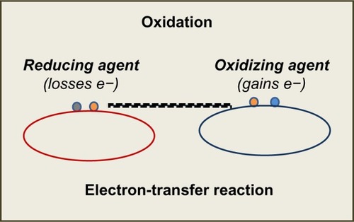
Oxygen (O2) is essential for life, as it plays a major role in the energy cycle of living organisms and is fundamental for the aerobic breathing of cells and tissues. From a chemical point of view, any reaction ending with a gain of electrons results in a reduction process, and in contrast, when concluding with losing electrons results in an oxidation reaction.Citation1
These concepts of reduction–oxidation (redox) are based on electron-transfer events. Compounds capable of accepting electrons are oxidizing agents, and those able to assign electrons are reducing agents (). Under normal conditions (during aerobic metabolism), the O2 undergoes tetravalent reduction to water (H2O). However, in situations that induce an incomplete reduction, very unstable and reactive species are formed, the so-called reactive oxygen species (ROS). In general, ROS are classified into two groups: radical species and nonradical species.Citation2
Figure 1 Redox reactions and Electron-transfer. Reducing and oxidizing agents.
A free radical (FR) is an atom or molecule that contains an unpaired electron in its outer orbital. This situation confers them instability and the need to lose/gain an electron in order to get stable. Among the nonradical oxygen species are hydrogen peroxide (H2O2), ozone (O3), and nitric oxide (NO). There is a typical sequential order in ROS generation, characterized by the production in the first place of the superoxide anion (O2•), followed by H2O2 and the hydroxyl radical (OH•).Citation3–Citation6
O2• is formed in multiple processes, and is the most abundant among the ROS. It possesses a half-life of milliseconds. Although O2• is not reactive enough to attack macromolecules directly, it can start an ROS chain formation. H2O2, a non-radical oxygen species, has the ability to generate other ROS. It is a small molecule lacking electrochemical charge, diffuses easily, and can pass through cell membranes, propagating its effects farther than other species. OH• is the most powerful and harmful of all oxygen species. However, its short half-life (10 seconds) is a limiting condition to the diffusion capacity. It is formed from O2 and H2O2 in the presence of transition metals (Fe/iron) during the sequential reactions of Haber–Weiss and Fenton:
Oxidative stress
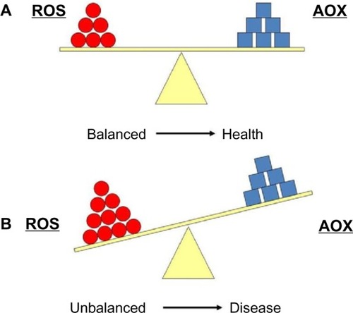
All biomolecules can be attacked by ROS: lipids, proteins, and nucleic acids. Among them, lipids are probably the most susceptible to undergo oxidation.Citation7 All cell membranes are rich in polyunsaturated fatty acids (PUFAs), which are easily injured by oxidizing agents. There are endogenous and exogenous defense mechanisms against oxidative attack. Oxidative stress (OS) and its biomarkers are the biochemical end point of the imbalance between ROS production and the ability of antioxidant (AOX) biological systems to counteract the effects of ROS metabolites.Citation6,Citation7
Cells maintain a reducing environment by the corresponding enzymes and the constant supply of metabolic energy. The OS effects depend on the intensity of such abnormalities and the cellular response. If the cell is unable to overcome and to recover its function, the exogenous and endogenous AOX defenses cannot counter it, and the cell can become extinct (). However, ROS have also been shown to function as signaling molecules.Citation8 This underlies relevant mechanisms that are essential for the integrity of living organisms and their aging process. Knowledge of the pathways that regulate ROS homeostasis is pivotal for relieving the deleterious effects of ROS in cells and tissues. Further research in this topic can provide strong evidence about specificity in ROS signaling in health and disease.Citation8
Figure 2 (A) Equilibrium between reactive oxygen species (ROS) production and antioxidant defense (AOX). (B) Imbalanced situation between ROS and AOX, which is associated with many pathologies.
Among the best known AOXs are the enzymes superoxide dismutase (SOD), catalase (CAT), and glutathione peroxidase (GSH-Px).Citation9 Since the discovery of SOD in 1969,Citation10 it has been assumed that ROS participate in the pathogenic mechanisms of various diseases and abnormal conditions. In 1956, Harman proposed the theory of FRs and aging,Citation11 which was supported by the basic theory of the relationship between the duration of life, metabolic rate, and O2 consumption by organisms. Gerschman and FennCitation12 and Gerschman et alCitation13,Citation14 demonstrated that FRs are toxic to our body.
OS has been associated with such pathologies as atherosclerosis, Parkinson’s and Alzheimer’s diseases, aging processes, dermatitis, cancer, asthma, neurodegeneration, infertility, atherosclerosis, hypertension, diabetes, and rheumatoid arthritis, among others.Citation7,Citation9,Citation15–Citation20 OS has also been implicated in ophthalmic processes, including ocular surface disorders, cataracts, glaucoma, diabetic retinopathy (DR), retinitis pigmentosa, toxic neuropathies, uveitis, and age-related macular degeneration (AMD).Citation13,Citation21–Citation28 However, the exact signaling pathways regulated by ROS, especially in age-related eye diseases, need further investigation. Delineating the specific signals modulated by ROS is of great diagnostic and therapeutic interest.
Biomarkers and surrogate end points of OS and its downstream effectors have been extensively studied in relation to ocular diseases. Ocular oxidative damage can be determined by direct/indirect methods in biological samples, including such techniques as enzymatic colorimetric assay or high-performance liquid chromatography. With these assays, oxidative activity can be measured: 1) malondialdehyde (MDA), 4-hydroxynonenal, oxidation of nucleic acids 8-hydroxy-2′-deoxyguanosine or 8-oxo-7,8-dihydro-2′-deoxyguanosine; 2) the isoprostanes and the prostaglandin-like compounds that are produced in vivo independently of cyclooxygenase enzymes; and 3) AOX capacity (total AOX capacity [TAC]) can also be determined, as well as enzymatic activities (SOD, CAT, GSH-Px, vitamins) and FR scavengers (GSH). All these oxidative and AOX activities can be assayed on various ophthalmological samples, including aqueous humor, vitreous body, and tear biopsies, and in the blood, urine, or saliva.Citation23–Citation26 Comparing the results of determinations of oxidative and AOX status may reflect the balance between them and the degree of OS by laboratory tests performed in samples from our ophthalmic patients.
Antioxidant mechanisms
The human body has some natural AOX defense systems to diminish the harmful action of continuous ROS production.Citation7,Citation9,Citation10,Citation21 These defenses are located in the cytoplasm, cellular membrane, and extracellular space, and can be classified as follows.
Enzymatic defense systems
These consist of intracellular mechanisms in which SOD, CAT, and GSH-Px eliminate ROS once formed. SODs are metalloproteins that accelerate the dismutation of superoxide to hydrogen peroxide. Protective action is associated with CATs and peroxidases in order to avoid the accumulation of hydrogen peroxide that would otherwise be converted into OH•. There are two main molecular types of SOD in humans: the cytosolic one (CuZnSOD), which contains copper and zinc, and the mitochondrial one (MnSOD), which contains manganese. CAT is located in the peroxisome and mitochondria, and its function consist of catalyzing the dismutation of hydrogen peroxide to water and molecular oxygen. It has been demonstrated that high concentrations of hydrogen peroxide stimulate CAT activity, while low concentrations are preferentially catalyzed by peroxidases. GSH-Px is located in cytosol and mitochondria. It is known that this works by eliminating hydroperoxides, transforming them into water. This reaction is associated with the transformation of reduced glutathion (GSH) into oxidized GSH (GSSG).Citation29,Citation30
Free radical scavengers
FR scavengers slow down the reactions of oxidation or “catch” FRs, transforming them into less aggressive compounds. They can be hydrosoluble and cytosolic (GSH and C vitamin) or liposoluble, located in the membrane (vitamin E and carotenoids). Vitamin C is the main hydrosoluble AOX, and may also regenerate tocopherol (vitamin E), which is one of the main liposoluble AOXs in cellular membranes, together with carotenoids (β-carotene, lutein, zeaxanthin, and lycopenes).Citation31 GSH is considered the most abundant AOX in cytosol, nucleus, and mitochondria, making the ratio of GSH:GSSG a good way to measure OS.
Chelating agents of transition metals
These are molecules that bind iron and copper, like the flavonoids. In this way, they avoid these metals to act in Fenton and Haber–Weiss reactions.Citation3,Citation4 Some authors prefer another classification according to the AOX source. In this manner, AOXs can be endogenous (synthesized by the own body), such as GSH, SOD, and CAT. But they also can be exogenous (only obtained from food), such as vitamins C and E, carotenoids, flavonoids and oligoelements. Besides these AOX defense systems, there are also repairing and exchanging mechanisms of damaged biomolecules.Citation9,Citation29,Citation31
Aging eyes as targets of oxidative stress
The predicted gain in life span in most countries is expected to lead progressively to an increase in the number of older people suffering age-related chronic diseases in the next decade. Moreover, within the cycle of life, aging can be defined as a progressive loss of the efficacy of physiological and biochemical processes occurring until death. Several theories have postulated the link between OS and aging, of which the most famous is the FR theory of aging,Citation11 which maintains that production of FRs and the subsequent FR damage increases with age.
The eye is an organ that is continuously exposed to ionizing radiation, pollutants, industrial smoke, and driving fumes, which makes the eyeballs extremely susceptible to oxidative attack. The retina is a photosensitive tissue that is affected by light rays and undergoes high metabolic activity. Furthermore, the retina contains a higher concentration of PUFAs than other body tissues.Citation32 Because of this, our retinas are vulnerable to the action of ROS. Also, those endogenous oxygen species generated into the eyes can induce ROS-related ocular disorders, as mentioned earlier. OS processes have been associated with a wide variety of eye diseases, such as ocular surface diseases,Citation21 cataracts,Citation22 glaucoma,Citation23 uveitis,Citation33 retinopathies, and AMD.Citation25,Citation26,Citation28,Citation33,Citation34 All of these pathologies are related to age, which is in agreement with the Harman theory of FRs in aging.Citation11
In this context, it is conceivable that the concentration of endogenous AOX sources suffers a significant decrease with age, and likewise, ROS production increases with aging. Moreover, OS processes associated with ocular pathologies contribute to their progression. Many of these diseases are prevalent pathologies leading to blindness. We review these visual impairment-inducing disorders in relation to OS, nutrition, and nutraceutic formulations.
Oxidative stress-related ocular pathologies
Ocular surface diseases
Dry-eye disorder (DED) is a recently introduced term that defines the ocular surface dysfunction leading to tear-film impairment and dry eyes. DEDs are complex diseases involving the lacrimal glands, eyelids, and tear film, as well as a variety of ocular surface tissues, including epithelial, inflammatory, immune, and goblet cells.Citation35–Citation38
DED displays high prevalence rates worldwide,Citation35,Citation39 and usually affects people aged 65 years and above. Moreover, DED is more common in women, as emphasized by the Women’s Eye Health Organization and other large studies,Citation40–Citation42 with an estimation of about 3.23 million American women suffering from DED.
OS plays a significant role in a wide spectrum of ocular conditions, including ocular surface pathologies.Citation43,Citation44 As a result of the imbalance between prooxidant and AOX sources, the ocular surface structures can be damaged. It has also been reported that tears provide key molecules for preventing OS injury to the cornea.Citation45 Evidence is growing that constant exposure to OS is involved in the initiation and progression of cellular injury that leads to ocular surface pathology in DED, conjunctivochalasis, ultraviolet light-induced ocular surface epithelial injury, and tobacco smoke-induced ocular surface epithelial damage. OS and its downstream signaling are linked to a series of cellular events. This strongly indicates that OS activates cell regulatory molecules that may alter the regenerative capacity of the corneal epithelial cell layer under dry-eye conditions, as reported by Nakamura et al in a blink-suppressed dry-eye rat model.Citation46 The authors speculated that an imbalance in the tear-film status, a synergistic effect between prolonged exposure to atmospheric oxygen, and insufficient supplementation of AOX, agents may have induced the overexpression of ROS production on the ocular surface. In this context, AOX supplements may help in counteracting the oxidative status in the altered ocular surface.
A significant increase in oxidative activity and a decrease in AOX defenses in ocular fluids and tissues, as well as in plasma samples, have been associated with DED patients.Citation43–Citation45 In fact DED has been recognized as an OS-induced process. In addition, it has recently been reported that chronic inflammation plays a crucial role in DED, as measured by the release of cytokines and chemokines that initiate or amplify the body’s immune and inflammatory response.Citation46–Citation48 Significantly increased levels of cytokines/chemokines have been detected in the tear fluid and conjunctiva of people with DED compared to normal eyes.Citation46–Citation50 Certain cytokines, such as interleukin 6, may induce cells to produce other compounds that play a role in the development of inflammation, such as prostaglandins, enzymes, and ROS.Citation51 Tsubota et alCitation52 recently suggested a new approach to the aging process and DED through controlling levels of calories and/or ROS in order to prevent DED.
OS can harm the ocular surface by inducing serious tissue damage to the cornea and conjunctiva, leading to visual impairment that may be counteracted, at least in part, by appropriate diet and nutritional supplements.
Glaucoma
Glaucoma is the second-most relevant cause of blindness worldwide. The disease is a progressive optic neuropathy, often caused by elevated intraocular pressure (IOP) consequent to abnormal high resistance to aqueous humor drainage via the trabecular meshwork and Schlemm’s canal.Citation53 Regulation of IOP depends on the balance of complex mechanisms involved in the production and outflow of aqueous humor, including the stability and survival of cellular phenotypes involved in this process and the correct maintenance of homeostasis.Citation54,Citation55 The most frequent glaucoma type is the chronic clinical manifestation known as the primary open-angle glaucoma (POAG), mainly affecting individuals aged 40 years or more.Citation53 In spite of elevated IOP, apoptosis has been recognized as the end point for retinal ganglion cell (RGC) death in any glaucoma type.Citation54–Citation56
Oxidative damage was found in the deoxyribonucleic acid (DNA) of POAG patients.Citation57 Moreover, a significant correlation between oxidative DNA damage and IOP was described in glaucomatous patients. It has also been shown that POAG patients display a genetic background rendering them susceptible to ROS-induced damage because of a more frequent deletion, compared to control individuals, of the gene encoding for GSH S-transferase, pivotal for the AOX defense mechanism.Citation57 It has to be considered that OS occurs in the trabecular meshwork and Schlemm’s canal, as well as in the posterior eye-segment structures (RGCs and optic nerve fibers). OS has also been linked to POAG by increasing flow resistance in the anterior chamber of the eye in the presence of high levels of hydrogen peroxide. Abundant AOX activity of the trabecular meshwork and aqueous humor of these patients has also been reported in POAG individuals.Citation23 Recent reports implicate OS, inflammation, ischemia, and excitotoxicity among the proapoptotic factors for RGCs in glaucomatous patients.Citation58 Corresponding markers to these etiopathogenic mechanisms have been subsequently described.Citation59 The most probable OS biomarkers for POAG are shown in .
Table 1 Oxidative stress biomarkers in primary open-angle glaucoma
However, RGC loss in glaucoma is still a mystery to scientists. In response to glaucomatous stimuli, cross talk between proapoptotic signals (caspase activation and mitochondrial dysfunction) and survival promoters (neurotrophins) determines the fate of RGCs.Citation54–Citation56 From a molecular viewpoint, it is difficult to address the role of OS in glaucoma standards of care. Future studies in OS and its downstream signaling may be useful for better managing this insidious disease
Retinal diseases
The retina is extremely vulnerable to ROS damage.Citation21,Citation32 Major reasons for this susceptibility are constant exposure to visible light, causing photooxidation, and high oxygen consumption, due to the great energy demanded.Citation32 In addition, the extraordinary concentration of PUFAs makes the retina prone to lipid oxidation.Citation60
Also, the retinal pigment epithelium (RPE) is subjected to OS. The ROS formed from phagocytosis and lipid peroxidation and/or focal light, together with high oxygen tension in the macula, point to the fact that OS is extremely important in the RPE. Moreover, the RPE phagocytic function provides an additional oxidative burden because of the constant shedding of the outer segments of photoreceptors.Citation61,Citation62
The major PUFA in the retina is the docosahexaenoic acid (DHA), highly concentrated in the outer segment of the photoreceptors. Retinal DHA is a target for ROS, but at the same time it plays a key role in the homeostasis and function of the photoreceptors and RPE under physiologic conditions.Citation63–Citation65 When OS increases, DHA activates prosurvival signaling via neuroprotectin D1, which is its major mediator.Citation63–Citation65 The presence of lipofuscin, a product of the degradation of photoreceptors, may also act as a photosensitizer,Citation66 leading to an increase of the risk of oxidative injury. However, studies in this field are steadily growing. Curcio et alCitation67 emphasized that the RPE secretes apolipoprotein B particles into Bruch’s membrane, and these accumulate with age, which in turn may form a lipid wall, a precursor of the basal linear deposit. Then, certain constituents of the described aggregates may interact with ROS, resulting in proinflammatory peroxidised lipids leading to upregulation of cytokines/chemokines, as well as provoking neovascularization.
However, in the RPE, OS is also capable of inducing protective pathways, such as the phosphatidylinositide 3-kinase/Akt and nuclear factor erythroid-2-related factor 2 pathways. Vascular endothelial growth factor and neuroprotectin D1 signaling acts also by protecting cells and tissues, as reported by Faghir and BazanCitation68 and KlettnerCitation69 in recent publications.
Many retinal disorders have been related to oxidative damage. The pathophysiology of some of them includes oxidative damage to the RPE, as in AMD or Stargardt disease.Citation64,Citation65 The mechanism of RPE injury has not been clearly described, but it may be related to A2E (a lipofuscin fluorophore).Citation69,Citation70–Citation74 AMD, a leading cause of severe visual loss, is a multifactorial disease, and thus its pathogenesis remains poorly understood. Nevertheless, the important role of OS in the development of AMD has been demonstrated. In normal conditions, the RPE constantly phagocytizes the photoreceptor outer-segment membranes, but in AMD an RPE dysfunction leads to the accumulation of undigested material, called lipofuscin.Citation69–Citation73 Lipofuscin is deposited into insoluble aggregates in the RPE cells, displaying potent photoinducible properties, contributing to an enhancement and propagation of OS.
Other retinal dystrophies, such as retinitis pigmentosa, have been related to oxidative damage and progressive cell loss.Citation74 Furthermore, the role of OS in mitochondrial neuroretinal diseases, such as Leber hereditary optic neuropathy, has also been documented.Citation75
Increased intraocular iron results in oxidative damage to the retina. The retinal localization of lipid peroxidation (LPO) sites induced by iron or nicotine adenine dinucleotide phosphate (NADPH) have been described by histochemical and immunocytochemical assays.Citation76,Citation77 Also, increased superoxide anions were found in photoreceptor inner segments of adult C57Bl/6 mice given intravitreous injections of FeSO4, and higher concentrations of the injected FeSO4 induced photoreceptor damage.Citation78 Iron overload was identified “postmortem” in the RPE as well as in the photoreceptors of a 72-year-old patient with advanced macular geographic atrophy.Citation79 Whether iron contributes to AMD pathogenesis or it is an AMD by-product remains unclear. Anyway, it can be speculated that AOX and iron chelators may be effective in preventing the development and progression of AMD.Citation80 In fact, it has been recently demonstrated that iron chelators protect experimental animals from OS.Citation81
The role of OS in diabetics and their eyes warrants particular attention. DR is the leading cause of blindness in developed countries. The presence of inappropriate glucose levels in the blood for years produces changes in retinal vessels that cause damage to the cells and progressively to the vision. These complications caused by the hyperglycemia are increased by excess generation of ROS and nitrogen species.Citation82 This occurs mainly when diabetes is not properly controlled. FRs may function as signaling molecular and intra/extracellular defense mechanisms, as well as phagocytosis regulators. They may also act as molecules involved in biogenic vascular regulation.Citation8 FR overproduction and/or insufficient removal results in vascular dysfunction, causing damage to cellular protein fragmentation, cross-linking and aggregation of amino acids, change in membrane lipids, and nucleic acid alteration by fragmentation, deletions, and mutations in the DNA chain. All these changes may trigger a nonphysiological cell death (necrosis), and constitute important OS biomarkers that can be measured in an appropriate and accurate form in diabetic patients, especially in the presence of chronic complications.Citation83 Many types of ROS have been implicated in diabetes (both of mitochondrial and nonmitochondrial origin), among them NADPH oxidase, xanthine oxidase, uncoupled endothelial nitric oxide synthase, lipoxygenase, cyclooxygenase, cytochrome P450 enzymes, and other hemoproteins.Citation84 Mitochondria are thought to be the main source of ROS generation in diabetics. Lipid peroxidation is one major pathogenic mechanism through which OS may damage the retina.Citation25,Citation32,Citation84,Citation85 Endothelial cells of retinal vessels and the RPE are constituents of the blood–retinal barrier. These vessels are stabilized by the presence of pericytes. During DR development, these pericytes disappear progressively, and the microvasculature undergoes progressive changes, such as altered endothelial cells, presence of capillary occlusions by endothelial cell proliferation, and neovascularization.Citation86 The hyperglycemia also causes biochemical alterations through OS, leading to an increase in apoptosis of the retinal endothelial cells.Citation87
Independently of manifestations of the ocular complications of diabetes, the main objectives for managing diabetics include the following challenges: 1) maximum nutritional and metabolic control ( and ), 2) normal blood glycemia levels to reduce the risk of complications, 3) adequate lipid and lipoprotein profiles to reduce the risk of cardiovascular disease, and 4) normal blood pressure levels to reduce cardiac and brain stroke risk.
Table 2 Biochemical parameters for optimal control in diabetic patients
Table 3 References of glycemic index respect to the glucose (100) in main nutrients.
Nutritional intervention in diabetes mellitus is essential for early identification and treatment of chronic diabetes complications by modifying the diet and to introduce adequate changes in lifestyle to prevent obesity, hypertension, dyslipidemia, neuropathy, cardiovascular disease, nephropathy, and retinopathy (see ). It is widely recommended to gather data from individual nutrition facts by considering anthropometry, social and cultural demands, working conditions, and lifestyle of the diabetic patient (see ).
Currently, OS is considered an important factor in the pathogenesis of DR.Citation81–Citation88 OS is measured indirectly by determining the oxidation of by-products of proteins and lipids, which reflects the extent of cellular damage. Most popular and frequently used is the determination of MDA by the thiobarbituric acid-reactive substance test and the detection of F2-isoprostanes that reflect oxidative damage to lipids.Citation25,Citation84,Citation86,Citation87 Also, concentration measurements of AOX enzymes and other compounds with the same functions (GHS, NADPH, coenzyme Q, and vitamins A, C, and E), are performed in some cases by means of high-performance liquid chromatography.Citation89
In Italy, Mancino et alCitation25 examined 19 patients with type 2 diabetes mellitus with DR and 14 nondiabetic, age-matched subjects forming a control group. Blood, aqueous humor, and vitreous body samples were collected at the time of surgery (cataract, vitrectomy). MDA concentrations and blood extracorporeal photopheresis were measured with high-performance liquid chromatography. The TAC of the samples was estimated with the oxygen radical absorbance-capacity method. The authors reported that the level of blood and vitreous MDA in the proliferative DR (PDR) subgroup was significantly higher compared to controls and to the no-PDR subgroup of patients. The PDR patients also had lower levels of TAC at the vitreous body and aqueous humor levels, but not at the blood level, compared to controls and nonproliferative DR (NPDR) patients. In all diabetic patients, the blood extracorporeal photopheresis values were significantly lower, compared to control subjects. These results support the hypothesis that OS and the decreased AOX defenses are associated with the progression of DR to its proliferative form, suggesting that AOX supply may be helpful for correcting OS and for blocking disease progression.
Gene expression can be influenced by the environment, including the exogenous and the endogenous world, which includes such factors as chemicals, light, temperature, metabolism, and hormones that can determine which genes are turned on and off, thereby impacting the way an organism develops and functions. Dolinoy et alCitation90 supplemented the diet of rat mothers with methyl-donating substances, such as folic acid and vitamin B12, and it was found that this type of intervention counteracted the reduction in DNA methylation caused by bisphenol A (a compound that is usually present in plastic drink bottles, including baby bottles). This epigenetic mechanism clearly shows how profoundly environment can affect gene expression in a long-lasting way.
Although diabetes mellitus is a multifactorial disease, ethnicity, sex, genomic factors, age, nutritional characteristics, lifestyle, and even environmental factors can change the genetics. Therefore, measuring solely on glycemic biomarkers may not be sufficient for controlling DR.
Studies on the role of nutritional supplements in eye diseases
For an optimal nutritional status, as well as for having a healthy body and for being at lesser risk of developing diseases, the dietary guidelines for Americans from the US Department of Health and Human ServicesCitation91 describe a healthy diet as one that: 1) emphasizes fruits, vegetables, whole grains, and fat-free (or low-fat) milk and milk products, 2) includes lean meats, poultry, fish, beans, eggs, and nuts, and 3) is low in saturated fats, trans fats, cholesterol, salt (sodium), and added sugars. However, if additional supplements are needed, these have to be appropriately traded and labeled to display full information about composition, nutritional properties, and safety.
The Mediterranean diet has been strongly associated with a reduction in overall risk for cardiovascular diseases, cancer, and neurodegenerative disorders.Citation92 Surprisingly, there are no studies specifically looking at an association between the Mediterranean diet and a presumptive benefit for aging eyes. However, the Mediterranean diet,Citation92–Citation95 with variations reflecting influences of the culture and landscape, which has proven to be rich in vitamins, carotenoids, and ω-3 FAs, seems to be a good tool for counteracting oxidative damage linked to age-related eye diseases.
The eyes are at risk of suffering oxidative damage due to a great deal of FAs in the retina, their high exposure to oxygen and light, environmental pollutants, and ultraviolet rays. In addition, the rate of ROS formation in the mitochondria increases through aging. Given this scenario, the most prevalent ocular diseases closely related to OS and the role of diet and nutritional supplements are now revisited.
Dry-eye disorders
The ocular surface epithelial cell layers (the conjunctiva and cornea) are the most important tissues for protecting the eyes from external injuries.Citation35,Citation37 Several studiesCitation44–Citation46,Citation50–Citation52,Citation96,Citation97 have been published on the role of OS and nutraceutic supplements in DED.
Nutraceutic formulations containing specific AOXs and ω-3 FAs have been assayed in relation to DED, with positive results on subjective symptoms as well as the clinical evolution of the disease, as confirmed by the Ocular Surface Disease Index (OSDI), a personal questionnaire of DED severity, the ophthalmologic examination, expression patterns of immune mediators in tear samples of mild-to-moderate dry eyes with respect to individuals with healthy ocular surface,Citation50 and in patients undergoing chronic glaucomatous therapy with antihypertensive eyedrops compared to control eyes.Citation51
In another European study, the French ALIENOR (Antioxydants, Lipides Essentiels, Nutrition et Maladies Oculaires) studyCitation96 analyzed several characteristics of DED, among other eye diseases, such as self-reported dry eye, current use of artificial tears, OSDI score, and the tear breakup-time test. In addition, the authors recorded some nutritional data and performed plasmatic determinations of vitamins and FAs. The results confirmed a slight increase with age of use of artificial tears and OSDI score, as well as the association of eye diseases with history of nutritional status. An update of this study has been recently published.Citation97
Glaucoma
Recent evidence demonstrates the important role of OS in glaucoma pathogenesis.Citation21,Citation23 Nutritional supplementation, including Ginkgo biloba, curcumin (derived from a spice), and resveratrol has been recommended for patients with normotensive glaucoma for its beneficial effect on the processes of inflammation and reduction of vascular flow and OS.Citation98,Citation99 A correlation was also found between AOX supplementation in patients with POAG (in the intermediate stage), a significant percentage of stabilization in the visual field, and in the analysis of optical fibers in relation to the progression of glaucoma.
Coleman et alCitation100 showed that a diet with a low intake of AOXs, particularly retinol and vitamin B1, is associated with a high risk of glaucoma. From a total of 1,155 women participating in a study of osteoporotic fractures, Kang et alCitation101 diagnosed 95 women with glaucoma. These patients were evaluated (by analysis of optic disk photographs and visual field) for the risk of glaucomatous progression according to the consumption of fruits and vegetables. The study showed less progression of glaucoma in women who frequently consumed carrots, cabbage, green leafy vegetables, and apricots versus those that did not. These results are really interesting, and make us look to the importance of nutrition and OS, as well as the AOX defenses in glaucoma. Moreover, dietary fat consumption in relation to POAG was analyzed in 76,199 women from the Nurses’ Health Study and 40,306 men from the Health Professionals Follow-up StudyCitation101 that were POAG-free. A high ratio of n-3 to n-6 PUFAs appears to increase the risk of POAG.
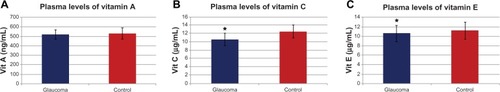
Zanon-Moreno et alCitation102,Citation103 proposed that genetic factors may play a role in modulating the effect of dietary AOX intake on glaucoma. The researchers firstly studied the association between selected polymorphisms in key transporter proteins related to vitamin C and vitamin A concentrations and POAG. A total of 300 subjects (150 POAG cases and 150 controls) from a Mediterranean population were recruited to determine the plasmatic concentrations of vitamins A and C and the single-nucleotide polymorphisms (SNPs) in genes related to these vitamin concentrations (rs176990 and rs190910 in the RBP1 gene, and rs10063949 and rs1279683 in the Na+-dependent L-ascorbic acid transporters 1 and 2, respectively [encoded by the SLC23A1 and SLC23A2 genes]). A significant association between the rs1279386 (A>G) SNP in SLC23A2 and POAG risk was detected. It was also found that POAG patients had lower plasma vitamin A and C concentrations than the controls (). SNPs in RBP1 were not associated with vitamin A concentrations or POAG risk. A significant association between the rs1279386 SNP in SLC23A2 and plasma vitamin C concentrations was found in relation to glaucoma risk. The rs10063949 SNP in SLC23A1 was not associated with either plasma vitamin C concentrations or POAG risk.Citation102 The same group analyzed the association of selected SNPs in genes related to vitamins C and E, and GSH-Px activity and POAG risk. In this study, 250 POAG cases and 250 controls were selected, also from the same Mediterranean area. Plasma concentrations of vitamins C and E were measured (), and GSH-Px activity was also determined. POAG patients had statistically significant (after correction for multiple testing) lower plasma vitamin C (P<0.001) and vitamin E (P<0.001) concentrations than the nonglaucomatous subjects. Higher plasma GSH-Px activity in POAG cases than in controls (P<0.001) was found. Also analyzed were specific SNPs (rs1279683 in the Na+-dependent L-ascorbic acid transporter 2 gene [SLC23A2], rs6994076 in the TTPA gene, rs737723 in the SEC14L2/TAP gene, and rs757228 in the GPX4 gene) in relation to glaucoma risk. A novel association between the rs737723 polymorphism and POAG risk was found that remained statistically significant after adjustment for multiple comparisons. A higher glaucoma risk in GG homozygous individuals was statistically confirmed in association with the rs1279683 polymorphism. Furthermore, a significant (P<0.05) gene–gene interaction between the SEC14L2/TAP and SLC23A2 polymorphisms in determining POAG risk was detected in those subjects who had both risk genotypes at the same time (P<0.01). Any association with POAG risk for the rs6994076 or rs757228 polymorphisms was identified. The rs1279683 and rs6994076 polymorphisms were significantly (P<0.001) associated with plasma vitamins C and E, respectively. However, the rs757228 polymorphism in the GPX4 gene was not associated with plasma GSH-Px activity.Citation103 In these two studies, novel associations between the specific SNPs and higher POAG risk were confirmed. The results also suggested a gene–gene interaction between both polymorphisms that increases POAG risk. These works support the role of vitamin-transporter proteins in the expression and availability of antioxidant vitamins, from both the natural food and nutraceutics supplementation.
Figure 3 (A–C) Plasma vitamin levels in primary open-angle glaucoma patients.
Age-related macular degeneration
AMD is the leading cause of severe vision loss in developed nations. AMD is caused by complex interactions between aging, genetics,Citation27,Citation28,Citation34 environmental factors, and comorbid diseases.Citation66,Citation67 Numerous studies have analyzed the relationship between the intake of nutritional supplements and AMD, and the most relevant are revisited herein. For a summary on these studies in AMD see .
Table 4 Relation of clinical-epidemiological studies on the role of micronutritional supplements in age-related macular degeneration
The Beaver Dam Eye StudyCitation104 found an inverse association between vitamin A and E intake and the incidence of large drusen, and between zinc intake and the incidence of macropigmentary changes in early AMD. The Rotterdam Eye StudyCitation105 concluded that a high dietary intake of β-carotene, vitamins C and E, and zinc was associated with a substantially reduced risk of AMD in elderly individuals. Clinical trials like the Age-Related Eye Disease Study (AREDS)Citation106 randomly assigned patients to daily oral tablets containing AOXs (vitamin C 500 mg, vitamin E 400 IU, and β-carotene 15 mg), zinc 80 mg, and copper 2 mg, or placebo. This clinical trial showed a statistically significant benefit of the combination of high-dose AOX vitamins and zinc, providing a moderate risk reduction of developing advanced AMD over a median of 6.3 years of follow-up in those at high risk of developing advanced AMD. Increased intake of ω-3 FAs, especially long-chain ω-3 FAs, such as DHA and eicosapentaenoic acid (EPA), has been associated with improving a number of chronic diseases, including AMD.Citation107,Citation108 Strong evidence for a beneficial role of ω-3 fatty acids in eye health was found in two cross-sectional studies: the US Twin Study of Age-Related Macular Degeneration (US Twin)Citation108 and AREDS.Citation106 Also, lutein and zeaxanthin seems to have a protective effect on the outer retina and RPE by absorbing short-wavelength light and minimizing oxidative damage, thus aiding cell-membrane stability. The Carotenoids in Age-Related Eye Disease Study (CAREDS)Citation109–Citation113 concluded that lutein- and zeaxanthin-rich diets may protect against intermediate AMD in female patients aged 75 years or more. The Australian Blue Mountains Eye StudyCitation114 analyzed the intake of ω-3 fatty acids in diet, as well as higher dietary lutein and zeaxanthin intake. Results displayed a lower incidence of developing forms of initial and advanced AMD. Furthermore, the AREDS reported in 2007 that dietary lutein and zeaxanthin intake was inversely associated with neovascular AMD, geographic atrophy, and large or extensive intermediate drusen.Citation115 Thus, the Dietary Ancillary Study of the Eye Disease Case-Control Study (DSEDCS)Citation116 revealed a relative risk for AMD estimated according to dietary indicators of AOX status, controlling for smoking and other risk factors, by using multiple logistic regression analyses in which participants with higher food intake with lutein and zeaxanthin showed a lower risk of AMD. In the French POLA Study,Citation117 levels of AOX nutrients in relation to AMD were analyzed, and data showed a lower rate of progression from moderate AMD to advanced AMD in one eye. The authors concluded that vitamin E may provide protection against AMD. Current data processing of this study strongly indicates that AMD develops mainly in subjects aged 80 years or older, and in subjects with early age-related maculopathy signs and symptoms. The North American National Health and Nutrition Examination Survey (NHANES)Citation118 was a cross-sectional nested case-control study matching subjects on age, sex, and race that was performed using data on adult participants in the third NHANES. Study outcomes included late AMD, defined as neovascular disease or geographic atrophy (5:1 matching), and a composite of both early AMD, defined as soft drusen or pigment irregularities with or without any drusen, and late AMD (1:1 matching). The main goal of this study was to analyze the blood levels of vitamin E and AMD. Data showed a lesser risk of AMD at initial stages with elevated levels of this vitamin. The Lutein and Antioxidant Supplementation Trial (Veterans LAST)Citation119 found a relationship between the intake of carotenoids and subjective signs of improvement of vision in 90 patients with AMD. Finally, the most recent results from the AREDS2,Citation120 a multicenter, randomized, double-masked, placebo-controlled Phase III study, merit special consideration in this review. It was conducted through 2006–2012 with 4,203 participants aged 50–85 years at risk for progression to advanced AMD. The clinical characteristics of the suitable participants were bilateral large drusen, or large drusen in one eye and advanced AMD in the fellow eye. The main purpose of the AREDS2 was to determine whether adding lutein + zeaxanthin, DHA + EPA, or both to the initial AREDS formulation decreases the risk of developing advanced AMD or not. After a median follow-up of 5 years, 1,940 study eyes (1,608 participants) progressed to advanced AMD. Statistical probabilities of progression to advanced AMD by 5 years of follow-up were 31% for placebo, 29% for lutein + zeaxanthin, 31% for DHA + EPA, and 30% for lutein + zeaxanthin and DHA + EPA. When compared with placebo in the primary analyses, no statistically significant reduction in the progression to advanced AMD was found. The evaluation of the effects of eliminating β-carotene from the original AREDS formulation (500 mg vitamin C, 400 IU vitamin E, and 15 mg β-carotene) and/or lowering the zinc (previously 80 mg zinc as zinc oxide, and 2 mg copper as cupric oxide) is an important point to consider. In fact, zinc decreased copper levels and caused copper-deficiency anemia. β-carotene elimination from the utilized formulation or the incorporation of a lower dose of zinc had no apparent effect of progression to advanced AMD. Important data reflected that more lung cancers were identified between the β-carotene versus no-β-carotene group (2.0%) versus 11 (0.9%) (nominal P=0.04), mostly in former smokers.
The main conclusion of the AREDS2120 was that addition of lutein + zeaxanthin, DHA + EPA, or both to the primary formulation did not further reduce risk of progression to advanced AMD. Moreover, AREDS2120 also concluded that lutein + zeaxanthin could appropriately substitute beta-carotene in AMD interventional studies with nutraceutics (to avoid the described risk of lung cancer in smokers).
Diabetic retinopathy
DR is the most common microvascular complication of diabetes mellitus and one of the leading causes of blindness worldwide. Among the studies on the role of AOXs and ω-3 fatty acids in the prevention of DR, the San Luis Valley Diabetes StudyCitation121 found no protective effect between the AOX nutrients β-carotene and vitamin C or E and DR. The Third National Health and Nutrition ExaminationCitation124 reported that serum levels of α-tocopherol and vitamin C were not associated with DR. It was also concluded in the Diabetes Control and Complications TrialCitation123 (which included over 1,000 type 1 diabetics) that those patients who followed a low-fat diet decreased the rate of progression of DR a third more than those patients not following the low-fat diet. As for type 2 diabetes, another study found less frequency of DR in subjects who took multivitamins or supplements that included vitamins C and E.Citation124 ω-3 PUFAs may also play a beneficial role in DR. In a recent study, the antiangiogenic properties of edible berries were shown.Citation125 BrownleeCitation126 proposed hyperglycemia as responsible for superoxide anion overproduction (occurring in the mitochondria). This may provide a new concept for future research and drug discovery.
Garcia-Medina et alCitation127 designed a follow-up study in the Mediterranean area of Spain with the main goal of evaluating the effect of AOX supplementation on 105 type 2 diabetics with NPDR over a 5-year period, in a similar manner to the follow-up schedule designed by the AREDS. A complete ophthalmic examination and plasmatic determinations of oxidative (MDA) and AOX parameters (total AOX status) were obtained as the baseline. One part of the cohort was randomly assigned to oral AOX supplementation at nutritional doses with the formulation defined in . The same examinations were performed with 97 diabetic patients who completed the 5-year follow-up period. Best-corrected visual acuity, DR score, MDA, and total AOX status values were compared at the beginning and the end of the follow-up. The authors found that best-corrected visual acuity did not change during the follow-up, irrespective of supplementation. However, the DR stage showed a retardation of progression in the subgroup with supplementation, but worsened in the subgroup with no AOX supplementation. Furthermore, the AOX-supplementation group maintained its AOX plasma status levels, which was related to decreased oxidative plasma activity. The authors concluded that oral AOX supplementation could be a useful adjunctive long-term therapy in the treatment of NPDR. Currently, a new study (the Valencia Diabetic Retinopathy Study [VDRS]) is being carried out to analyze the role of a combined formulation of AOX and ω-3 fatty acids in the risk and progression of DR by evaluating clinical manifestations, biochemical parameters, nutritional facts, and lifestyle, as well as subjective sensations in 350 participants made up of type 1 diabetics, type 2 diabetics, and healthy controls, through a 3-year follow-up. Preliminary data confirmed previous results from our group and others, with a significant increase in plasmatic MDA levels (P<0.01) and decreased total AOX activity (P<0.001) in both diabetic types with respect to the controls.
Table 5 Combined formulation of antioxidants utilized in the 5-year follow-up study on type 2 diabetic patients and diabetic retinopathy
Adverse effects of vitamins and oligoelements in human health
It is of maximum interest to ascertain if nutraceutic formulations with vitamins, minerals, and PUFAs may avoid, delay, or modify age-related ocular diseases. All nutraceutic formulations have to be balanced and based on scientific evidence. With the megadoses of vitamins and minerals that have been extensively suggested by different authors, it has to be considered that secondary effects can appear.
Firstly, it has to be said that vitamin E supplementation has been associated with increased risk of heart failure.Citation128 In addition, β-carotene and vitamin A intake have been reported in relation with a significant increased incidence of cancer and mortality rate in smokers and workers exposed to asbestos.Citation129–Citation131 Zinc sulfate supplementation was associated with an increased risk of genitourinary disorders during the AREDS.Citation96,Citation120 Zinc decreased copper levels and copper deficiency induced anemia. For this reason, copper should be taken with high-dose zinc.
Liposoluble vitamins, particularly vitamin A and vitamin K and some of its by-products, can induce toxicity. Excess in vitamin A has been associated with birth defects and increased risk of bleeding. Patients with liver disease and high alcohol intake may be at risk for hepatotoxicity from vitamin A supplementation.Citation132 Severe toxicity induced by hypervitaminosis A includes eye and liver damage, irritability, headache, visual disturbances, skin redness and peeling, hypercalcemia, delirium, and coma.
Vitamin C mediates the absorption of iron, a prooxidant that can be stored by the organism, and that has been associated with a major risk of cardiovascular diseases and formation of hydroxyl radicals through the reactions of Haber–Weiss and Fenton.Citation3,Citation4 Previous reports confirm that a high intake of vitamin C and vitamin E slowed the process of cellular aging and increased life expectancy. However, new findings in experimental animals suggest that high-dose supplementation may actually reduce lifespan by 11% and 26% for vitamins E and C respectively for voles in the cold, and by 17% and 18% for vitamins E and C respectively for voles in the warm. This study also confirms that major differences exist in the effects of high-dose AOXs on oxidative damage and life span across species.Citation133
Appropriate vitamin E dosing can be confusing. Current guidelines provide recommended dietary allowances and upper tolerable threshold for vitamin E in milligrams. It is usually recognized that most commercial products remain labeled in international units.Citation134 It was suggested that vitamin E supplementation may potentiate the effects of warfarin (Coumadin); therefore, taking vitamin E along with the latter can increase the risk of bruising and bleeding.
In spite of these reports, it is important to say that the Food Standards AgencyCitation135 showed that there were only eleven reported reactions from nutritional supplements over an 11 year follow-up period. The majority was in the lowest category of harm. When compared with other foods or medicines, food supplements in general displayed better results.
Final comments
Micronutrient supplementation remains a subject of debate worldwide. Still, many ophthalmologists remain skeptical about nutraceutic supplements and their role at the initiation or progression of age-related eye diseases. However, it is becoming scientifically more evident that optimal nutrition status helps in preventing OS and subsequent OS-related ocular disorders.
The main epidemiological studies have focused basically on formulations including vitamins C and E, carotenoids, flavonoids, selenium, and zinc. Perhaps we have to include other micronutrients that could influence certain processes related to eye health. More data from studies in different populations may improve our knowledge about the specific changes in diet and lifestyle that could positively influence the development of ocular conditions that are becoming more common as the population ages. A greater number of epidemiological studies on diet and the use of nutraceutics in Spain and other European countries, as well as the repercussion for on ocular diseases, have to be done to maximize eye health in the elderly.
Today, there is general agreement that ROS are involved in the physical, biochemical, molecular, and pathological changes associated with aging. Oxidative damage to lipids, proteins, and nucleic acids accumulates and increases with age, whereas AOX defenses decrease in parallel. These facts are closely related to such age-related diseases as DED, POAG, AMD, and DR. As one of the modifiable lifestyle factors is nutrition, elderly people should be informed about the importance of a healthy diet and the exact role of micronutritional supplement intake, since it has a positive effect on the body and may help to decrease the risk of ocular diseases and visual impairment associated with aging.
Disclosure
Roberto Gallego-Pinazo is a consultant for Novartis, Alcon, and Zeiss. Maria I Lopez-Galvez is a consultant for Allergan, Alimera, Novartis. The other authors report no conflicts of interest in this work.
References
- KrebsHASome aspects of the regulation of fuel supply in omnivorous animalsAdv Enzyme Regul1972103974204569539
- CheesemanKHSlaterTFAn introduction to free radical biochemistryBr Med Bull1993494814938221017
- HaberFWeissJÜber die Katalyse des Hydroperoxydes [On the catalysis of hydroperoxides]Naturwissenschaften193220948950
- FentonHJThe oxidation of tartaric acid in presence of ironJ Chem Soc Trans189465899910
- GoldsteinSMeyersteinDCzapskiGThe Fenton reagentsFree Radic Biol Med1993154354458225025
- AmesBNShigenagaMKOxidants are a major contributor to agingAnn N Y Acad Sci199266385961482105
- SiesHOxidative stress: introductory remarksSiesHOxidative StressLondonAcademic Press198517
- D’AutréauxBToledanoMBROS as signalling molecules: mechanisms that generate specificity in ROS homeostasisNat Rev Mol Cell Biol2007881382417848967
- HalliwellBFree radicals and antioxidants: updating a personal viewNutr Rev20127025726522537212
- McCordJMFridovichISuperoxide dismutase. An enzymic function for erythrocuprein (hemocuprein)J Biol Chem1969244604960555389100
- HarmanDAgeing: a theory based on free radical and radiation chemistryJ Gerontol1956229830013332224
- GerschmanRFennWAscorbic acid content of adrenal glands of rat in oxygen poisoningAm J Physiol19521766813124487
- GerschmanRNadigPWSnellACNyeSWEffect of high oxygen concentrations on eyes of newborn miceAm J Physiol1954179639639
- GerschmanRGilbertDLNyeSWFennWInfluence of x-irradiation on oxygen poisoning in miceProc Soc Exp Biol Med195486272913177582
- AgilADuránRBarreroFPlasma lipid peroxidation in sporadic Parkinson’s. Role of the L-dopaJ Neurol Sci2006240313616219327
- ChoiJSullardsMCOlzmannJAOxidative damage of DJ-1 is linked to sporadic Parkinson and Alzheimer diseasesJ Biol Chem2006281108161082416517609
- AlalufSMuir-HowieHHuHLEvansAGreenMRAtmospheric oxygen accelerates the induction of a post-mitotic phenotype in human dermal fibroblasts: the key protective role of glutathioneDifferentiation20006614715511100905
- AlexandrovaMBochevPMarkovaVDynamics of free radical processes in acute ischemic stroke: influence on neurological status and outcomeJ Clin Neurosci20041150150615177393
- AmesBNShigenagaMKHagenTMOxidants, antioxidants and degenerative diseases of agingProc Natl Acad Sci U S A199390791579228367443
- ConnerEMGrishamMBInflammation, free radicals, and antioxidantsNutrition1996122742778862535
- WilliamsDLOxidative stress and the eyeVet Clin North Am Small Anim Pract20083817919218249249
- SpectorAOxidative stress-induced cataract: mechanism of actionFASEB J19959117311827672510
- Zanon-MorenoVMarco-VenturaPLleo-PerezAOxidative stress in primary open angle glaucomaJ Glaucoma20081726326818552610
- Pinazo-DuránMDAzorinIMontoliuCGuerriCRenau-PiquerasJFree radicals are involved in the alcoholic optic neuropathyAlcohol Clin Exp Res199822A84
- MancinoRDi PierroDVaresiCLipid peroxidation and total antioxidant capacity in vitreous, aqueous humor, and blood samples from patients with diabetic retinopathyMol Vis2011171298130421633716
- MeloPZanon-MorenoVAlvesCJOxidative stress response in the adult rat retina and plasma after repeated administration of methamphetamineNeurochem Int20105643143619948197
- BeattySKohHPhilMHensonDBoultonMThe role of oxidative stress in the pathogenesis of age-related macular degenerationSurv Ophthalmol20004511513411033038
- MargrainTHBoultonMMarshallJSlineyDHDo blue light filters confer protection against age-related macular degeneration?Prog Retin Eye Res20042352353115302349
- RahmanKStudies on free radicals, antioxidants and co-factorsClin Interv Aging2007221923618044138
- CarochoMFerreiraICA review on antioxidants, prooxidants and related controversy: natural and synthetic compounds, screening and analysis methodologies and future perspectivesFood Chem Toxicol201351152523017782
- BirbenESahinerUMSackesenCErzurumSKalayciOOxidative stress and antioxidant defenseWorld Allergy Organ J2012591923268465
- DolyMDroy-LefaixMTBraquetPOxidative stress in diabetic retinaEXS1992622993071450593
- YadavUCKalariyaNMRamanaKVEmerging role of antioxidants in the protection of uveitis complicationsCurr Med Chem20111893194221182473
- PrasharSPandavSSGuptaANathRAntioxidant enzymes in RBCs as a biological index of age related macular degenerationActa Ophthalmol (Copenh)1993712142188333268
- PerryHDDry eye disease: pathophysiology, classification, and diagnosisAm J Manag Care200814S79S8718452371
- TavaresFPFernandesRSBernardesTFBonfioliAASoaresEJDry eye diseaseSemin Ophthalmol201025849320590418
- LabbéABrignole-BaudouinFBaudouinC[Ocular surface investigations in dry eye]J Fr Ophtalmol2007307697 French17287676
- ScheinODMuñozBTielschJMBandeen-RocheKWestSPrevalence of dry eye among the elderlyAm J Ophthalmol19971247237289402817
- MuñozBWestSKRubinGSCauses of blindness and visual impairment in a population of older Americans: the Salisbury Eye Evaluation StudyArch Ophthalmol200011881982510865321
- MossSEKleinRKleinBELong-term incidence of dry eye in an older populationOptom Vis Sci20088566867418677233
- SchaumbergDASullivanDABuringJEDanaMRPrevalence of dry eye syndrome among US womenAm J Ophthalmol200313631832612888056
- GipsonIKSpurr-MichaudSArgüesoPTisdaleANgTFRussoCLMucin gene expression in immortalized human corneal-limbal and conjunctival epithelial cell linesInvest Ophthalmol Vis Sci2003442496250612766048
- ZuclichJAConnollyJSOcular damage induced by near-ultraviolet laser radiationInvest Ophthalmol Vis Sci197615760764822714
- HiguchiATakahashiKHirashimaMKawakitaTTsubotaKSelenoprotein P controls oxidative stress in corneaPLoS One20105e991120360971
- UchinoYKawakitaTIshiiTIshiiNTsubotaKA new mouse model of dry eye disease: oxidative stress affects functional decline in the lacrimal glandCornea201231Suppl 1S63S6723038038
- NakamuraSShibuyaMNakashimaHInvolvement of oxidative stress on corneal epithelial alterations in a blink-suppressed dry eyeInvest Ophthalmol Vis Sci2007481552155817389484
- VanDerMeidKRSuSPKrenzerKLWardKWZhangJZA method to extract cytokines and matrix metalloproteinases from Schirmer strips and analyze using LuminexMol Vis2011171056106321552500
- Enríquez-de-SalamancaACastellanosESternMETear cytokine and chemokine analysis and clinical correlations in evaporative-type dry eye diseaseMol Vis20101686287320508732
- BaudouinCBrignoleFBecquetFPisellaPJGoguelAFlow cytometry in impression cytology specimens. A new method for evaluation of conjunctival inflammationInvest Ophthalmol Vis Sci199738145814649191610
- Pinazo-DuránMDGalbis-EstradaCPons-VázquezSCantú-DibildoxJMarco-RamírezCBenítez-del-CastilloJEffects of a nutraceutical formulation based on the combination of antioxidants and ω-3 essential fatty acids in the expression of inflammation and immune response mediators in tears from patients with dry eye disordersClin Interv Aging2013813914823430672
- Galbis-EstradaCPinazo-DuránMDCantú-DibildoxJMarco-RamírezCDiaz-LlopisMBenítez-del-CastilloJPatients undergoing antihypertensive treatment with antihypertensive eye drops responded positively with respect to their ocular surface disorder to oral supplementation with antioxidants and essential fatty acidsClin Interv Aging2013871171923818768
- TsubotaKKawashimaMInabaTThe antiaging approach for the treatment of dry eyeCornea201231Suppl 1S3S823038031
- WeinrebRNKhawPTPrimary open-angle glaucomaLancet20043631711172015158634
- OsborneNNWoodJPChidlowGBaeJHMelenaJNashMSGanglion cell death in glaucoma: what do we really know?Br J Ophthalmol19998398098610413706
- HuangWDobberfuhlAFilippopoulosTTranscriptional up-regulation and activation of initiating caspases in experimental glaucomaAm J Pathol200516767368116127148
- Pinazo-DuránMDGallego-PinazoRZanón-MorenoVSerranoMGlaucoma genetics – regulation of cell surviving and death in the retinaRumelktSGlaucoma: Basic and Clinical ConceptsRijeka, CroatiaInTech2011207224
- SaccàSCIzzottiAOxidative stress and glaucoma: injury in the anterior segment of the eyeProg Brain Res200817338540718929123
- TezelGOxidative stress in glaucomatous neurodegeneration: mechanisms and consequencesProg Retin Eye Res20062549051316962364
- Pinazo-DuránMDZanón-MorenoVGarcía-MedinaJJGallego-PinazoREvaluation of presumptive biomarkers of oxidative stress, immune response and apoptosis in primary open-angle glaucomaCurr Opin Ophthalmol20131398107
- BazanNGSurvival signaling in retinal pigment epithelial cells in response to oxidative stress: significance in retinal degenerationsAdv Exp Med Biol200657253154017249620
- KennedyCJRakoczyPEConstableIJLipofuscin of the retinal pigment epithelium: a reviewEye (Lond)199597637718849547
- CaiJNelsonKCWuMSternbergPJrJonesDPOxidative damage and protection of the RPEProg Retin Eye Res20001920522110674708
- MukherjeePKMarcheselliVLSerhanCNBazanNGNeuroprotectin D1: a docosahexaenoic acid-derived docosatriene protects human retinal pigment epithelial cells from oxidative stressProc Natl Acad Sci U S A20041018491849615152078
- BazanNGNeuroprotectin D1 (NPD1): a DHA-derived mediator that protects brain and retina against cell injury-induced oxidative stressBrain Pathol20051515916615912889
- CalandriaJMMukherjeePKde Rivero VaccariJCZhuMPetasisNABazanNGAtaxin-1 poly(Q)-induced proteotoxic stress and apoptosis are attenuated in neural cells by docosahexaenoic acid-derived neuroprotectin D1J Biol Chem2012287237262373922511762
- KleinRPetoTBirdAVannewkirkMRThe epidemiology of age related macular degenerationAm J Ophthalmol200413748649515013873
- CurcioCAJohnsonMRudolfMHuangJDThe oil spill in ageing Bruch membraneBr J Ophthalmol2011951638164521890786
- FaghirZBazanNGPI3K/Akt and mTOR/p70S6K pathways mediate neuroprotectin D1-induced retinal pigment epithelial cell survival during oxidative stress-induced apoptosisExp Eye Res20109071872520230819
- KlettnerAOxidative stress induced cellular signaling in RPE cellsFront Biosci (Schol Ed)2012439241122202067
- KoenekoopRThe gene for Stargardt disease, ABCA4, is a major retinal gene: a mini-reviewOphthalmic Genet200324758012789571
- NowakJZOxidative stress, polyunsaturated fatty acids-derived oxidation products and bisretinoids as potential inducers of CNS diseases: focus on age-related macular degenerationPharmacol Rep20136528830423744414
- CastilhoAAveleiraCALealECHeme oxygenase-1 protects retinal endothelial cells against high glucose- and oxidative/nitrosative stress-induced toxicityPLoS One20127e4242822879979
- SparrowJRZhouJBen-ShabatSVollmerHItagakiYNakanishiKInvolvement of oxidative mechanisms in blue-light-induced damage to A2E-laden RPEInvest Ophthalmol Vis Sci20024341222122711923269
- MalekiSGopalakrishnanSGhanianZOptical imaging of mitochondrial redox state in rodent model of retinitis pigmentosaJ Biomed Opt2013181600423291617
- ZhuoYLuoHZhangKLeber hereditary optic neuropathy and oxidative stressProc Natl Acad Sci U S A2012109198821988323197830
- Pinazo-DuránMDVerdejoCAzorínIRenau-PiquerasJIborraFJColocalization of aldehyde dehydrogenases and Fe/NADPH-induced lipid peroxidation in tissue sections of rat retinaOphthalmic Res200032616810754436
- Galbis-EstradaCPons-VázquezSGallego-PinazoRGlutathione-dependent formaldehyde dehydrogenase (ADH3) and low km mitochondrial aldehyde dehydrogenase (ALDH2). New evidence for differential expression in the rat retina in response to oxidative stressFree Radic Res201246778422117533
- RogersBSSymonsRCKomeimaKDifferential sensitivity of cones to iron-mediated oxidative damageInvest Ophthalmol Vis Sci20074843844517197565
- DentchevTHahnPDunaiefJLStrong labeling for iron and the iron-handling proteins ferritin and ferroportin in the photoreceptor layer in age-related macular degenerationArch Ophthalmol20051231745174616344450
- HeXHahnPIacovelliJIron homeostasis and toxicity in retinal degenerationProg Retin Eye Res20072664967317921041
- SongDSongYHadziahmetovicMZhongYDunaiefJLSystemic administration of the iron chelator deferiprone protects against light-induced photoreceptor degeneration in the mouse retinaFree Radic Biol Med201253647122579919
- ArdenGBSivaprasadSHypoxia and oxidative stress in the causation of diabetic retinopathyCurr Diabetes Rev2011729130421916837
- SonSMRole of vascular reactive oxygen species in development of vascular abnormalities in diabetesDiabetes Res Clin Pract200677Suppl 1S65S7017467110
- GuptaMMChariSLipid peroxidation and antioxidant status in patients with diabetic retinopathyIndian J Physiol Pharmacol20054918719216170987
- VerdejoCMarcoPRenau-PiquerasJPinazo-DuranMDLipid peroxidation in proliferative vitreoretinopathiesEye (Lond)19991318318810450379
- RobisonWGJrJacotJLKatzMLGloverJPRetinal vascular changes induced by the oxidative stress of alpha-tocopherol deficiency contrasted with diabetic microangiopathyJ Ocul Pharmacol Ther20001610912010803422
- Martín-GallánPCarrascosaAGussinyéMDomínguezCBiomarkers of diabetes-associated oxidative stress and antioxidant status in young diabetic patients with or without subclinical complicationsFree Radic Biol Med2003341563157412788476
- ValkoMLeibfritzDMoncolJCroninMTMazuzMTelsenJFree radicals and antioxidants in normal physiological functions and human diseaseInt J Biochem Cell Biol200739448416978905
- PácalLVarvařovskáJRušavýZParameters of oxidative stress, DNA damage and DNA repair in type 1 and type 2 diabetes mellitusArch Physiol Biochem201111722223021338322
- DolinoyDCHuangDJirtleRLMaternal nutrient supplementation counteracts bisphenol A-induced DNA hypomethylation in early developmentProc Natl Acad Sci U S A2007104130561306117670942
- US Department of Health and Human ServicesDietary guidelines for Americans, 2010 Available from: http://www.dietaryguidelines.govAccessed December 19, 2013
- KeysAMenottiAKarvonenMJThe diet and 15-year death rate in the seven countries studyAm J Epidemiol19861249039153776973
- KeysAMediterranean diet and public health: personal reflectionsAm J Clin Nutr1995611321S1323S7754982
- SofiFCesariFAbbateRGensiniGFCasiniAAdherence to Mediterranean diet and health status: meta-analysisBMJ2008377a134418786971
- Serra-MajemLRomanBEstruchRScientific evidence of interventions using the Mediterranean diet: a systematic reviewNutr Rev200664S27S4716532897
- DelcourtCKorobelikJFBarberger-GateauPNutrition and age-related eye diseases: the ALIENOR (Antioxydants, LIpides Essentiels, Nutrition et Maladies OculaiRes) studyJ Nutr Health Aging20091485486121125205
- MaletFLe GoffMColinJDry eye disease in French elderly subjects: the Alienor StudyActa Ophthalmol Epub672013
- QuarantaLBettelliSUvaMGSemeraroFTuranoRGandolfoEEffect of Ginkgo biloba extract on preexisting visual field damage in normal tension glaucomaOphthalmology2003110235936212578781
- RitchRPotential role for Ginko biloba extract in the treatment of glaucomaMed Hypotheses20005422123510790757
- ColemanALStoneKLKodjebachevaGGlaucoma risk and the consumption of fruits and vegetables among older women in the study of osteoporotic fracturesAm J Ophthalmol20081451081108918355790
- KangJHPasqualeLRWillettWCDietary fat consumption and primary open-angle glaucomaAm Soc Clin Nutr20047957555764
- Zanon-MorenoVAsensio-MarquezEMCiancotti-OliverLEffects of polymorphisms in vitamin E-, vitamin C-, and glutathione peroxidase-related genes on serum biomarkers and associations with glaucomaMol Vis20131923124223401652
- Zanon-MorenoVCiancotti-OlivaresLAsencioJAssociation between a SLC23A2 gene variation, plasma vitamin C levels, and risk of glaucoma in a Mediterranean populationMol Vis2011172997300422171153
- VandenLangenbergGMMares-PerlmanJAKleinRKleinBEBradyWEPaltaMAssociations between antioxidant and zinc intake and the 5-year incidence of early age-related maculopathy in the Beaver Dam Eye StudyAm J Epidemiol19981482042149676703
- Van LeeuwenRBoekhoornSVingerlingJRDietary intake of antioxidants and risk of age-related macular degenerationJAMA20052943101310716380590
- Age-Related Eye Disease Study Research GroupA randomized, placebo-controlled, clinical trial of high-dose supplementation with vitamins C and E, beta carotene, and zinc for age-related macular degeneration and vision loss: AREDS report no 8Arch Ophthalmol20011191417143611594942
- ConnorKMSanGiovanniJPLofqvistCIncreased dietary intake of omega-3-polyunsaturated fatty acids reduces pathological retinal angiogenesisNat Med20071386887317589522
- SeddonJMGeorgeSRosnerBCigarette smoking, fish consumption, omega-3 fatty acid intake, and associations with age-related macular degeneration: the US Twin Study of Age-Related Macular DegenerationArch Ophthalmol2006124995100116832023
- SanGiovanniJPChewEYAgrónEThe relationship of dietary omega-3 long-chain polyunsaturated fatty acid intake with incident age-related macular degeneration. AREDS report no. 23Arch Ophthalmol20081261274127918779490
- KrinskyNILandrumJTBoneRABiologic mechanisms of the protective role of lutein and zeaxanthin in the eyeAnnu Rev Nutr20032317120112626691
- SembaRDDagnelieGAre lutein and zeaxanthin conditionally essential nutrients for eye health?Med Hypotheses20036146547213679014
- SchweigertFJReimannJMicronutrients and their relevance for the eye – function of lutein, zeaxanthin and omega-3 fatty acidsKlin Monbl Augenheilkd2011228537543 German20740395
- MoellerSMParekhNTinkerLAssociations between intermediate age-related macular degeneration and lutein and zeaxanthin in the Carotenoids in Age-Related Eye Disease study (CAREDS): ancillary study of the Women’s Health InitiativeArch Ophthalmol20061241151116216908818
- TanJSWangJJFloodVRochtchinaESmithWMitchellPDietary antioxidants and the long-term incidence of age-related macular degeneration: the Blue Mountains Eye StudyOphthalmology200811533434117664009
- Age-Related Eye Disease Study Research GroupSanGiovanniJPChewEYThe relationship of dietary carotenoid and vitamin A, E, and C intake with age-related macular degeneration in a case control study: AREDS report no 22Arch Ophthalmol20071251225123217846363
- SeddonJMAjaniUASperdutoRDDietary carotenoids, vitamins A, C, and E, and advanced age-related macular degeneration. Eye Disease Case-Control Study GroupJAMA1994272141314207933422
- DelcourtCCristolJPTessierFLégerCLDescompsBPapozLAge-related macular degeneration and antioxidant status in the POLA study. POLA Study Group. Pathologies Oculaires Liées à l’AgeArch Ophthalmol19991171384139010532448
- Mares-PerlmanJAFisherAIKleinRLutein and zeaxanthin in the diet and serum and their relation to age-related maculopathy in the third national health and nutrition examination surveyAm J Epidemiol200115342443211226974
- RicherSStilesWStatkuteLDouble-masked, placebo-controlled, randomized trial of lutein and antioxidant supplementation in the intervention of atrophic age-related macular degeneration: the Veterans LAST study (Lutein Antioxidant Supplementation Trial)Optometry20047521623015117055
- Age-Related Eye Disease Study 2 Research GroupLutein + zeaxanthin and omega-3 fatty acids for age-related macular degeneration: the Age-Related Eye Disease Study 2 (AREDS2) randomized clinical trialJAMA20133092005201523644932
- Mayer-DavisEJBellRAReboussinBARushingJMarshallJAHammanRFAntioxidant nutrient intake and diabetic retinopathy: the San Luis Valley Diabetes StudyOphthalmology1998105226422709855158
- WeinerDETighiouartHReynoldsRSeddonJMKidney function, albuminuria and age-related macular degeneration in NHANES IIINephrol Dial Transplant2011263159316521339308
- No authors listedDiabetes Control and Complications Trial (DCCT): results of feasibility study. The DCCT Research GroupDiabetes Care1987101192882967
- MillenAEGruberMKleinRKleinBEPaltaMMaresJARelations of serum ascorbic acid and alpha-tocopherol to diabetic retinopathy in the Third National Health and Nutrition Examination SurveyAm J Epidemiol200315822523312882944
- RoySKhannaSAlessioHMAnti-angiogenic property of edible berriesFree Radic Res20023691023103112448828
- BrownleeMBiochemistry and molecular cell biology of diabetic complicationsNature201141481382011742414
- Garcia-MedinaJJPinazo-DuranMDGarcia-MedinaMZanon-MorenoVPons-VazquezSA 5-year follow-up of antioxidant supplementation in type 2 diabetic retinopathyEur J Ophthalmol20112163764321218388
- LonnEBoschJYusufSEffects of long-term vitamin E supplementation on cardiovascular events and cancer: a randomized controlled trialJAMA20052931338134715769967
- Rodríguez-CastillaVCuadradoCPozoSMoreirasOConcentraciones plasmáticas de carotenos y vitaminas antioxidantes en personas de edad avanzada: influencia del tabaquismo. Proyecto HALE [Plasmatic concentrations of carotenoids and antioxidant vitamins in elder people: influence of smoking. Hale Project]Clin Investig Arterioscler200517101111
- No authors listedThe effect of vitamin E and beta carotene on the incidence of lung cancer and other cancers in male smokers. The Alpha Tocopherol, Beta Carotene Cancer Prevention Study GroupN Engl J Med1994330102910358127329
- OmennGSGoodmanGEThornquistMDEffects of a combination of beta carotene and vitamin A on lung cancer and cardiovascular diseaseN Engl J Med1996334115011558602180
- FosmireGJZinc toxicityAm J Clin Nutr1990512252272407097
- BlockKIKochACMeadMNTothyPKNewmanRAGyllenhaalCImpact of antioxidant supplementation on chemotherapeutic toxicity: a systematic review of the evidence from randomized controlled trialsInt J Cancer2008151231227123918623084
- SelmanCMcLarenJSCollinsARDuthieGGSpeakmanJRDeleterious consequences of antioxidant supplementation on lifespan in a wild-derived mammalBiol Lett2013915
- Food and Nutrition Board, Institute of MedicineCopper. Dietary reference intakes for vitamin A, vitamin K, boron, chromium, copper, iodine, iron, manganese, molybdenum, nickel, silicon, vanadium, and zincWashington, D.C.National Academy Press2001224257