
Abstract
Background/hypothesis
Whole body exercise (WBE) changes lymphocyte subset percentages in peripheral blood. Resistive breathing, a hallmark of diseases of airway obstruction, is a form of exercise for the inspiratory muscles. Strenuous muscle contractions induce oxidative stress that may mediate immune alterations following exercise. We hypothesized that inspiratory resistive breathing (IRB) alters peripheral blood lymphocyte subsets and that oxidative stress mediates lymphocyte subpopulation alterations following both WBE and IRB.
Patients and methods
Six healthy nonathletes performed two WBE and two IRB sessions for 45 minutes at 70% of VO2 maximum and 70% of maximum inspiratory pressure (Pimax), respectively, before and after the administration of antioxidants (vitamins E, A, and C for 75 days, allopurinol for 30 days, and N-acetylcysteine for 3 days). Blood was drawn at baseline, at the end of each session, and 2 hours into recovery. Lymphocyte subsets were determined by flow cytometry.
Results
Before antioxidant supplementation at both WBE end and IRB end, the natural killer cell percentage increased, the T helper cell (CD3+ CD4+) percentage was reduced, and the CD4/CD8 ratio was depressed, a response which was abolished by antioxidants only after IRB. Furthermore, at IRB end, antioxidants promoted CD8+ CD38+ and blunted cytotoxic T-cell percentage increase. CD8+ CD45RA+ cell percentage changes were blunted after antioxidant supplementation in both WBE and IRB.
Conclusion
We conclude that IRB produces (as WBE) changes in peripheral blood lymphocyte subsets and that oxidative stress is a major stimulus predominantly for IRB-induced lymphocyte subset alterations.
Introduction
Peripheral blood leukocyte subpopulations respond rather stereotypically to whole body exercise (WBE). During WBE, neutrophil, lymphocyte, and monocyte counts increase, followed by a reduction in lymphocyte count after exercise due to redistribution and apoptosis.Citation1 Regarding specific lymphocyte subpopulations, exercise is mainly associated with an increase in CD8+ T-lymphocyte and CD56+ CD16+ natural killer (NK) cell percentage and a subsequent decrease in CD4+ T-lymphocyte percentage.Citation1 Alterations in lymphocyte count have been associated with suppressed immune function following intense exercise.Citation2
Reactive oxygen species (ROS) generated during intense WBE are among the potential modulators of this responseCitation3 though their exact role has not been established. Highly intense exercise induces lymphocyte apoptosis via an ROS-dependent pathway.Citation4 Oxidative stress may induce DNA damage of immunocompetent cells after prolonged and strenuous exercise (marathon run).Citation5 Yet antioxidant supplementation (N-acetylcysteine [NAC]) showed no effect in exercise-induced proliferation and activity of lymphocyte subsets in trained athletes.Citation6 As in WBE, free radicals in the form of ROS and reactive nitrogen species are generated during increased contractile activity of the inspiratory muscles, mainly the diaphragm.Citation7,Citation8
Resistive breathing is encountered in obstructive airway diseases, such as asthma and chronic obstructive pulmonary disease (COPD), especially during exacerbations.Citation9 Inspira-tory resistive breathing (IRB) is a form of exercise of the inspiratory muscles and is associated with intense respiratory muscle contractions. When strenuous enough, IRB produces diaphragmatic fatigue and diaphragmatic structural injury and enacts as an immune challenge initiating cytokine upregulation in the diaphragmCitation10,Citation11 and the plasma.Citation12,Citation13 IRB has also been shown to induce pulmonary inflammation and lung injury in experimental animal models.Citation14,Citation15
The immune system’s role and the influence of the excessive production of oxidative derivatives in obstructive pulmonary diseases are active research fields.Citation16–Citation18 In continuation of our previous experimentsCitation10–Citation15 in humans and animals where resistive breathing, as a model of airway obstruction, produced oxidative stress-dependent systematic and pulmonary inflammation, we decided to investigate its effects on the cells that orchestrate various immune responses, the lymphocytes. We thus hypothesized that IRB, as WBE, induces lymphocyte subpopulation changes in the peripheral blood and that oxidative stress modulates this response. We also hypothesized that oxidative stress modulates the peripheral blood lymphocyte subpopulation responses to WBE. To test our hypotheses, we conducted WBE and IRB sessions of equal duration before and after in vivo supplementation of antioxidants in healthy nonathlete volunteers.
Subjects and methods
Subjects
Six healthy male volunteers, who were not involved in strenuous manual labor due to their profession, free of any history of asthma and other relevant respiratory conditions, with a mean age of 33 years (28–37 years) were studied. They did not participate in regular exercise training or sports activities and had not had febrile illness in the 3 months before or throughout the duration of the experiment. The subjects were instructed to refrain from intense physical activity or regular exercise training during the study period, to adopt their usual dietary pattern, and were also allowed to have normal daily activities. Once per week, each subject visited the laboratory, was supplied with doses of antioxidants for 7 days, and returned any unused ones. On the same day of the visit, each subject was asked for changes in everyday habits and for symptoms of illness. Each participant was receiving reminder calls every 2 days to ensure compliance with antioxidant supplementation. The Ethics Committee of our institution, Evangelismos Hospital, approved the study protocol, and all the subjects provided written informed consent.
Overall study design
The six subjects performed two sessions of IRB and WBE before and after the administration of antioxidants. Each participant performed an IRB session followed by a WBE session after 15 days, in order to eliminate the possible influences of one session on the other (ie, any muscle injury incurring during one session persisting and thus affecting the immune response of the next session),Citation1 and then, antioxidant supplementation began. The same IRB session was performed after 60 days of antioxidant supplementation followed by the same WBE after 15 days totaling 75 days of antioxidant supplementation and 90 days of overall experimental duration. Therefore, each participant served as a self-control eliminating between subject variability. Experiments were performed at summer time minimizing the chance of (viral) respiratory tract infections, which in fact did not happen to any of our subjects.
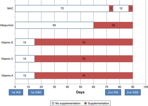
After the 1st WBE session, participants started receiving a combination of antioxidants, including 200 mg vitamin E, 50,000 IU vitamin A, and 1,000 mg vitamin C per day, for a total of 75 days. Allopurinol, 600 mg/day, was administered for a total of 30 days starting at day 61 (14 days before the 2nd IRB session) and NAC, 1,800 mg/day, for 3 days before the 2nd IRB and the 2nd WBE sessions. Antioxidants were supplemented in one dose in the morning with breakfast except NAC which was administered in three doses (600 mg each). In the morning of testing, a full dose of antioxidants and 600 mg of NAC were supplemented 2 hours before the session. This design was chosen instead of a randomized crossover design because of the nature of the antioxidants: vitamins with long-lasting effects (especially vitamin E) after the completion of the supplementation.Citation19 The duration and combination of antioxidants were based on our previous experiments with WBECitation19 and IRB.Citation13
The flowchart of the study design is as follows:
Day 1: 1st IRB session
Day 15: 1st WBE session
Day 16: Initiation of vitamin A (50,000 IU/day), vitamin E (200 mg/day), and vitamin C (1,000 mg/day) in one dose in the morning
Day 61: Initiation of allopurinol (600 mg/day) one dose in the morning
Day 72: Initiation of NAC (1,800 mg/day in three doses of 600 mg)
Day 75: 2nd IRB session; supplementation in the morning 2 hours before session of vitamin A (50,000 IU), vitamin E (200 mg), vitamin C (1,000 mg), allopurinol (600 mg), and NAC (600 mg morning dose); termination of NAC supplementation
Day 87: Initiation of NAC (1,800 mg/day in three doses of 600 mg)
Day 90: 2nd WBE session; supplementation in the morning 2 hours before session of vitamin A (50,000 IU), vitamin E (200 mg), vitamin C (1,000 mg), allopurinol (600 mg), and NAC (600 mg morning dose); termination of all antioxidant supplementation. illustrates the flow chart of the study design.
Figure 1 Antioxidant supplementation time line.
Abbreviations: IRB, inspiratory resistive breathing; NAC, N-acetylcysteine; WBE, whole body exercise.
Preliminary testing
Preliminary testing was conducted 2 weeks before the initiation of the experimental sessions (1st IRB session).
WBE
The maximum WBE capacity of each subject was assessed at the initial visit to the laboratory. Participants got accustomed to an electrically braked cycle ergometer (Medical Graphics CPX/D; Medical Graphics Corporation, Saint Paul, MN, USA) and were instructed to maintain a constant pedal speed of 60 rpm. Increments of 10 watts every minute were applied until each subject reached a maximum work load (volitional exhaustion) in order to determine the maximum oxygen consumption rate (VO2 max) in accordance with our previous experiments. Heart rate and blood pressure were continuously monitored.Citation19
IRB
The IRB sessions were also performed in accordance with previous experiments.Citation9,Citation13,Citation20 Subjects were placed in the sitting position and were accustomed to the procedure. A mouthpiece was adjusted to a T-piece adapter, and two one-way valves (Hans Rudolph, Inc., Shawnee, KS, USA) were attached to each side in order to determine an inspiration and expiration port. A tube with an adjustable orifice was attached to the inspiratory port in order to increase inspiratory resistance, while the expiratory port was left without load. A rigid tube connected the mouthpiece with a pressure transducer (Validyne, Los Angeles, CA, USA), and the measurements were recorded on a 16-channel electrostatic recorder (Gould ES 1000; Gould Instruments, Cleveland, OH, USA) and displayed on an oscilloscope (Tektronix 2213; Beaverton, OR, USA). To prevent glottis closure during the run, a small hole, 2 mm in diameter, was opened in the mouthpiece. The maximum inspiratory pressure (Pimax) was measured while the subject performed maximal inspiratory effort from functional residual capacity against an occluded inspiratory port. The Pimax was considered the most negative mouth pressure that could be sustained for at least 1 second. Multiple efforts (up to 5 after introductory training on how to perform the maneuver) were performed, and the most negative pressure was considered each subject’s Pimax. Pimax maneuvers were separated by 2 minutes each to avoid potentiation. Preliminary resistive breathing sessions were performed for each subject in order to select the amount of resistance that was required as to achieve inspiratory pressures at 70% of the maximum inspiratory pressure. Excessive dyspnea and hypoxia were criteria for the premature termination of the session. Throughout the IRB run, arterial blood oxygen saturation, arterial blood pressure, and heart rate were monitored.
Main experimental trials
Testing was always performed at 10 am after a night rest of 8 hours in order to avoid possible influences of circadian rhythm variations, and a breakfast rich in carbohydrates was administrated 2 hours before testing (with a full dose of antioxidants and 600 mg of NAC). Each session’s duration was 45 minutes, and blood was drawn at rest (baseline), end (of session), and 120 minutes into recovery, through an intravenous catheter inserted in a forearm vein.
WBE
WBE was conducted at 70% of VO2 max for 45 minutes after a 10-minute warm-up period of increasing intensity (10 W/min). Heart rate and blood pressure were continuously monitored.Citation19
IRB
Subjects begun the IRB run through the precustomized resistive load and were constantly encouraged to achieve pressures of 70% of their Pimax. Subjects were breathing in their own pattern, and the IRB run was terminated after 45 minutes. Throughout the IRB run, arterial blood oxygen saturation, arterial blood pressure, and heart rate were monitored.
Blood samples
Blood samples were collected into sterile syringes and were transferred to precooled sterile ethylenediaminetetraacetic acid tubes or sodium heparin tubes for flow cytometric evaluation (used within 2 hours from venipuncture).
Flow cytometric acquisition and analysis
Fluorescently labeled monoclonal antibodies against cell surface markers characteristic of lymphocyte subpopulations were used for staining. Counting was carried out with flow cytometry (Epics® Elite Esp.; Coulter Electronics, Beckman Coulter, Inc., Brea, CA, USA). We measured CD3, CD19, CD16, CD56, CD8, CD4, CD45RO, CD45RA, HLA-DR, and CD38. The percentages of total T cells (CD3+), B cells (CD19+), NK cells (CD3− CD16+ CD56+), T cytotoxic cells (CD3+ CD8+), T helper cells (CD3+ CD4+), memory T cells (CD45RO+, CD4+, or CD8+), naïve T cells (CD45RA+, CD4+, or CD8+), and markers of activation of CD8+ cells (HLA-DR+ and CD38+) were also measured.
Malondialdehyde (MDA) by high-performance liquid chromatography (MDA-HPLC) assay
The MDA levels as an index of oxidative stress and the efficacy of antioxidant administration were determined at the end of the WBE or IRB sessions, respectively. The MDA-HPLC method was mainly based on the procedure by Nielsen et al.Citation21 The reagents potassium phosphate monobasic, potassium hydroxide, phosphoric acid (H3PO4), sodium hydroxide (NaOH), HPLC-grade methanol, and thiobarbituric acid (TBA) were purchased from Sigma-Aldrich Co. (St Louis, MO, USA). Malondialdehyde tetrabutylammonium salt (96% purity) was also purchased from Sigma-Aldrich Co. and used for standards’ preparation.
For sample preparation, 100 µL of plasma was added to 700 µL of 1% H3PO4 and 200 µL of 42 mmol/L TBA. The mixture was heated for 30 minutes at 100°C and then ice-cooled for 2 minutes. A 250 µL of the aliquot was separated and added to 250 µL of 1 M NaOH in methanol (1:6). After centrifugation (5 minutes at 10,000 g) and filtration, 20 µL of the supernatant was injected in the HPLC apparatus at a flow rate of 1 mL/min, column temperature and pressure at 26°C and 2,200 psi, respectively. The absorbance was read at 532 nm.
For the MDA standard curve preparation, a stock solution of 100 µM of MDA was prepared in 0.01 mmol/L HCl. Dilutions from stock solution of 10–0.5 µmol/L were then performed. The same treatment described for plasma was used for the standard curve reaction.
Statistical analysis
Changes in the percentage of lymphocytes subpopulations over time (rest [baseline], end of WBE or IRB session, and 120 minutes into recovery) were compared by the nonparametric Friedman’s analysis of variance (ANOVA). The Wilcoxon matched pair test was used for post hoc analysis. The percentage of each cell subpopulation at iso-time points and the differences in cell percentages over time, before and after antioxidant supplementation, were compared with the Wilcoxon matched pair test. A p-value of 0.05 was initially considered as statistically significant and was accordingly adjusted using the sharper Bonferroni-type procedure for multiple comparisons described by Hochberg and Benjamini.Citation22 Data are presented as mean values ± standard errors of the mean.
Results
MDA levels at end-exercise or IRB sessions
MDA levels at end exercise were 2.67±0.54 µmol/L before antioxidant supplementation and decreased to 1.86±0.42 µmol/L after antioxidant supplementation (p=0.023). MDA levels at end IRB were 2.14±0.41 µmol/L before antioxidant supplementation and decreased to 1.17±0.28 µmol/L after antioxidant supplementation (p=0.034).
Heart rate and arterial pressure measurements
Heart rate and mean arterial pressure were significantly higher in WBE compared with IRB (Wilcoxon matched pair test: p=0.0001 and p=0.005, respectively).
CD3+ cells (T-lymphocytes)
WBE
T-lymphocyte percentages did not change significantly in the WBE session regardless of antioxidant administration ().
Table 1 Lymphocyte subpopulation percentage changes during WBE
IRB
T-lymphocyte percentages did not change significantly in the IRB session regardless of antioxidant administration ().
Table 2 Lymphocyte subpopulation percentage changes during IRB
CD3+ CD4+ cells (T helper cells)
WBE
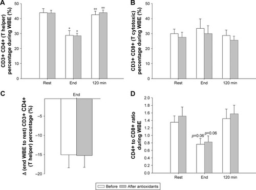
Both before and after antioxidant supplementation, T helper cell percentage was significantly reduced at exercise end compared with rest (p=0.04 for each) and was restored to rest values at recovery (before antioxidant supplementation, Friedman’s ANOVA x2=8.4, p=0.015; after antioxidant supplementation x2=7.6, p=0.02; ). The degree of T helper cell percentage decrease at end-WBE compared with rest was not affected by antioxidants ().
Figure 2 CD4+ and CD8+ lymphocyte percentage during WBE.
Abbreviation: WBE, whole body exercise.
IRB
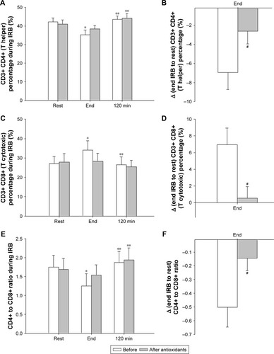
Before antioxidant supplementation, the percentage of T helper cells was significantly reduced at the end of resistive breathing compared with rest and were restored to pre-IRB levels at 120 minutes into recovery (Friedman’s ANOVA x2=6.3, p=0.042; Wilcoxon matched pair test, rest-end p=0.027, end-120 minutes p=0.043). Antioxidants attenuated T helper cell percentage decrease at IRB session’s end. (Friedman’s ANOVA x2=8.3, p=0.016; Wilcoxon matched pair test rest-end: p=0.07, end-120 minutes: p=0.027; ). The magnitude of T helper cell percentage decrease at end-IRB compared with rest was significantly reduced after antioxidant supplementation (Wilcoxon matched pair test p=0.046; ).
Figure 3 CD4+ and CD8+ lymphocyte percentage during IRB.
Abbreviation: IRB, inspiratory resistive breathing.
CD3+ CD8+ cells (cytotoxic T cells)
WBE
The percentage of cytotoxic T cells did not significantly change at either time point, before and after antioxidant supplementation ().
IRB
The percentage of cytotoxic T cells increased significantly after IRB and returned to pre-IRB levels at 120 minutes into recovery (Friedman’s ANOVA x2=7.0, p=0.03; Wilcoxon matched pair test rest-end p=0.046, end-120 minutes p=0.03). Antioxidant supplementation blunted the cytotoxic T cell percentage changes (Friedman’s ANOVA x2=3.0, p=0.22; ). The IRB end-to-rest difference of cytotoxic T cell percentage was significantly greater before antioxidant supplementation compared with after antioxidant supplementation (p=0.05; ).
CD4/CD8 ratio
WBE
The CD4/CD8 ratio changed significantly over time before (Friedman’s ANOVA x2=6.5, p=0.038) and after antioxidant supplementation (Friedman’s ANOVA x2=6.0, p=0.049). Both before and after antioxidant supplementation, the ratio tended to be reduced at exercise end compared with baseline (Wilcoxon matched pair test, p=0.06; ).
IRB
The CD4/CD8 ratio before antioxidant supplementation was significantly decreased at IRB session end and was restored to baseline values at 120 minutes into recovery (Friedman’s ANOVA x2=9.0, p=0.011; Wilcoxon matched pair test rest-end p=0.028, end-120 minutes p=0.028). After antioxidant supplementation, no significant decrease at IRB end was observed compared with rest, although the ratio was increased at 120 minutes into recovery compared with IRB end (Friedman’s ANOVA x2=8.33, p=0.015; Wilcoxon matched pair test, rest-end p=0.17, end-120 minutes p=0.028). The end-to-rest difference of CD4/CD8 ratio was attenuated after antioxidant supplementation compared with before antioxidant supplementation (p=0.05; ).
CD4+ CD45RA+ (Th cells expressing CD45RA)
WBE
CD45RA+ Th cell percentage was not affected by WBE either before or after antioxidants ().
IRB
The percentages of CD45RA+ Th cells did not significantly change either before or after antioxidant supplementation ().
CD4+ CD45RO+ (Th cells expressing CD45RO)
WBE
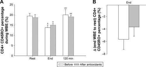
The percentages of Th cells expressing CD45RO were reduced postexercise (end) and returned to preexercise levels (rest) at 120 minutes into recovery, both before and after antioxidant supplementation (before: Friedman’s ANOVA x2=7.60, p=0.022; Wilcoxon matched pair test rest-end p=0.043; after: Friedman’s ANOVA x2=7.00, p=0.030; Wilcoxon matched pair test rest-end p=0.046; ). The WBE end-to-rest difference of CD4+ CD45RO+ cells was not affected by antioxidants ().
Figure 4 CD4+ CD45RO+ cell percentage during WBE.
Abbreviation: WBE, whole body exercise.
IRB
The percentages of Th cell-expressing CD45RO did not change during IRB, neither before nor after antioxidant supplementation ().
CD8+ CD45RA+ (cytotoxic T cells expressing CD45RA)
WBE
The percentage of CD45RA+ cytotoxic T cells increased postexercise (end) and returned to preexercise levels (rest) at 120 minutes into recovery before antioxidant supplementation (Friedman’s ANOVA x2=7.60, p=0.022; Wilcoxon matched pair test; rest-end p=0.043, end-120 minutes p=0.043). In contrast, no significant change was observed after antioxidant supplementation (Friedman’s ANOVA x2=4.00, p=0.14). The iso-time comparison revealed a significant increase at rest percentages after antioxidant supplementation (Wilcoxon matched pair test p=0.043; ). The WBE-end to rest increase was not affected by antioxidant supplementation ().
Figure 5 CD8+ CD45RA+ cell percentage during WBE and IRB.
Abbreviations: IRB, inspiratory resistive breathing; WBE, whole body exercise.
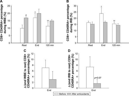
IRB
IRB increased the percentage of CD8+ CD45RA+ T cells after IRB session end that returned to rest values at 120 minutes into recovery (Friedman’s ANOVA x2=9.3, p=0.009; Wilcoxon matched pair test rest-end p=0.03, end-120 minutes p=0.03). After antioxidant supplementation, CD8+ CD45RA+ T cell percentage changes were blunted (Friedman’s ANOVA x2=5.20, p=0.07; ). The IRB end to rest increase tended to be reduced after antioxidant supplementation (Wilcoxon matched pair test p=0.07; ). The antioxidant-induced upregulation of CD45RA+ percentage at rest of WBE () was not observed at rest before IRB ().
CD8+ CD45RO+ (cytotoxic T cells expressing CD45RO)
WBE
The percentage of CD45RO+ cytotoxic T cells did not change after WBE irrespective of antioxidant supplementation ().
IRB
The CD8+ CD45RO+ remained unchanged during IRB both before and after antioxidant supplementation ().
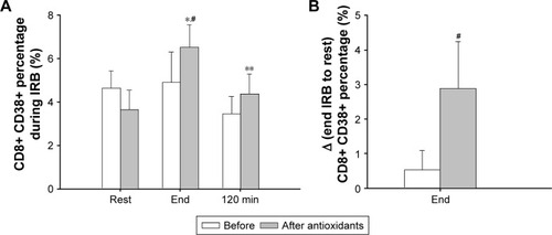
CD8+ CD38+ cells
WBE
CD8+ CD38+ percentage changes before and after antioxidant supplementation were not significant (Friedman’s ANOVA x2=2.21, p=0.33; Friedman’s ANOVA x2=1.0, p=0.60, respectively). The iso-time comparison revealed that antioxidants upregulated CD8+ CD38+ percentages significantly at 120 minutes into recovery (Wilcoxon matched pair test p=0.043; ).
IRB
Before antioxidant supplementation, the percentage of CD8+ CD38+ cells was not affected from IRB (Friedman’s ANOVA x2=3.5, p=0.17). After antioxidant supplementation, an increase in CD8+ CD38+ cell percentage at IRB end was observed compared with rest. CD8+ CD38+ cell percentage returned to pre-IRB levels at 120 minutes into recovery (Friedman’s ANOVA x2=8.4, p=0.014; Wilcoxon matched pair test rest-to-end p=0.043, end-120 minutes p=0.027; ). Iso-time comparisons revealed that the end IRB percentage was higher after antioxidant supplementation (Wilcoxon matched pair test p=0.043; ). Indeed, the increase in CD8+ CD38+ cell percentage after antioxidant supplementation was significantly greater than that before antioxidant supplementation (Wilcoxon matched pair test p=0.04; ).
Figure 6 CD8+ CD38+ cell percentage during IRB.
Abbreviation: IRB, inspiratory resistive breathing.
CD8+ HLA-DR+ cells
WBE
CD8+ HLA-DR+ cell percentage did not change significantly over time neither before nor after antioxidant supplementation ().
IRB
CD8+ HLA-DR+ percentage changes were not significant before and after antioxidant supplementation ().
CD3− CD16+ CD56+ (NK cells)
WBE
NK cell percentages significantly changed over time, regardless of antioxidant supplementation (before antioxidant supplementation: Friedman’s ANOVA x2=8.4, p=0.015; after antioxidant supplementation: Friedman’s ANOVA x2=9.3, p=0.009). Both before and after antioxidant supplementation, NK cell percentage increased postexercise (end) compared with rest (p=0.04 and p=0.02, respectively) and went back to preexercise levels at 120 minutes into recovery (). The difference in NK cell percentage postexercise (end) relative to rest did not differ before and after antioxidant supplementation ().
Figure 7 NK cell percentage during WBE and IRB.
Abbreviations: IRB, inspiratory resistive breathing; NK, natural killer; WBE, whole body exercise.

IRB
Before antioxidant supplementation, NK cell percentage increased at IRB end (Friedman’s ANOVA x2=6.3, p=0.042; end IRB to rest, p=0.028). Antioxidant supplementation blunted the IRB-induced increase in NK cell percentage (Friedman’s ANOVA x2=4.3, p=0.11; ). The end-to-rest difference of NK cell percentages was significantly greater before antioxidant supplementation compared with after antioxidant supplementation (p=0.027; ).
CD19+ (B cells)
WBE
Both before and after antioxidant supplementation, B-cell percentage decreased postexercise (end) and was restored to rest levels at 120 minutes into recovery (Friedman’s ANOVA, before antioxidants: x2=7.6, p=0.02; after antioxidants, x2=9.3, p=0.009; ). The reduction of B-cell percentage at end WBE relative to rest was attenuated after antioxidant supplementation, but this effect did not reach statistical significance, compared with before antioxidant supplementation (p=0.06; ).
Figure 8 B-lymphocyte percentage during WBE.
Abbreviation: WBE, whole body exercise.
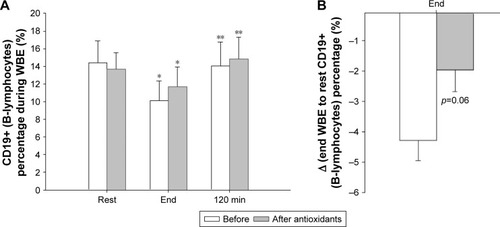
IRB
IRB did not produce any changes in the percentage of B cells regardless of antioxidant supplementation ().
Discussion
The major finding of our study is that IRB induces significant alterations in peripheral blood lymphocyte subpopulations as does WBE, and this effect is blunted by antioxidants mainly in resistive breathing. During IRB, NK and cytotoxic T-cell percentages increase, T helper cell percentage is reduced, and CD4/CD8 ratio is depressed under the influence of oxidative stress. Two hours into recovery in WBE and IRB, the percentage of most lymphocyte subsets returns to rest levels.
Lymphocyte subset response to WBE and IRB
During exercise, all lymphocytes are recruited to the intra-vascular space, yet each subset responds differently.Citation1
NK cells are the most sensitive subset to exercise showing a 10-fold increase in the last minute of a short bout (20 minutes) of intense (85% of VO2 max) exercise, while T helper and B cells increase only by 50%–100%.Citation23 Women exercising at 60% of VO2 max for 45 minutes demonstrated an increase in lymphocyte count at exercise end lasting for 1.5 hours with NK cells accounting for two thirds of and with T cells the remaining one third of lymphocytosis.Citation24 Exercise intensity positively correlates with the postexercise increase of the NK cell percentage.Citation25,Citation26
Our findings during WBE confirm these observations. We measured NK cells (CD56+ CD16+) that are considered more responsive to exercise.Citation23,Citation27 After a WBE session with an intensity of 70% of VO2 max, NK cell percentage nearly doubled and was restored to preexercise levels at 2 hours into recovery.Citation26,Citation28 Interestingly, antioxidants had no effect on the aforementioned alterations. To the best of our knowledge, our study is the first to describe NK cell response after resistive breathing. IRB led to a significant rise of NK cell percentage (at end) that returned to rest levels after 2 hours as in WBE. Antioxidants blunted this response, which suggests that oxidative stress is a stimulus for the NK cell increase secondary to resistive breathing.
CD8+ lymphocytes (cytotoxic T cells) are also mobilized and recruited into the vascular compartment in proportion to exercise intensity. Campbell et al have shown an almost 5 times increase in the percentage of cytotoxic T cells after a short session of intense exercise.Citation23 The most actively cytotoxic T cells, the reexpressing CD45RA (CD3+ CD8+ CD45RA+), demonstrate the highest exercise responsiveness.Citation23 Accordingly, the CD45RA+ cell percentage markedly increased after WBE in our experiment. We show here for the first time that the CD45RA+ cell percentage increases during IRB. Another novel finding of our study is that antioxidants abolished the CD45RA+ cell response to IRB and WBE, suggesting that oxidative stress is mediating this response.
CD4+ T helper cells exhibit a more “modest” response to exercise increasing by 50%–100%;Citation23 thus, a depressed CD4/CD8 ratio is expected postexercise.Citation1,Citation24,Citation29 We found that T helper cell percentage was depressed at WBE and IRB end probably due to a higher recruitment of cytotoxic and NK cells in the endovascular compartment, resulting in a depressed CD4/CD8 ratio. Antioxidants attenuated T helper cell percentage and CD4/CD8 reduction only at IRB end. We also found a significant decrease in the percentage of CD4+ CD45RO+ cells at WBE end regardless of antioxidant administration.
In agreement with the literature, antioxidant supplementation tended to upregulate CD8+ CD38+ percentage.Citation30 At 120 minutes into recovery of WBE, CD8+ CD38+ percentage was increased after antioxidant supplementation compared with before antioxidant supplementation. During IRB, CD8+ CD38+ percentage changes were observed only after antioxidant supplementation.
B-lymphocytes (CD19+) seem to be less responsive to exercise. Studies have shown that B-lymphocytes are relatively unaffected by exercise,Citation31,Citation32 whereas others showed an increase during repeated bouts of acute exercise and a reduction in the recovery period.Citation33 We found a significant decrease in their percentage at WBE end and restoration to baseline values 2 hours into recovery regardless of antioxidant administration. By contrast, no change in B-lymphocyte percentage was observed during IRB.
Lymphocyte migration control and redistribution
Lymphocytosis in peripheral blood is a stereotypical result of environmental stress such as surgery, burn, trauma, sepsis, and exercise.Citation34 The immediate postexercise lymphocytosis reflects the transition from reservoirs to target organs where lymphocytes may encounter pathogen invasion or antigen exposure. Consequently, the recovery lymphocytopenia or return to preexercise values is a result of the evolution completion of the migration process. Thus, changes in the percentage of lymphocyte subsets in the vascular compartment at different time points of a stress event (eg, exercise) are part of their functional redistribution to various body compartments. Migration is regulated by adhesion molecules and cell migration cytokines (chemokines), yet the exact control mechanism (mechanisms) remains (remain) relatively elusive. Adrenergic stimulation and, to a lesser extent, steroids and apoptosis have been suggested.Citation35,Citation36
Peripheral blood lymphocyte count and catecholamines are directly related, namely the lymphocytosis during/after stress is dose-dependent on the amount of adrenaline and noradrenaline that are secreted.Citation37 In animal models, exogenous administration of epinephrine increased peripheral blood lymphocytes as observed in exercise.Citation36 NK cells present the highest expression of β2-adrenergic receptors followed by cytotoxic T cells; consequently, the aforementioned subgroups are proportionally responsive to exercise intensity.Citation38 IRB at 70% of Pimax represents a smaller adrenergic response to WBE (at 70% of VO2 max). Interestingly, heart rate and mean arterial pressure (which are indirect measures of adrenergic stimulation) were significantly higher in WBE compared with IRB. Indeed, doubling the work of breathing does not increase sympathetic activation in healthy subjects and COPD patients.Citation39 IRB-induced lymphocyte subset alterations may be influenced by factors other than (a weak) sympathetic activation.
Oxidative stress and exercise
The link between exercise and oxidative stress is firmly established.Citation7,Citation8,Citation40–Citation42 Working muscles are considered the main source of oxidative products (electron leakage from mitochondria, oxidases, myostatin, phospholipase A2, etc.), but not the only one.Citation43 Exercise-induced muscle injury may activate through cytokine production (IL-1β and tumor necrosis factor-alpha [TNF-α]) neutrophils and macrophages, cells capable of producing large amounts of ROS through oxidative burst, their indigenous defense mechanism.Citation44 Endothelial cells and catecholamines are also implicated in ROS generation.Citation43 Oxidative stress derivatives are regulators of cell function and signaling molecules integrated in exercise physiology.Citation45 Our group has shown that oxidative stress is a stimulus for the cytokine induction after both IRBCitation13 and WBECitation19 and that ROS stimulate IL-6 production from skeletal myotubes.Citation46
We have administrated a cocktail of antioxidants in nonathletes that has already proven effective in attenuating the plasma cytokine response after WBECitation19 and IRB.Citation13 The rationale behind the composition of the mixture is thoroughly analyzed in our previous work.Citation13,Citation19 Vitamin E is an efficient lipid-soluble free radical scavenger protecting biomembranes, while vitamin C is a water-soluble antioxidant that along with vitamin A can directly scavenge singlet oxygen, superoxide, and hydroxyl radicals in both intra- and extracellular fluid. Allopurinol inhibits the ROS-generating xanthine oxidase, and NAC forms L-cysteine, which is further metabolized to glutathione, a key biological antioxidant molecule.Citation13 The mixture was effective in reducing lipid peroxidation as evidenced by the reduced MDA levels detected postexercise (end) or post-IRB (end) after antioxidant administration. Treatment with allopurinol efficiently inhibits blood glutathione oxidation and lipid peroxidation after strenuous exercise in COPD patients.Citation47 We have used a cocktail of antioxidants with the rationale that multiple mechanisms and sources of oxidative stress exist within cells; thus, a single antioxidant would be unlikely to be effective. We have chosen a relatively long period of administration on the premise that some antioxidants like vitamin E require a long time to become incorporated into membranes.Citation48 On the other hand, as we have already mentioned, the mixture of antioxidants that we used prevented us from adopting a randomized crossover design due to their residual effects, ie, the plasma concentration of vitamin E reached peak values after 15 days of supplementation and maintained this plateau for the next 15 days,Citation49 and the serum concentration of carotenoids peaked at 24–48 hours after a single dose and returned to baseline after 7 days.Citation50 Other investigators who used only one antioxidant in trained athletes failed to produce alterations in lymphocyte subsets.Citation6 We used untrained healthy volunteers and not athletes who present an upregulated antioxidant capacity secondary to training.Citation43,Citation51 Our results suggest that an oxidative stress-dependent pathway regulates lymphocyte redistribution during IRB and possibly in obstructive lung diseases.
The mechanisms by which oxidative stress may regulate lymphocyte redistribution during IRB are not known and are not the scope of this study. However, some speculations are worth pursuing. Lymphocyte migration to and out of lymphoid and nonlymphoid organs presupposes a stepwise chemokine-controlled interaction with the endothelium. The transmigration through the endothelium includes the capture and roll at the vessel wall mediated primarily by selectins and integrins (rolling and adhesion). Activation and transendothelial migration are mediated by adhesion molecules that are present in the membranes of lymphocytes and endothelial cells, the intracellular adhesion molecule 1 (ICAM-1) and vascular intracellular adhesion molecule 1 (VCAM-1).Citation35 ICAMs are upregulated by IL-6, IL-1β, and TNF-α, the three cytokines that were abolished after antioxidant supplementation in IRB.Citation13 The ICAM-1 gene is induced by IL-6 and TNF-α mediated via activator of transcription-3 (Stat3) and nuclear factor-κB.Citation52 TNF-α and IL-1β also induced the expression of ICAM-1 and VCAM-1 by endothelial cells.Citation53 It is therefore tempting to speculate that IL-6, TNF-α, and IL-1β precipitate lymphocyte migration independently of the stress neurohormonal stimulation through the upregulation of adhesion chemokines that are essential in lymphocyte transendothelial migration. In our experiment, antioxidant supplementation may have blunted IRB-induced cytokine release and consequently lymphocyte redistribution. In contrast, WBE lymphocyte subset alterations may be dominated by stress-induced neurohormonal mechanisms that are less influenced by antioxidants and cytokines.
Comparison between WBE and IRB
Although we have used IRB as a model to isolate the effects of respiratory muscle activation, we cannot exclude respiratory muscle activation during WBE. It is known that WBE results in increased inflammation and oxidative stress. One potential source of the immune response observed during WBE may relate to the respiratory muscle metaboreflex/fatigue, which accompanies aerobic exercise. It is known that WBE produces diaphragmatic fatigue when intensity exceeds 85% of VO2 max.Citation54 We have chosen a submaximal exercise intensity of 70% of VO2 max, which is not likely to induce respiratory muscle fatigue. We did not aim for diaphragmatic fatigue through our IRB session, and even with high Pi target (of 70% Pimax), we instructed subjects to breathe in their own pattern with a relatively high respiratory rate (20–25) and inspiratory time/total inspiratory and expiratory time ratio <0.6 using all respiratory muscles, not just the diaphragm. Therefore, all our subjects completed a 45-minute session of IRB, and Pimax at the end of the session was ±10% of initial Pimax. St Croix et al noticed an increase in muscle nerve sympathetic activity (peroneal nerve) and small increases in mean arterial blood pressure and heart rate only when they used a protocol of resistive breathing designed to produce diaphragmatic fatigue (Pi =60% Pimax, inspiratory time/total inspiratory and expiratory time ratio =0, 7, Vt =2x resting Vt and RR =15 breaths/min for 7±3 minutes until exhaustion).Citation55 Sheel et al triggered a similar metaboreflex reducing the blood flow in the femoral artery in a similar protocol of resistive breathing that produced diaphragmatic fatigue identified through changes in mouth twitch pressure in response to bilateral phrenic nerve stimulation, while mean arterial pressure remained unchanged.Citation56 In contrast, increased voluntary ventilation and inspiratory effort even with a Pi close to Pimax (95%) did not produce a sympathetic response.Citation55,Citation56 Taken together, although we cannot exclude the presence of respiratory muscle metaboreflex/fatigue with certainty, we believe that either our IRB protocol or our WBE protocol was not likely to induce such a metaboreflex.
Clinical implications
Obstructive pulmonary diseases such as COPD and asthma are characterized by increased airway resistance that is aggravated during exacerbations and may lead to respiratory failure and death. Our group has focused on the consequences of airway obstruction per se and has created an experimental model of breathing through increased airway resistance (resistive breathing) that was applied on both humans and animals.
In each subject, we applied inspiratory resistive load in order to achieve breathing inspiratory pressures close to 70% of their Pimax. The load we used was extrapolated from data of patients with severe COPD. Direct measurements of the actual load that respiratory muscles face during an exacerbation are extremely difficult since physiological experiments are not conducted in life-threating clinical conditions. However, indirect estimates can be made. In patients with COPD who require mechanical ventilation, respiratory muscles face similar load as in severe exacerbation. Our group showed that in COPD patients requiring invasive mechanical ventilation, the peak/maximum inspiratory pressure value was 0.62±0.15,Citation57 and failure to wean from mechanical ventilation was accompanied by Pimean/Pimax of 0.490±0.09.Citation58
We applied only inspiratory resistance, and this is a limitation of our study. One may argue that our human model is similar to extrathoracic obstruction (upper airway obstruction), where the resistive load is primary inspiratory, and that in COPD and asthma the obstruction is intrathoracic and the resistance is higher during expiration. Nevertheless, during asthma and COPD exacerbations, excess bronchoconstriction induces air-trapping and large negative intrathoracic pressures during inspiration to counteract the intrinsic positive end-expiratory pressure and the increased airway resistance.Citation18 Therefore, our model of IRB, where respiratory muscles contract strenuously and lungs are exposed to large negative intrathoracic pressure gradients, mimics key components of the respiratory pathophysiology of obstructive lung disease, especially during exacerbations. This IRB may induce cytokine upregulationCitation13 and, as shown in the current study, immune alterations.
Increased numbers of T-lymphocytes are observed in lung parenchyma, distal and proximal airways of COPD patients with a predominance of CD8+ over CD4+ cells.Citation16 T-cell numbers are correlated to the extent of alveolar destruction and the severity of airflow obstruction, especially CD8+ cells that induce apoptosis of alveolar cells and cytolysis through the release of perforins, granzyme B, and TNF-α.Citation59,Citation60 COPD patients also show altered peripheral blood lymphocyte subsets that are also correlated to the degree of airway obstruction. The percentage of peripheral blood CD8+ T-lymphocytes was higher in COPD patients compared with nonsmoking healthy controls. Furthermore, patients with low FEV1 had significantly higher CD8+ T-lymphocyte percentage in peripheral blood.Citation61 Smokers with COPD and predominantly small airway disease had higher CD8+ T-lymphocyte percentage in peripheral blood than “healthy” smokers.Citation62
We have shown here for the first time that a brief simulation of airway obstruction (IRB) in healthy humans increases CD8+ T-lymphocytes percentages in peripheral blood in an oxidative stress-dependent pathway. It is tempting to speculate that bouts of increased airway resistance, such as those observed during IRB or COPD exacerbation, increase CD8+ T-lymphocytes in the peripheral circulation and thence to the lung, thus contributing to the increased CD8+ T-lymphocyte levels observed. This would lead to bouts of CD8+ T-cell-mediated lung destruction and would partly explain why COPD patients with frequent exacerbations have more rapid decline in lung function.Citation63,Citation64
NK cell percentages in the peripheral blood and the lung do not differ between stable COPD patients and healthy subjects.Citation59,Citation61,Citation65 However, peripheral blood NK cell cytotoxic activity is impaired in COPD patients.Citation65 In our experiment, IRB resulted in an increase in the NK cells, which was reversed after the end of IRB. This response to the “mechanical stressor” of increased airway resistance (ie, IRB) would be adaptive in the case of COPD exacerbation (which is also associated with increased airway resistance). This is because COPD exacerbations are usually infective in origin, and the increased NK cells would lead to increased immune surveillance against the infective pathogens. However, in the case of COPD exacerbations, the relief of increased airway resistance with bronchodilators would decrease the NK cell percentage in the blood and would lead to “relative immunosuppression” in the face of the offending infective agent causing exacerbation.
Conclusion
We have shown for the first time that IRB induces lymphocyte subset alterations through an oxidative stress-dependent pathway. Lymphocytes are key immune cells that regulate both innate and adaptive immunity. Increased airway resistance may not only induce inflammation and lung injury, but also precipitate, through oxidative stress, peripheral immune alterations.
Acknowledgments
The authors would like to acknowledge the contribution of Dimitri Stathopoulos for proofreading the final draft of this paper. This study was funded by the Thorax Foundation, Athens, Greece.
Disclosure
The authors report no conflicts of interest in this work.
References
- PedersenBKHoffman-GoetzLExercise and the immune system: regulation, integration, and adaptationPhysiol Rev20008031055108110893431
- NiemanDCImmune response to heavy exertionJ Appl Physiol (1985)1997825138513949134882
- KrugerKMoorenFCExercise-induced leukocyte apoptosisExerc Immunol Rev20142011713424974724
- KrugerKFrostSMostEVolkerKPallaufJMoorenFCExercise affects tissue lymphocyte apoptosis via redox-sensitive and Fas-dependent signaling pathwaysAm J Physiol Regul Integr Comp Physiol20092965R1518R152719261913
- TsaiKHsuTGHsuKMOxidative DNA damage in human peripheral leukocytes induced by massive aerobic exerciseFree Radic Biol Med200131111465147211728819
- NielsenHBSecherNHKappelMPedersenBKN-acetylcysteine does not affect the lymphocyte proliferation and natural killer cell activity responses to exerciseAm J Physiol19982754 Pt 2R1227R12319756554
- VassilakopoulosTHussainSNVentilatory muscle activation and inflammation: cytokines, reactive oxygen species, and nitric oxideJ Appl Physiol (1985)200710241687169517185492
- BorzoneGZhaoBMerolaAJBerlinerLClantonTLDetection of free radicals by electron spin resonance in rat diaphragm after resistive loadingJ Appl Physiol (1985)19947728128188002533
- VassilakopoulosTRoussosCZakynthinosSThe immune response to resistive breathingEur Respir J20042461033104315572550
- VassilakopoulosTDivangahiMRallisGDifferential cytokine gene expression in the diaphragm in response to strenuous resistive breathingAm J Respir Crit Care Med2004170215416115117743
- SigalaIZacharatosPBouliaSNitric oxide regulates cytokine induction in the diaphragm in response to inspiratory resistive breathingJ Appl Physiol (1985)2012113101594160322961265
- VassilakopoulosTZakynthinosSRoussosCStrenuous resistive breathing induces proinflammatory cytokines and stimulates the HPA axis in humansAm J Physiol19992774 Pt 2R1013R101910516239
- VassilakopoulosTKatsaounouPKaratzaMHKollintzaAZakynthinosSRoussosCStrenuous resistive breathing induces plasma cytokines: role of antioxidants and monocytesAm J Respir Crit Care Med200216612 Pt 11572157812406849
- ToumpanakisDKastisGAZacharatosPInspiratory resistive breathing induces acute lung injuryAm J Respir Crit Care Med201018291129113620622034
- LoverdosKToumpanakisDLitsiouEThe differential effects of inspiratory, expiratory, and combined resistive breathing on healthy lungInt J Chron Obstruct Pulmon Dis2016111623163827499619
- BarnesPJInflammatory mechanisms in patients with chronic obstructive pulmonary diseaseJ Allergy Clin Immunol20161381162727373322
- LeidingerPKellerAHeiselSNovel autoantigens immunogenic in COPD patientsRespir Res2009102019284601
- Global Initiative for Chronic Obstructive Lung Disease (GOLD)Global Strategy for Diagnosis, Management, and Prevention of COPD – 2016 Available from: http://goldcopd.org/global-strategy-diagnosis-management-prevention-copd-2016/Accessed November 3, 2016
- VassilakopoulosTKaratzaMHKatsaounouPKollintzaAZakynthinosSRoussosCAntioxidants attenuate the plasma cytokine response to exercise in humansJ Appl Physiol (1985)20039431025103212571133
- KeastDCameronKMortonARExercise and the immune responseSports Med1988542482673287548
- NielsenFMikkelsenBBNielsenJBAndersenHRGrandjeanPPlasma malondialdehyde as biomarker for oxidative stress: reference interval and effects of life-style factorsClin Chem1997437120912149216458
- HochbergYBenjaminiYMore powerful procedures for multiple significance testingStat Med1990978118182218183
- CampbellJPRiddellNEBurnsVEAcute exercise mobilises CD8+ T lymphocytes exhibiting an effector-memory phenotypeBrain Behav Immun200923676777519254756
- NiemanDCNehlsen-CannarellaSLDonohueKMThe effects of acute moderate exercise on leukocyte and lymphocyte subpopulationsMed Sci Sports Exerc19912355785852072836
- NiemanDCMillerARHensonDAEffects of high- vs moderate-intensity exercise on natural killer cell activityMed Sci Sports Exerc19932510112611348231757
- TvedeNKappelMHalkjaer-KristensenJGalboHPedersenBKThe effect of light, moderate and severe bicycle exercise on lymphocyte subsets, natural and lymphokine activated killer cells, lymphocyte proliferative response and interleukin 2 productionInt J Sports Med19931452752828365836
- TimmonsBWCieslakTHuman natural killer cell subsets and acute exercise: a brief reviewExerc Immunol Rev20081482319203081
- YuDTClementsPJPearsonCMEffect of corticosteroids on exercise-induced lymphocytosisClin Exp Immunol1977282326331301451
- FryRWMortonARCrawfordGPKeastDCell numbers and in vitro responses of leucocytes and lymphocyte subpopulations following maximal exercise and interval training sessions of different intensitiesEur J Appl Physiol Occup Physiol19926432182271563367
- Sandoval-MontesCSantos-ArgumedoLCD38 is expressed selectively during the activation of a subset of mature T cells with reduced proliferation but improved potential to produce cytokinesJ Leukoc Biol200577451352115618297
- ShekPNSabistonBHBuguetARadomskiMWStrenuous exercise and immunological changes: a multiple-time-point analysis of leukocyte subsets, CD4/CD8 ratio, immunoglobulin production and NK cell responseInt J Sports Med19951674664748550256
- NataleVMBrennerIKMoldoveanuAIVasiliouPShekPShephardRJEffects of three different types of exercise on blood leukocyte count during and following exerciseSao Paulo Med J2003121191412751337
- NielsenHBSecherNHChristensenNJPedersenBKLymphocytes and NK cell activity during repeated bouts of maximal exerciseAm J Physiol19962711 Pt 2R222R2278760224
- Hoffman-GoetzLPedersenBKExercise and the immune system: a model of the stress response?Immunol Today19941583823877916952
- KrugerKMoorenFCT cell homing and exerciseExerc Immunol Rev200713375418198659
- KrugerKLechtermannAFobkerMVolkerKMoorenFCExercise-induced redistribution of T lymphocytes is regulated by adrenergic mechanismsBrain Behav Immun200822332433817910910
- BoschJABerntsonGGCacioppoJTDhabharFSMaruchaPTAcute stress evokes selective mobilization of T cells that differ in chemokine receptor expression: a potential pathway linking immunologic reactivity to cardiovascular diseaseBrain Behav Immun200317425125912831827
- TurnerJEBoschJAAldredSMeasurement of exercise-induced oxidative stress in lymphocytesBiochem Soc Trans20113951299130421936805
- RaupachTBahrFHerrmannPLüthjeLHasenfussGAndreasSInspiratory resistive loading does not increase sympathetic tone in COPDRespir Med2010104110711319619996
- ReidMBInvited review: redox modulation of skeletal muscle contraction: what we know and what we don’tJ Appl Physiol (1985)200190272473111160074
- JacksonMJPyeDPalomeroJThe production of reactive oxygen and nitrogen species by skeletal muscleJ Appl Physiol (1985)200710241664167017082364
- FerreiraLFReidMBMuscle-derived ROS and thiol regulation in muscle fatigueJ Appl Physiol (1985)2008104385386018006866
- SteinbacherPEcklPImpact of oxidative stress on exercising skeletal muscleBiomolecules20155235637725866921
- PeakeJSuzukiKNeutrophil activation, antioxidant supplements and exercise-induced oxidative stressExerc Immunol Rev20041012914115633591
- ThannickalVJFanburgBLReactive oxygen species in cell signalingAm J Physiol Lung Cell Mol Physiol20002796L1005L102811076791
- KosmidouIVassilakopoulosTXagorariAZakynthinosSPapapetropoulosARoussosCProduction of interleukin-6 by skeletal myotubes: role of reactive oxygen speciesAm J Respir Cell Mol Biol200226558759311970911
- HeunksLMViñaJvan HerwaardenCLFolgeringHTGimenoADekhuijzenPNXanthine oxidase is involved in exercise-induced oxidative stress in chronic obstructive pulmonary diseaseAm J Physiol19992776 Pt 2R1697R170410600916
- CannonJGMeydaniSNFieldingRAAcute phase response in exercise. II. Associations between vitamin E, cytokines, and muscle proteolysisAm J Physiol19912606 Pt 2R1235R12401905495
- MeydaniMFieldingRACannonJGBlumbergJBEvansWJMuscle uptake of vitamin E and its association with muscle fiber typeJ Nutr Biochem1997827478
- JohnsonEJRussellRMDistribution of orally administered beta-carotene among lipoproteins in healthy menAm J Clin Nutr19925611281351609749
- SachdevSDaviesKJProduction, detection, and adaptive responses to free radicals in exerciseFree Radic Biol Med200844221522318191757
- WungBSNiCWWangDLICAM-1 induction by TNFalpha and IL-6 is mediated by distinct pathways via Rac in endothelial cellsJ Biomed Sci20051219110115864742
- O’CarrollSJKhoDTWiltshireRPro-inflammatory TNFalpha and IL-1beta differentially regulate the inflammatory phenotype of brain microvascular endothelial cellsJ Neuroinflammation20151213126152369
- JohnsonBDBabcockMASumanOEDempseyJAExercise-induced diaphragmatic fatigue in healthy humansJ Physiol19934603854058487201
- CroixCMStMorganBJWetterTJDempseyJAFatiguing inspiratory muscle work causes reflex sympathetic activation in humansJ Physiol2000529Pt 249350411101657
- SheelAWDerchakPAPegelowDFDempseyJAThreshold effects of respiratory muscle work on limb vascular resistanceAm J Physiol Heart Circ Physiol20022825H1732H173811959638
- ZakynthinosSGVassilakopoulosTRoussosCThe load of inspiratory muscles in patients needing mechanical ventilationAm J Respir Crit Care Med19951524 Pt 1124812557551378
- VassilakopoulosTZakynthinosSRoussosCThe tension-time index and the frequency/tidal volume ratio are the major pathophysiologic determinants of weaning failure and successAm J Respir Crit Care Med199815823783859700110
- MajoJGhezzoHCosioMGLymphocyte population and apoptosis in the lungs of smokers and their relation to emphysemaEur Respir J200117594695311488331
- SaettaMDi StefanoATuratoGCD8+ T-lymphocytes in peripheral airways of smokers with chronic obstructive pulmonary diseaseAm J Respir Crit Care Med19981573 Pt 18228269517597
- de JongJWvan der Belt-GritterBKoeterGHPostmaDSPeripheral blood lymphocyte cell subsets in subjects with chronic obstructive pulmonary disease: association with smoking, IgE and lung functionRespir Med199791267769122514
- KimWDKimWSKohYAbnormal peripheral blood T-lymphocyte subsets in a subgroup of patients with COPDChest2002122243744412171814
- DonaldsonGCSeemungalTABhowmikAWedzichaJARelationship between exacerbation frequency and lung function decline in chronic obstructive pulmonary diseaseThorax2002571084785212324669
- MakrisDMoschandreasJDamianakiAExacerbations and lung function decline in COPD: new insights in current and ex-smokersRespir Med200710161305131217112715
- PrietoAReyesEBernsteinEDDefective natural killer and phagocytic activities in chronic obstructive pulmonary disease are restored by glycophosphopeptical (inmunoferon)Am J Respir Crit Care Med200116371578158311401877