
Abstract
COPD is characterized by persistent respiratory symptoms and airflow limitation, caused by a mixture of small airway disease and pulmonary emphysema. Programmed cell death has drawn the attention of COPD researchers because emphysema is thought to result from epithelial cell death caused by smoking. Although apoptosis has long been thought to be the sole form of programmed cell death, recent studies have reported the existence of a genetically programmed and regulated form of necrosis called necroptosis. Autophagy was also previously considered a form of programmed cell death, but this has been reconsidered. However, recent studies have revealed that autophagy can regulate programmed cell death, including apoptosis and necroptosis. It is also becoming clear that autophagy can selectively degrade specific proteins, organelles, and invading bacteria by a process termed “selective autophagy” and that this process is related to the pathogenesis of human diseases. In this review, we outline the most recent studies implicating autophagy, selective autophagy, and necroptosis in COPD. Strategies targeting these pathways may yield novel therapies for COPD.
Introduction
COPD involves persistent respiratory symptoms and airflow limitation. The chronic airflow limitation is caused by a mixture of small airway disease and pulmonary emphysema, usually due to significant exposure to noxious particles or gases. Cigarette smoke (CS) is the most common identifiable risk factor for COPD, with smokers known to have a greater COPD mortality rate than non-smokers.Citation1 Pulmonary emphysema is thought to result from epithelial cell death caused by smoking; therefore, COPD researchers have devoted considerable attention to programmed cell death.
Apoptosis was previously recognized as the sole form of programmed cell death, whereas necrosis was considered as uncontrolled cell death induced by extreme physical or chemical stress. However, emerging studies have demonstrated the existence of a genetically programmed and regulated form of necrosis, termed “necroptosis,” defined as necrotic cell death dependent on the receptor-interacting protein kinase 3 (RIPK3).Citation2–Citation4 RIPK3, RIPK1, and mixed-lineage kinase domain-like protein (MLKL) form a multiprotein complex called the “necrosome,” in which MLKL is an essential necroptosis inducer that acts downstream of RIPK3.Citation5,Citation6 Unlike apoptosis, which is thought to be a weak inducer of inflammation with little release of damage-associated molecular patterns (DAMPs) from dying cells, necroptosis is considered a strong inducer of inflammation that releases massive amounts of DAMPs.Citation7
Autophagy is a highly conserved process through which cells can recycle organelles and proteins by degrading them in lysosomes.Citation8 Autophagy proceeds through sequential steps beginning with the generation of autophagosomes from an isolation membrane, followed by elongation to form a mature autophagosome that captures cytosolic cargo. While the term “autophagic cell death” has long been used to refer to a type of cell death associated with excessive cytoplasmic vacuolization,Citation9 autophagy is considered to primarily act as a protective mechanism that may prevent cell death by maintaining cellular integrity through regenerating metabolic precursors and clearing subcellular debris.Citation10 Thus, currently, autophagic cell death is not considered as a form of programmed cell death,Citation11 but is ascribed to cell death with, rather than by, autophagy. In contrast, it is becoming clear that autophagy can regulate other cell death pathways. In cells of childhood acute lymphoblastic leukemia, induction of autophagy-dependent necroptosis is required to overcome glucocorticoid resistance.Citation12 In endothelial cells, autophagy inhibition rescues palmitic acid-induced necroptosis.Citation13 Thus, accumulating evidence suggests that autophagy may regulate necroptosis in the pathogenesis of human diseases.
While autophagy has long been considered as simply a non-specific homeostatic cellular process, increasing evidence suggests that it represents a more selective process than originally anticipated.Citation14 Selective autophagy delivers a wide range of autophagic cargo from protein aggregates to whole organelles and even intracellular microbes to the lysosome for degradation. It is thought that ubiquitin-positive substrates, such as protein aggregates, mitochondria, and invading bacteria not dealt with the proteasome system, are selectively degraded by autophagy.Citation15 Ubiquitination seems to function as a general tag for selective autophagy in mammalian cells. Various selective autophagy subtypes have been recognized and named after their specific targets, for example, aggregated proteins: aggrephagy,Citation16 mitochondria: mitophagy,Citation17 pathogens: xenophagy,Citation18 and cilia: ciliophagy.Citation19 The study of selective autophagy is an emerging field that is expected to provide new insights into the pathogenesis of human lung diseases.Citation14 In this review, we examine the growing evidence favoring the contribution of autophagy, selective autophagy (), and necroptosis () to COPD pathogenesis and discuss the dual nature of these processes in the lungs. A better understanding of the protective and injurious effects of these processes in disease pathogenesis will help design personalized therapies for COPD treatment.
Table 1 Roles of selective autophagy in COPD
Table 2 Role of necroptosis in COPD
Molecular mechanisms of autophagy, selective autophagy, and necroptosis
Autophagy is an evolutionarily conserved catabolic process by which cytoplasmic materials are delivered to and degraded in the lysosome.Citation10 Often referred to simply as autophagy, macroautophagy is the best characterized form of autophagy, involving the engulfment of cytoplasmic contents and organelles through a complex reorganization of subcellular membranes to form a new organelle: the autophagosome. In the 1990s, genetic studies in yeast identified a series of autophagy-related (ATG) genes that regulate the macroautophagic process.Citation20,Citation21 Autophagosome elongation requires two ubiquitin-like conjugation systems, the ATG5-12 conjugation system and the ATG8 conjugation system, which are regulated by various ATG proteins. Microtubule-associated protein light chain 3 (LC3), a homolog of yeast ATG8, is one of the most well-known ATG proteins and is often used as a specific marker to evaluate autophagy in vitro and in vivo.Citation22
Selective autophagy serves to selectively degrade mitochondria and other specific organelles, bacteria, and protein aggregates using the autophagic machinery.Citation23 To evaluate the inclusive list of molecular mechanisms involved in selective autophagy currently in the literature is beyond the scope of this review; therefore, we focus on mitophagy, the autophagy-dependent elimination of mitochondria, in this review. The proposed model for mitophagy is that damaged and depolarized mitochondria stabilize PTEN-induced putative kinase protein 1 (PINK1), which in turn recruits the E3 ubiquitin ligase Parkin. Subsequently, Parkin ubiquitinylates various mitochondrial outer membrane proteins, including the mitofusins MFN1 and MFN2,Citation24 voltage-dependent anion channels (VDACs),Citation25 and mitochondrial rho GTPase (MIRO),Citation26 and induces mitophagy by recruiting autophagic receptors such as p62.
A multiprotein complex, the necrosome, is formed by RIPK3, RIPK1, and MLKL and regulates necroptosis. Among these proteins, MLKL is an essential necroptosis inducer that acts downstream of RIPK3. Oligomerization and intramolecular autophosphorylation of RIPK3 lead to the recruitment and phosphorylation of MLKL, which exposes a 4-helical bundle domain.Citation27 Recent studies suggest two functions for MLKL: it serves as a platform at the plasma membrane for the recruitment of Ca2+ or Na+ ion channelsCitation28,Citation29 and serves as a direct pore-forming complex recruited by the binding of the amino terminus of its four-helical bundle domain to negatively charged phosphatidylinositolphosphates.Citation30–Citation32
Autophagy: regulation and function in COPD
Ning et alCitation33 examined comprehensive gene expression profiles in GOLD-2 vs GOLD-0 smokers, which suggested that the autophagy-related protein ATG8/microtubule-associated protein-1 LC3 was a candidate of molecular target in COPD. Further investigation demonstrated pivotal functional roles for autophagy proteins in CS-induced emphysema.Citation34,Citation35 In COPD lung tissues, autophagic vacuoles (autophagosomes/autolysosomes) were increased compared to those in control tissues as observed by electron microscopy, a gold-standard method for autophagy determination, whereas little vacuole formation was evident in control tissues.Citation35 Expression of the active form of LC3B, LC3B-II, as well as Atg4B, Atg5, Atg12, and Atg7 was significantly increased in COPD lungs.Citation35 Genetic depletion of the essential autophagy mediators, LC3B and Beclin 1, ameliorated CS extract (CSE)-induced epithelial cell death.Citation34 To determine whether increased autophagosome formation was correlated with autophagic activity in the lungs of CS-treated mice, we conducted in vivo autophagic flux assays.Citation19,Citation36 Analysis of LC3B steady-state levels in a lysosome-enriched fraction of lung homogenates revealed a time-dependent increase in leupeptin-sensitive LC3B degradation in vivo, which persisted until 24 hours after CS exposure, supporting the conclusion that CS causes a cumulative increase in autophagic flux in lung tissues.Citation19 These data suggest that the CS-induced autophagic pathway may regulate epithelial cell death associated with emphysematous airspace enlargement in chronic CS-exposed mice and in patients with COPD.
While CS induces autophagy in pulmonary epithelial cells, Monick et alCitation37 reported defective autophagy in CS-exposed macrophages. Such a deficit in autophagy was also found in the alveolar macrophages of smokers, suggesting that impaired delivery of bacteria to lysosomes may lead to recurrent infections in patients with COPD.Citation37 Moreover, Fujii et alCitation38 evaluated autophagy-regulated senescence in bronchial epithelial cells treated with CSE. While 3-methyladenine, an autophagy inhibitor, enhanced CSE-induced senescence in primary human bronchial epithelial cells (HBECs), Torin-1, an autophagy inducer, suppressed CSE-induced HBEC senescence.Citation38 The authors found that baseline autophagic activity in HBECs from patients with COPD was significantly higher than that in HBECs from non-smokers and non-COPD smokers; however, autophagy induction in HBECs in response to CSE exposure was significantly lower in COPD patients than in non-smokers and non-COPD smokers.Citation38 These findings suggest that the autophagic response is insufficient in the lungs of patients with COPD, which leads to accelerated epithelial cell senescence.
Interestingly, mTOR signaling also has been linked to CS-induced COPD/emphysema.Citation39 mTOR is an evolutionarily conserved serine-threonine kinase that acts as a sensor of environmental and cellular nutrition and energy status that also plays an important role in regulating autophagy.Citation40 Rtp801, a stress-related protein triggered by adverse environmental conditions, was overexpressed in human emphysematous lungs and in lungs of mice exposed to CS.Citation39 Rtp801 stabilized the assembly of the mTOR inhibitory complex TSC1-TSC2, resulting in exacerbation of oxidative stress-induced cell death.Citation39 The mTOR inhibitor rapamycin reduced alveolar inflammation in wild-type mice exposed to CS, whereas it increased the number of apoptotic and inflammatory cells in room air-exposed wild-type mice and abrogated the protective effects of Rtp801 knockout in mice exposed to CS. These studies highlight that the timing and lung cell targets of mTOR inhibition may be essential to define its beneficial and pathological roles in COPD.
Thus, accumulating evidence demonstrates that autophagy plays previously unforeseen roles in COPD pathogenesis and that it can have both protective and injurious effects on COPD progression. Although there is no unifying explanation for the discrepancies between the results of various studies, the timing and lung cell targets for autophagy may be essential to define its beneficial vs pathologic roles. A better understanding of the balance between cytoprotective and pro-death functions of autophagy in response to CS will be required for the therapeutic targeting of this process in COPD.
Selective autophagy: mitophagy and ciliophagy in COPD
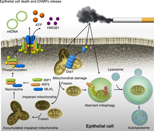
Recently, we reported that mitophagy regulates necroptosis, which contributes to the pathogenesis of COPD ().Citation41 Mitophagy selectively eliminates mitochondria by employing the autophagic machinery.Citation42 Parkin and PINK1 are key regulators of mitophagy.Citation42 CSE causes significant mitochondrial depolarization and induces mitophagy in lung epithelial cells.Citation41 We demonstrated that the mitochondrial division/mitophagy inhibitor Mdivi-1 protected against CS-induced cell death by reducing the phosphorylation of MLKL, a substrate for RIP3 in the necroptosis pathway.Citation41 Mice genetically deficient in PINK1 were protected against mitochondrial dysfunction, airspace enlargement, and mucociliary clearance disruption during CS exposure.Citation41 Our results suggest that CS-activated mitophagy may alter mitochondrial membrane integrity and induce mitophagy and necroptosis in pulmonary epithelial cells. Furthermore, recent studies have suggested that CS-induced mitophagy may regulate cellular senescence in COPD pathogenesis.Citation43 Genetic blocking of mitophagy resulted in enhanced CS-induced mitochondrial ROS production and cellular senescence in HBECs. The precise mechanism by which a cell “decides” to undergo either mitophagy-induced necroptosis or senescence remains unclear. One hypothesis is that mitophagy may lead to either senescence or necroptosis depending on the degree of cellular injury.Citation43
Figure 1 Mitophagy and necroptosis in COPD.
Abbreviations: CS, cigarette smoke; DAMP, damage-associated molecular pattern; MLKL, mixed-lineage kinase domain-like protein; PINK1, PTEN-induced putative kinase protein 1.
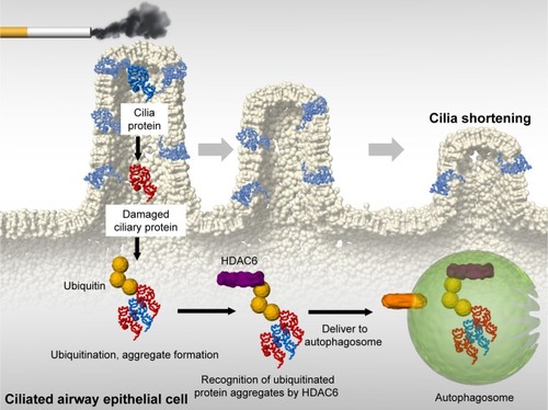
We also reported that ciliophagy, the consumption of cilia components by autophagy, regulates cilia length during CS exposure ().Citation19 We showed that autophagy-impaired (Becn1+/− or Map1lc3B−/−) mice, as well as tracheal epithelial cells isolated from these mice, display reduced CS-induced cilia shortening.Citation19 We identified cytosolic deacetylase histone deacetylase 6 (HDAC6) as a critical regulator of ciliophagy during CS exposure.Citation19 Importantly, analysis of human COPD specimens demonstrated epigenetic deregulation of HDAC6 by hypomethylation and increased protein expression in the airway.Citation19 These data suggest that ciliophagy, an HDAC6-dependent selective autophagy pathway, may represent a novel pathway critical to cilia homeostasis in response to CS exposure.
Figure 2 Ciliophagy in COPD.
Abbreviations: CS, cigarette smoke; HDAC6, histone deacetylase 6.
It remains unclear how cells utilize non-selective and selective autophagy pathways in response to CS exposure. We speculate that CS induces non-selective autophagy coincident with injury to organelles (eg, mitochondria, cilia), which would be degraded by selective autophagy. Thus, non-selective and selective autophagy may likely proceed simultaneously in a single cell. In addition, either process may be impacted by cell type and stimulus intensity. Thus, future studies are needed to elucidate the relationship between both types of autophagy in response to CS exposure.
Necroptosis: a critical regulator of cell death in COPD
It is becoming clear that autophagy and selective autophagy can regulate apoptosis and necroptosis in COPD.Citation34,Citation41 Chen et alCitation34 showed that dynamic interactions of the autophagy protein LC3B with Cav-1 and Fas regulate CS-induced lung epithelial cell apoptosis, leading to emphysematous airspace enlargement. Recently, we reported that CS-induced mitophagy may alter mitochondrial membrane integrity, leading to necroptosis induction ().Citation41 CSE-induced cell death in pulmonary epithelial cells was effectively reduced by the treatment with necrox-5, a necrosis inhibitor with antioxidant activity that localizes primarily in the mitochondria, or with necrostatin-1, a necroptosis inhibitor. CSE treatment resulted in increased phosphorylation of MLKL, a substrate for RIP3 in the necroptosis pathway, which was decreased in PINK1-knockdown cells.Citation41 Similarly, we observed that the mitochondrial division/ mitophagy inhibitor Mdivi-1 inhibited MLKL phosphorylation induced by CSE in pulmonary epithelial cells. These results suggest that mitophagy regulates CSE-induced necroptosis in pulmonary epithelial cells. Furthermore, we detected high levels of RIP3 near emphysematous regions in the lungs of mice exposed to CS for 3 months.Citation41 Importantly, we observed stronger PINK1 and RIP3 expression in the epithelial cells of patients with COPD than in that of control subjects. Confocal imaging confirmed higher and coincident expression of PINK1 and RIP3 in COPD compared to that in healthy lungs. These observations in human clinical samples strongly suggest that our experimental data associating mitophagy with necroptosis are relevant to COPD. Moreover, we have reported that sphingolipids regulate lung epithelial cell mitophagy and necroptosis during CS exposure.Citation44 Inhibition of ceramide-generating acid sphingomyelinase reduces both CS-induced PINK1 phosphorylation and necroptosis.Citation44 Our data provide a mechanistic explanation for how CS induces mitophagy-driven necroptosis and further support the fact that the dysregulated sphingolipid metabolism is implicated in the pathogenesis of structural cell injury in COPD.
COPD is also characterized by chronic inflammation of the airways, lung tissue, and pulmonary blood vessels as a result of exposure to CS. COPD patients show chronic neutrophilic inflammation in the airways, accompanied by aberrant tissue repair and remodeling.Citation45 While apoptosis has often been described as a major physiological process for emphysema, apoptosis is not generally accompanied by inflammation because of no or limited release of DAMPs. Therefore, it has been proposed that additional mechanisms underlie CS-induced apoptosis leading to airway inflammation in COPD. In contrast, there is a general consensus that necroptosis directly triggers inflammation through a massive release of DAMPs from the disintegrating cells.Citation7 Pouwels et alCitation46 revealed that CS-induced necroptosis and the release of DAMPs trigger neutrophilic airway inflammation in mice. Exposure to CS increased the levels of DAMPs and numbers of neutrophils in bronchoalveolar lavage fluid in mice, and this effect was statistically reduced on treatment with the necroptosis inhibitor necrostatin-1.Citation46 More recently, Wang et alCitation47,Citation48 reported a novel regulatory mechanism of necroptosis-mediated inflammation. Endoplasmic reticulum chaper-one GRP78 promoted a CSE-induced inflammatory response and mucus hyperproduction in airway epithelial cells, likely through the upregulation of necroptosis and subsequent activation of the nuclear factor-κB and activator protein-1 pathways.Citation48 In contrast, the mTOR suppresses the CS-induced inflammatory cytokines interleukin-6 and interleukin-8 through the nuclear factor-kB pathway, likely through the modulation of autophagy, apoptosis, and necroptosis.Citation47 These results suggest that necroptosis might be a promising therapeutic target for emphysema and inflammation.
Conclusion
Accumulating evidence demonstrates that autophagy, selective autophagy, and necroptosis exert previously unknown functions during COPD pathogenesis. While autophagic cell death is not currently considered a form of programmed cell death, autophagy proteins can regulate programmed cell death, including necroptosis and apoptosis, in a context-specific fashion. Furthermore, the autophagic pathway shares a number of signal molecules with programmed cell death pathways. In COPD, cell death has been mainly evaluated in terms of clinical phenotypes of emphysema (eg, loss of alveolar surface area); however, it is now predicted that cell death can regulate further biological processes, such as inflammation. In particular, necroptosis is a promising target for regulating COPD inflammation, as it directly triggers inflammation through a massive release of DAMPs. Therefore, careful consideration and further research are needed for designing strategies to manipulate these pathways as valid therapeutic interventions.
Author contributions
KM, SM, TS, and YG designed the strategy and goal of this review. KM drafted the article. All authors read and approved the final manuscript. All authors contributed to data analysis, drafting and revising the article, gave final approval of the version to be published, and agree to be accountable for all aspects of the work.
Acknowledgments
The authors are grateful to Augustine MK Choi for collaboration and suggesting the topic treated in this article.
Disclosure
The authors report no conflicts of interest in this work.
References
- KohansalRMartinez-CamblorPAgustíABuistASManninoDMSorianoJBThe natural history of chronic airflow obstruction revisited: an analysis of the Framingham offspring cohortAm J Respir Crit Care Med2009180131019342411
- ChoYSChallaSMoquinDPhosphorylation-driven assembly of the RIP1-RIP3 complex regulates programmed necrosis and virus-induced inflammationCell200913761112112319524513
- HeSWangLMiaoLReceptor interacting protein kinase-3 determines cellular necrotic response to TNF-alphaCell200913761100111119524512
- ZhangDWShaoJLinJRIP3, an energy metabolism regulator that switches TNF-induced cell death from apoptosis to necrosisScience2009325593833233619498109
- SunLWangHWangZMixed lineage kinase domain-like protein mediates necrosis signaling downstream of RIP3 kinaseCell20121481–221322722265413
- ZhaoJJitkaewSCaiZMixed lineage kinase domain-like is a key receptor interacting protein 3 downstream component of TNF-induced necrosisProc Natl Acad Sci U S A2012109145322532722421439
- PasparakisMVandenabeelePNecroptosis and its role in inflammationNature2015517753431132025592536
- KlionskyDJEmrSDAutophagy as a regulated pathway of cellular degradationScience200029054971717172111099404
- SchweichelJUMerkerHJThe morphology of various types of cell death in prenatal tissuesTeratology1973732532664807128
- MizushimaNKomatsuMAutophagy: renovation of cells and tissuesCell2011147472874122078875
- KroemerGLevineBAutophagic cell death: the story of a misnomerNat Rev Mol Cell Biol20089121004101018971948
- BonapaceLBornhauserBCSchmitzMInduction of autophagy-dependent necroptosis is required for childhood acute lymphoblastic leukemia cells to overcome glucocorticoid resistanceJ Clin Invest201012041310132320200450
- KhanMJRizwan AlamMWaldeck-WeiermairMInhibition of autophagy rescues palmitic acid-induced necroptosis of endothelial cellsJ Biol Chem201228725211102112022556413
- MizumuraKChoiAMRyterSWEmerging role of selective autophagy in human diseasesFront Pharmacol2014524425414669
- KirkinVMcewanDGNovakIDikicIA role for ubiquitin in selective autophagyMol Cell200934325926919450525
- YamamotoASimonsenAThe elimination of accumulated and aggregated proteins: a role for aggrephagy in neurodegenerationNeurobiol Dis2011431172820732422
- LemastersJJSelective mitochondrial autophagy, or mitophagy, as a targeted defense against oxidative stress, mitochondrial dysfunction, and agingRejuvenation Res2005813515798367
- LevineBEating oneself and uninvited guests: autophagy-related pathways in cellular defenseCell2005120215916215680321
- LamHCCloonanSMBhashyamARHistone deacetylase 6-mediated selective autophagy regulates COPD-associated cilia dysfunctionJ Clin Invest2013123125212523024200693
- TsukadaMOhsumiYIsolation and characterization of autophagy-defective mutants of Saccharomyces cerevisiaeFEBS Lett19933331–21691748224160
- KlionskyDJCreggJMDunnWAA unified nomenclature for yeast autophagy-related genesDev Cell20035453954514536056
- KabeyaYMizushimaNYamamotoAOshitani-OkamotoSOhsumiYYoshimoriTLC3, GABARAP and GATE16 localize to autophagosomal membrane depending on form-II formationJ Cell Sci2004117Pt 132805281215169837
- StolzAErnstADikicICargo recognition and trafficking in selective autophagyNat Cell Biol201416649550124875736
- GeggMECooperJMChauKYRojoMSchapiraAHTaanmanJWMitofusin 1 and mitofusin 2 are ubiquitinated in a PINK1/parkin-dependent manner upon induction of mitophagyHum Mol Genet201019244861487020871098
- GeislerSHolmströmKMSkujatDPINK1/Parkin-mediated mitophagy is dependent on VDAC1 and p62/SQSTM1Nat Cell Biol201012211913120098416
- WangXWinterDAshrafiGPINK1 and Parkin target Miro for phosphorylation and degradation to arrest mitochondrial motilityCell2011147489390622078885
- HildebrandJMTanzerMCLucetISActivation of the pseudokinase MLKL unleashes the four-helix bundle domain to induce membrane localization and necroptotic cell deathProc Natl Acad Sci U S A201411142150721507725288762
- CaiZJitkaewSZhaoJPlasma membrane translocation of trimerized MLKL protein is required for TNF-induced necroptosisNat Cell Biol2014161556524316671
- ChenXLiWRenJTranslocation of mixed lineage kinase domain-like protein to plasma membrane leads to necrotic cell deathCell Res201424110512124366341
- SuLQuadeBWangHSunLWangXRizoJA plug release mechanism for membrane permeation by MLKLStructure201422101489150025220470
- WangHSunLSuLMixed lineage kinase domain-like protein MLKL causes necrotic membrane disruption upon phosphorylation by RIP3Mol Cell201454113314624703947
- DondelingerYDeclercqWMontessuitSMLKL compromises plasma membrane integrity by binding to phosphatidylinositol phosphatesCell Rep20147497198124813885
- NingWLiCJKaminskiNComprehensive gene expression profiles reveal pathways related to the pathogenesis of chronic obstructive pulmonary diseaseProc Natl Acad Sci U S A200410141148951490015469929
- ChenZHLamHCJinYAutophagy protein microtubule-associated protein 1 light chain-3B (LC3B) activates extrinsic apoptosis during cigarette smoke-induced emphysemaProc Natl Acad Sci U S A201010744188801888520956295
- ChenZHKimHPSciurbaFCEgr-1 regulates autophagy in cigarette smoke-induced chronic obstructive pulmonary diseasePLoS One2008310e331618830406
- HaspelJShaikRSIfedigboECharacterization of macroau-tophagic flux in vivo using a leupeptin-based assayAutophagy20117662964221460622
- MonickMMPowersLSWaltersKIdentification of an autophagy defect in smokers’ alveolar macrophagesJ Immunol201018595425543520921532
- FujiiSHaraHArayaJInsufficient autophagy promotes bronchial epithelial cell senescence in chronic obstructive pulmonary diseaseOncoimmunology20121563064122934255
- YoshidaTMettIBhuniaAKRtp801, a suppressor of mTOR signaling, is an essential mediator of cigarette smoke-induced pulmonary injury and emphysemaNat Med201016776777320473305
- KimYCGuanKLmTOR: a pharmacologic target for autophagy regulationJ Clin Invest20151251253225654547
- MizumuraKCloonanSMNakahiraKMitophagy-dependent necroptosis contributes to the pathogenesis of COPDJ Clin Invest201412493987400325083992
- YouleRJNarendraDPMechanisms of mitophagyNat Rev Mol Cell Biol201112191421179058
- ItoSArayaJKuritaYPARK2-mediated mitophagy is involved in regulation of HBEC senescence in COPD pathogenesisAutophagy201511354755925714760
- MizumuraKJusticeMJSchweitzerKSSphingolipid regulation of lung epithelial cell mitophagy and necroptosis during cigarette smoke exposureFaseb J20183241880189029196503
- CurtisJLFreemanCMHoggJCThe immunopathogenesis of chronic obstructive pulmonary disease: insights from recent researchProc Am Thorac Soc20074751252117878463
- PouwelsSDZijlstraGJvan der ToornMCigarette smoke-induced necroptosis and DAMP release trigger neutrophilic airway inflammation in miceAm J Physiol Lung Cell Mol Physiol20163104L377L38626719146
- WangYLiuJZhouJSMTOR suppresses cigarette smoke-induced epithelial cell death and airway inflammation in chronic obstructive pulmonary diseaseJ Immunol201820082571258029507104
- WangYZhouJSXuXCEndoplasmic reticulum chaperone GRP78 mediates cigarette smoke-induced necroptosis and injury in bronchial epitheliumInt J Chron Obstruct Pulmon Dis20181357158129445274