
Abstract
Background
Zingiber spectabile, commonly known as Beehive Ginger, is used as an ethnobotanical plant in many countries as an appetizer or to treat stomachache, toothache, muscle sprain, and as a cure for swelling, sores and cuts. This is the first report of isolation of Streptomyces strain from the root of this plant. Strain Universiti Kebangsaan 25 (SUK 25) has a very high activity to produce secondary metabolites against methicillin-resistant Staphylococcus aureus (MRSA), which is associated with high morbidity and mortality rates due to acquired multidrug resistance genes and causes medication failure in some clinical cases worldwide. Phylogenetic analysis based on the 16S ribosomal RNA gene sequence exhibited that the most closely related strain was Streptomyces omiyaensis NBRC 13449T (99.0% similarity).
Aim
This study was conducted to carry out the extraction, identification, and biological evaluation of active metabolites isolated from SUK 25 against three MRSA strains, namely, MRSA ATCC 43300, MRSA ATCC 33591, and MRSA ATCC 49476.
Materials and methods
The production of secondary metabolites by this strain was optimized through Thronton’s media. Isolation, purification, and identification of the bioactive compounds were carried out using reversed-phase high-performance liquid chromatography, high-resolution mass spectrometry, Fourier transform infrared, and one-dimensional and two-dimensional nuclear magnetic resonance.
Results
During screening procedure, SUK 25 exhibited good antimicrobial potential against several strains of MRSA. The best biological activity was shown from fraction number VII and its subfractions F2 and F3 with minimum inhibitory concentration values at 16 µg/mL and 8 µg/mL, respectively. These two subfractions were identified as diketopiperazine cyclo-(tryptophanyl-prolyl) and chloramphenicol.
Conclusion
On the basis of obtained results, SUK 25 isolated from Z. spectabile can be regarded as a new valuable source to produce secondary metabolites against bacteria, especially MRSA.
Introduction
Natural products are naturally derived metabolites and/or by-products from plants, animals, or microorganisms.Citation1,Citation2 Natural products, in particular from microbes, are important sources of novel compounds for pharmaceuticals. Approximately 300,000 plant species growing in unexplored area on the earth are host to one or more endophytes.Citation3,Citation4 One of these plants is Zingiber spectabile (common name Beehive Ginger), a member of the ginger family, which is used as an ethnobotanical plant in many countries, especially in South-East Asia. Ginger is used as a remedy ingredient to treat a wide range of diseases including cancerCitation5 and arthritis,Citation6,Citation7 and it is also used as an antioxidant. It is used as an appetizer or to treat stomachache, toothache, muscle sprain, and as a cure for swelling, sores and cuts.Citation8
The Streptomyces species is a Gram-positive, aerobic, and spore-forming bacteria belonging to the order Actinomycetales that belongs to the phylum Actinobacteria.Citation9 It has a high DNA G-C content around 52%–70%.Citation10–Citation13 It is well known for its ability to produce secondary metabolites with diverse chemical structures and different activity against numerous pathogenic microorganisms. Several studies have reported that ~23,000 biologically active secondary metabolite compounds were produced by microorganisms, among which >10,000 of these compounds were produced by actinobacteria. This represents 45% of all bioactive microbial metabolites discovered. Streptomyces species alone produces around two-thirds of these compounds.Citation14,Citation15
Endophytic Streptomyces sp. Strain Universiti Kebangsaan (SUK 25) was isolated from the root of Z. spectabile plant.Citation16 It appeared to have a good inhibitory activity against methicillin-resistant Staphylococcus aureus (MRSA).Citation17 This bacterium is the major cause of hospital-associated infections (HAI-MRSA), and the resistance is the result of a supplemental penicillin-binding protein (PBP2a) encoded by the chromosomal mecA gene.Citation18,Citation19 The development of pathogenic bacteria resistance against antibiotic has become a serious problem worldwide. From this view, attempts have been made to extract, identify, and evaluate the active metabolites from SUK 25 against MRSA, and two compounds, namely, cyclo-(tryptophanyl-prolyl), which is a type of cyclic dipeptides diketopiperazines (DKPs), and chloramphenicol (CAP) have been isolated from this strain. Cyclic dipeptides DKPs or 2,5-DKPs, also known as dipeptide anhydrides (DPKs), are relatively simple compounds. Therefore, they are among the most common peptide derivatives found in nature.Citation20 DPKs have been known since the beginning of the 20th century. They are found endogenously in many organisms and in large amounts in some foods and beverages.Citation21 DPKs are extensively gained by extraction from natural sources such as plants, animals, and some microorganisms or by means of synthetic methods.Citation22 CAP (previously called chloromycetin) is an antibiotic belonging to the family of nitroaromatic compounds; it was reported as a broad-spectrum antibiotic produced by Streptomyces species.Citation23 The aim of this study was to screen the antagonistic activity of Streptomyces SUK 25 isolated from the root of Z. spectabile against multidrug-resistant pathogen, MRSA.
Materials and methods
Plant specimen and extract preparation
This study carried out the isolation, identification, and purification of active secondary metabolites from SUK 25, which were isolated from the root of the plant Z. spectabile. Root samples of Z. spectabile were collected in 2009 from Bukit Panchor Country Park, Penang Island, Northern Peninsular Malaysia (5.14°N latitude and 100.54°E). This plant was selected based on its ethnobotanical property. Specimen voucher number was given, and the plant sample was submitted to Universiti Kebangsaan Malaysia Herbarium collection. The pure colonies of endophytic actinomycetes obtained were subcultures onto International Streptomyces Project 2 (ISP2) agar and incubated at 28°C.Citation16 The production of secondary metabolites by SUK 25 was optimized through Thronton’s media with some modification.Citation17,Citation24
Chemicals and media
All the chemicals used for extraction, column chromatography (CC), high-performance liquid chromatography (HPLC), and liquid chromatography–mass spectrometry (LC–MS) were HPLC grade purchased from EMD Millipore, Billerica, MA, USA; thin-layer chromatography (TLC) was performed on percolated plates (20×20 cm) silica gel 60-F254 purchased from EMD Millipore; and Sephadex LH20 was purchased from GE Healthcare Bio-Sciences AB, Uppsala, Sweden. Mueller Hinton Broth (MHB) and Mueller Hinton Agar (MHA) were purchased from Difco, France.
In vitro antibacterial activity
Primary screening (cross-streak plate method)
The antibacterial activity was carried out by cross-streak plate method as described by Williston et alCitation25 and also by Alexander.Citation26 A single streak line of the SUK 25 was made on nutrient agar and then incubated at 28°C for 7 days. The cultures of MRSA ATCC 43300, ATCC 33591, and ATCC 49476 were standardized to 0.5 McFarland standards using sterile MHB, corresponding to the reading of 0.08–0.1 at A625 and then streaked at right angles to the original streak of SUK 25 and incubated at 37°C. The diameter of inhibition zone (IZ, mm) was measured after 24 hours. The control plate was also maintained without inoculating SUK 25.
Secondary screening (disk diffusion assay method)
The crude extract from SUK 25 was subjected to secondary screening using disk diffusion assay method, in accordance with the Clinical and Laboratory Standards Institute (CLSI) guidelines. MRSA ATCC 43300 was inoculated into 10 mL MHB and incubated at 37°C for 2–6 hours. Turbidity was standardized to 0.5 McFarland (CLSI, 2011). The crude extract (1 mg/mL) of SUK 25 was dissolved in 10% methanol, and 30 µg/disk was loaded on a blank disk (6 mm diameter; Whatman™, Gred AA; Sigma–Aldrich Co., St Louis, MO, USA) and then dried in the hood. After that, the disk was placed on the MHA already lawned with MRSA. This culture was incubated overnight at 37°C. After overnight culture, the IZ in millimeters was measured for each plate, and vancomycin (30 µg/disk) (Thermo Fisher Scientific, Waltham, MA, USA) was used as a positive control.
Fermentation, production, and extraction of secondary metabolites
For isolation and identification of bioactive metabolites, the Streptomyces sp. SUK 25 was cultured on ISP2 agar media, followed by incubation at 28°C for 14 days. Then a few blocks of ISP2 media containing pure and good growth of SUK 25 were then transferred into 250 mL of modified Thronton’s medium is composed of (g/L) K2HPO4 1.0, KNO3 0.5, MgSO4·2H20 0.2, CaCl2·H2O 0.1, NaCl 0.1, FeCl3 0.01, asparagine 0.5, and glucose 1.5 instead of mannitol at pH 7.4.Citation24 Then it was incubated at 28°C for 3 days with a gentle shake at 140 rpm using an orbital shaker. An aliquot of 60 mL of spore suspension of SUK 25 was aseptically transferred into 600 mL of Thronton’s broth as seeded media. After 12 days of incubation at 28°C, fermentation filtrates were extracted with three half-volumes of ethyl acetate (EtOAC), and the crude extracts were separated and dried using rotary evaporator at 40°C and 240 mbar.Citation27
Phylogenetic tree full of 16S rDNA gene sequencing DNA extraction
Genomic DNA was isolated according to Kieser et al,Citation28 and the purity and concentration were determined using NanoDrop spectrophotometer. Polymerase chain reaction (PCR) was performed according to Coombs and FrancoCitation29 using two sets of gene primer, 27f (5′-AGAGTTTGATCMTGGCTCAG-3′) with 765r (5′-CTGTTTGCTCCCCACGCTTTC-3′) and 704f (5′-CTGGCGGTGAAATGCGTAGA-3′) with 1492r (5′-AAGGAGGTGWTCCARCC-3′), and the PCR product was sent to 1st BASE for sequencing. Sequencing data were analyzed using software BioEdit Version 7 and compared to the GenBank databases (BLAST) and EzTaxon Biocloud. Phylogenetic tree was built using the software Molecular Evolutionary Genetics Analysis (MEGA4) using the neighbor-joining (NJ) method.Citation30
Chemotaxonomy analysis of ll-diaminopimelic acid cell wall of SUK 25
Chemotaxonomy features were analyzed through a type of ll-diaminopimelic acid (DAP) cell wall as described by Staneck and Roberts.Citation31 Fifteen milligrams of mycelia dry weight of SUK 25 grown in ISP2 for 14 days was added to 1 mL of 6N hydrochloric acid, and vortexed and autoclaved for 15 minutes at 121°C. After cooling, the hydrolysate was filtered using a filter paper (Whatman no 1) before being taken to dryness in a heating block at 95°C. Then, 1 mL of dH2O was added, and these procedures were repeated for two times. The filtrate was evaporated to dryness in an oven overnight. After evaporation, 1 µL of hydrolysate and 0.01 M DAP standard (Sigma-Aldrich Co.) in 0.2 M NaOH, which contained both the meso- and L isomers, was spotted on cellulose aluminum plate (EMD Millipore). Descending chromatography was performed in the solvent system containing methanol:dH2O:6N HCl:piridin (80:26:4:10), respectively, sprayed with ninhydrin (0.2% in acetone), and heated at 100°C for 3 minutes to reveal the spot.
Separation and purification of antimicrobial compounds by CC and TLC
Fractionations and purifications of the dry crude extract were carried out using reversed-phase open CC and TLC. A 700 mg dry crude extract was dissolved in 10 mL methanol. This mixture was then subjected to separation through size-exclusion CC containing 150 mg Sephadex LH20, using column size 2×40 cm. Gradient elution started with 100% chloroform, and the polarity of methanol was increased until 100% (100:0, 95:5, 90:10, 85:15, 80:20, 70:30, 60:40, 50:50, and 0:100 v:v). Further purification using silica gel column was carried out using column size 2×40 cm. After CC, the fractions were applied to TLC plate and developed using mixture of hexane, ethyl acetate, and methanol (4:4:2 v:v) as mobile phase. The separated compounds were visualized under short ultraviolet (UV) at λ254 nm (absorbance) and long wavelength UV at λ365 nm (fluorescence), and then sprayed with 10% H2SO4 in ethanol and heated at 80°C–100°C for 10 minutes. The compounds that had similar Rf in TLC were mixed together.
Bioautography on disk diffusion
Disk diffusion assay method for all fractions that were purified from open CC was carried out in order to test the activity of each fraction separately against MRSA ATCC 43300.
Determination of the minimum inhibitory concentration and minimum bactericidal concentration
The minimum inhibitory concentrations (MICs) of the compounds were determined according to the method described by the CLSI.Citation32,Citation33 Twofold serial dilutions of each fraction dissolved in 10% MeOH were made with MHB in a 96-well microtiter plate to give concentrations ranging from 0.12 µg/mL to 1,000 µg/mL. Fifty microliters of test bacterial suspension was inoculated in each well to give a final concentration of 1×10⁵ CFU/mL. Bacteria inoculum in MHB was used as a positive control, whereas the tested compounds in MHB were used as a negative control. Determination of minimum bactericidal concentrations (MBCs) was performed by inoculating 20 µL of each dilution with apparent no bacterial growth in MIC wells and streak on MHA plates. Then, the plates were incubated for 24 hours at 37°C. The presence of colonies was considered an evidence of bacteriostatic action, while the absence of colonies indicated bactericidal activity.
Purification of compounds using reversed-phase high-performance liquid chromatography
The crude extract of each fraction isolated from open CC was analyzed using an Agilent 1200 HPLC system equipped with a C-18 column (4.6×250 mm, 5 µm). The mobile phase consisted of H2O:Acetonitrile (ACN) with 0.1% trifluoroacetic acid (TFA) added to both solvents, and a gradient elution step was applied as shown in Table S1. The flow rate was 1 mL/min with an injection volume of 30 µL from 1 mg/mL, and the elution was monitored by the UV absorption at 210 nm, 240 nm, and 360 nm. The preparative HPLC protocol was carried out as same as analytical HPLC, only the quantity of injection and flow rate was injected to C-18 separation column (size 22×150 mm, 5 µm), where the flow rate was 10 mL/min with an injection volume of 100 µL each time from 7 mg/mL. Fractions were collected from a fraction collector.
Structure determination of antimicrobial compounds
FT-IR spectrophotometer, optical rotation, and melting point
Fourier transform infrared (FT-IR) spectrophotometer (Spectrum one FTIR spectrometer, PerkinElmer Inc., Waltham, MA, USA) using potassium bromide (KBr) disk method was carried out to determine the absorption bands of compounds. Optical rotation was measured on an Autopol VI automatic polarimeter (Rudolph Research analysis, USA). Melting point was measured on Stuart (Bibby Scientific, UK).
Nuclear magnetic resonance
The structure of compounds was determined using nuclear magnetic resonance (NMR) spectroscopy FT–NMR spectroscopy 600 MHz with Cryoprobe AVANCE III (Berker, USA). The probe temperature was maintained at 298 K, and a standard 5 mm NMR tube was used. Four milligrams of purified solid compound was dissolved in 200 µL MeOH-d4 (99.8 atom% D; Sigma-Aldrich Co.). Experiments that included one-dimensional (1D) 1H, 13C and two-dimensional (2D) NMR as sequences used in structural elucidation involving correlation spectroscopy (COSY), nuclear overhauser effect spectroscopy, and heteronuclear multiple-bond correlation spectroscopy (HMBC) were carried out to Berker standard pulse sequences. 1H and 13C chemical shifts (δ) are expressed in parts per minute (ppm) relative to solvent peak (MeOH-d4: 1H δ 5.18 ppm and 13C δ 47.7 ppm).
High-resolution mass liquid chromatography–mass spectrometry (HR-MS)
HR-MS was performed on an Agilent 6410 triple quadrupole mass spectrometer (Agilent Technologies, Santa Clara, CA, USA) equipped with an electrospray ionization interface negative mode coupled to an Agilent 1200 HPLC (Agilent Technologies). Column Agilent Zorbax Eclipse Plus (150×2.1 mm, 5 µm) HR-MS (Agilent Technologies) was used with column temperature 30°C, flow rate was 0.3 mL/min, injection volume was 20 µL, and mobile phase: pH 8.5 H2O (A) and acetonitrile (B). The retention time (RT) was measured in minutes. The flow rate was as shown in Table S1.
Statistical analysis
Statistical analysis of the data was performed using SPSS. One-way analysis of variance complemented by Tukey’s post hoc test was performed, and all data were reported as the mean ± standard deviation of the mean. All values were considered significant at P<0.05.
Results
The present work describes a comprehensive methodology for the screening, purification, and characterization of bioactive secondary metabolites isolated from endophytic Streptomyces SUK 25.
Screening for antibacterial activity
Primary screening
Primary screening of SUK 25 was carried out to determine the potential of this strain to production of antimicrobial secondary metabolites against MRSA. SUK 25 showed good activity against MRSA ATCC 43300, MRSA ATCC 33591, and MRSA ATCC 49476 as a primary screening. The distance between the edge of the tested bacterial growth and the SUK 25 bacterial growth was measured and recorded in millimeters. The IZ was 12 mm from the streaked right angle to the original streak of SUK 25 as shown in Figure S1.
Secondary screening by disk diffusion assays
SUK 25 was subjected to secondary screening using a sterile disk 6 mm in diameter; 30 µg/disk of either compounds isolated from SUK 25 or vancomycin was used as a positive control. The plates were incubated at 37°C overnight. The mean diameters of triplicate samples of IZ were 20±1.2 mm and 18±1.3 mm for crude extract and vancomycin, respectively. The effect of this fraction was larger than vancomycin as shown in Figure S2.
Bioautography
The results of bioautography using the disk diffusion method of purified fractions from CC against MRSA are listed in . Thirteen fractions were isolated. Only fraction numbers I, II, and VII using chloroform (CHCl3) as an eluting solvent revealed activity against MRSA ATCC 43300 with a diameter of IZ at 14 mm, 15 mm, and 22 mm, respectively. However, only fraction numbers IX and X showed activity when MeOH was used as an elution solvent; the diameter of IZ was at 13 mm and 14 mm, respectively, as shown in and Figure S3.
Table 1 Isolation and purification details of secondary metabolite compounds, diameter of inhibition zone, MIC values (µg/mL), and MBC values (µg/mL) of active fractions against MRSA ATCC 43300
Determination of the MIC and MBC
MIC is defined as the lowest concentration of an antimicrobial compound that inhibited the visible growth of a sensitive stain after the appropriate incubation period. MICs of all fractions are presented in . MICs of fraction numbers VII, F2, F3, and vancomycin against MRSA ATCC 43300 were 8 µg/mL, 16 µg/mL, 8 µg/mL and 2 µg/mL, respectively.
MBC was recorded as the lowest extract concentration, killing 99.9% of the bacterial inoculum after 24 hours incubation at 37°C. The MBCs of all active fractions were in the range of 32–128 µg/mL. MBCs of fraction numbers VII, F2, and F3, standard CAP, and vancomycin were 64 µg/mL, 32 µg/mL, 64 µg/mL, 64 µg/mL, and 4 µg/mL, respectively. The MICs and MBCs of all active fractions are shown in . From the bioautography, MICs, MBCs, and preparative HPLC results, fraction number VII (including subfractions F2 and F3) was selected for further analysis, where these subfractions exhibited strong anti-MRSA biological activity.
Phylogenetic tree of 16S rDNA sequences of SUK 25
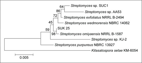
Genomic DNA was extracted, and the 16S ribosomal RNA (rRNA) gene sequence was amplified by PCR using universal bacterial 16S rRNA gene primers as described.Citation29 SUK 25 isolate sequences were compared with isolates in GenBank with Blast N software through the website www.ncbi.nlm.nih.gov. Sequences were aligned and edited using Bioedit software Version 7. Phylogenetic analysis was performed using MEGA 4.0 software to generate phylogenetic trees. The results showed that SUK 25 (1,450 nucleotides) is closely related to Streptomyces omiyaensis NBRC 13449T with 99% similarities ().
Figure 1 Neighbor-joining tree showing the relationship of strain SUK 25 based on a 16S rRNA gene sequences (1,450 nucleotides with closely related members of the genus Streptomyces omiyaensis NBRC 13449T and Kitasatospora setae KM-6054 as the outgroup).
Chemotaxonomy analysis of DAP cell wall of SUK 25
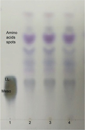
For TLC analysis, all the triplicate samples of SUK 25 strains were found to support the identification they obtained in the DAP isomer, chromatographically similar to the standard from Sigma-Aldrich Co. The DAP spots were seen as gray-green fading to yellow, with the L-DAP isomer moving ahead of the meso isomer with hydrolysates; amino acid spots appeared purple or red and migrated ahead of the DAP spotCitation31 as shown in .
Figure 2 TLC analysis of whole-cell hydrolysate of SUK 25.
Abbreviations: TLC, thin-layer chromatography; SUK 25, strain Universiti Kebangsaan 25; DAP, diaminopimelic acid.
Chemical characterization of isolated compounds
Analytical and preparative HPLC, melting point, optical rotations, and HR-MS
Different separation techniques such as TLC, CC, analytical and reversed-phase high-performance liquid chromatography (RP-HPLC), melting point, optical rotations, FT-IR, and HR-MS were carried out to obtain and identify the fraction FVII, which yielded two subfractions, namely, compound (F2) and compound (F3). HPLC purification peak is shown in Figures S3 and S4.
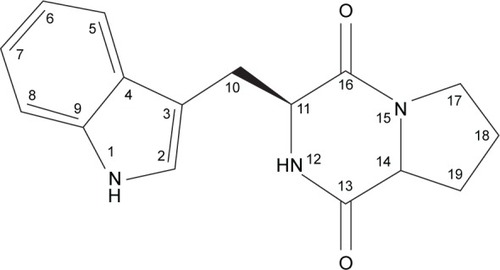
Compound F2: cyclo-(l-tryptophanyl-l-prolyl) (3S, 8aR)-3-(1H-indol-3ylmethyl) hexahydropyrrolo [1, 2-a] pyrazine-1, 4-dione
Analytical HPLC of this fraction showed RT at 11.64 minutes with a UV absorbing band at 254 nm, by using RP-HPLC method as shown in Figure S1. The F2 was isolated as a colorless solid, gave a dark red coloration with anisaldehyde/sulfuric acid, and became pink with Ehrlich’s reagent and blue with chlorine/o-anisidine as indication of a peptide with Rf =0.47 (CHCl3:MeOH; 95:5, v/v). The melting point of this fraction was between 165°C–168°C. The optical rotations of this compound [α]Citation25 C (0.001 g) was (−46.819°) using methanol as a solvent. The m/z value of this compound was 283.1321 [M+] at RT 6.63 minutes. The mass spectrum confirmed the structure based on the proposed fragmentation mechanism pathway. The molecular formula is C16H17N3O2. The 1H and 13C signals were assigned on the basis of chemical shifts, spin–spin coupling constants, splitting patterns, and signal intensities. The 1H NMR spectrum of F2 showed a broad 1H singlet at δ 8.20 characteristic for an NH of an indole moiety, five aromatic protons at δ 7.59, 7.40, 7.24, 7.14 (m, 2H) confirming the indole skeleton substituted at 3-position. In the aliphatic region, another broad 1H singlet of an acidic proton at δ 5.75 due to NH of an amide, two oxygenated or aminated methines at δ 4.39 (dd) and 4.08 (t), three different resonances each of 1H, 2H, and 1H, respectively, at δ 3.76 (dd), 3.64 (m), and 2.97 (dd) were seen. In addition, three multiplets of 4H between δ 2.36 and 1.90 were representative for two methylene groups. The 13C NMR spectrum exhibited four signals in the region between 22.48 and 45.33 ppm attributed to four CH2. The rest of the spectrum displayed 12 signals, among them seven were CH (123.4–110.0) and five were quaternary carbons (169.5–111.6), two of them belong to amide carbonyls (169.5 and 165.7) as shown in Figure S3. The prediction data from NMR suggested that this subfractionation is similar with the NMR data of cyclo-(l-tryptophanyl-l-prolyl), also known as brevianamide F.34 1H and 13C NMR data are shown in and . A search in AntiBase revealed that F2 was known as l,l-cyclo-(tryptophanyl-prolyl) or cyclo-(l-Trp-l-Pro), which was further confirmed by comparison with the previous literature by Kobayashi and PalumboCitation35 and Amar et al.Citation36
Table 2 1D-1H NMR and 13C NMR spectrum data of FVII-F2 cyclo-(l-tryptophanyl-l-prolyl)
Figure 3 The chemical structure elucidation of cyclo-(l-tryptophanyl-l-prolyl).
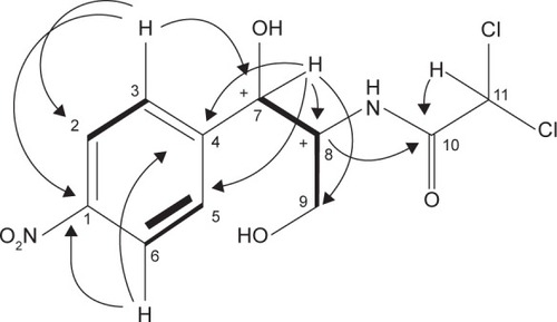
Compound F3: CAP 2,2-dichloro-N-[(1R, 2R)-2-hydroxy-1-(hydroxymethyl)-2-(4-nitrophenyl) ethyl]-acetamide
Analytical HPLC of this fraction showed RT at 14.3 minutes with UV absorbing band at 273 nm as shown in Figures S1 and S2. By using RP-HPLC, the F3 was isolated as a grayish-white or yellowish-white crystalline powder, gave a dark red coloration with anisaldehyde/sulfuric acid, and became pink with Ehrlich’s reagent and blue with chlorine/o-anisidine as indication of a peptide with Rf =0.75 (CHCl3:MeOH; 95:5, v/v). The melting point of this fraction was between 150°C and 152°C. The optical rotation of this compound [α]Citation25 C (0.001 g) was +31.493°, whereas the optical rotation of standard CAP [α]Citation25 C (1 g) was +17.496° using ethyl acetate. F3 and standard CAP were characterized by HR-MS spectra using negative ionization mode due to the better response in relation to the positive ionization mode. The m/z value of this compound was 322 [M−H]–at RT 11.9 minutes. The mass spectrum confirmed the structure based on the proposed fragmentation mechanism pathway. The molecular formula is C11H12Cl2N2O5 and was supported by 1H and 13C NMR data as shown in and Figures S4–S8. The 1H and 13C signals were assigned on the basis of chemical shifts, spin–spin coupling constants, splitting patterns, and signal intensities and by using 1H–1H COSY, 1H–13C heteronuclear single quantum coherence (HSQC), and 1H–13C HMBC experiments. The 1H and 13C chemical shifts of compound FVII-F3 are given in and . Chemical shifts were calibrated internally against the residual signal of the solvent in which the sample was dissolved in MeOH-d4 with δ H at 5.18 and C at 47.7. 1H NMR (600 MHz, MeOH-d4) of FVII-F3 showed 12 protons, at 8.3 (2H, m, aromatic-H), 7.67 (2H, m, aromatic-H), 6.3 (1H, s), 5.18 (1H, s), 4.16 (1H, s), and 3.62–3.8 (2H, dd, J=6.0 Hz; 12.0 Hz). The 13C and HSQC spectra show eleven carbon signal peaks at 165.2, 148.6, 126.9, 126, 150.2, 69.9, 57.10, and 60.80 (six aromatic carbons). The 2D 1H–1H and 1H–13C experiments, and especially the long-range 1H–13C couplings observed in the HSQC, COSY, and HMBC spectrum as shown in Figures S9–S11, permitted the connectivity between all the groups of the molecule to be established.
Table 3 1D-1H NMR and 13C NMR spectrum data of FVII-F3 chloramphenicol
Figure 4 The chemical structure elucidation of chloramphenicol using 2D-NMR HMBC and COSY correlations of FVII-F3 compound.
Abbreviations: 2D-NMR, two-dimensional nuclear magnetic resonance; HMBC, heteronuclear multiple-bond correlation; COSY, correlations spectroscopy.
Fourier transform infrared spectroscopy
The FT-IR spectrum for the two compounds exhibited absorption bands, which indicated the presence of NH, aromatic C–H stretching, carbonyl group, and C=O groups in the structure, respectively, at Vmax 3,345 cm−1 and 3,257 cm−1 (NH2 group and O–H stretching), 2,901 cm−1 (aromatic C–H stretching), 1,684 cm−1 (carbonyl group), 1,343 cm−1 (C–H bending), 652 cm−1 (carbon-chlorine bonds), 1,092 and 1,121 (C–N stretch), and 972 cm−1 (H–C–H – asymmetric stretch) as shown in Table S2 and Figures S12 and S13.
Discussion
The aim of this study was to screen the antagonistic activity of Streptomyces SUK 25 isolated from roots of Z. spectabile against drug-resistant pathogen, MRSA. The primary screening results indicated that SUK 25 has the ability to produce secondary metabolites in nutrient agar, which diffused through the agar that prevented and inhibited the growth of three MRSA strains, namely, MRSA ATCC 43300, MRSA ATCC 33591, and MRSA ATCC 49476. The IZ was ~12 mm from the edge of growth of SUK 25. A secondary screening using disk diffusion method had 20 mm IZ against MRSA ATCC 43300, which is similar with previous study conducted by Morakchi et al.Citation37 Cho et alCitation38 also reported the antibacterial activity of Streptomyces sp. CS392 against MRSA and vancomycin-resistant enterococci. Marine Streptomyces sp. VITBRK2 also has the ability to produce active secondary metabolites against MRSA and vancomycin-resistant enterococci strains.Citation39 Furthermore, strain Streptomyces MUSC 135T, which was isolated from a soil sample collected from a mangrove forest located on the east coast of Peninsular Malaysia, exhibited a broad-spectrum bacitracin against MRSA ATCC BAA-44.Citation40 In addition, Streptomyces sp. HUST012 was isolated from the stems of the medicinal plant Dracaena cochinchinensis Lour. This strain produced secondary metabolites, namely, SPE-B11.8 and SPE-B5.4, which showed antimicrobial activity against MRSA ATCC 25923 with MIC 62.5 and 0.04, respectively.Citation41
The results of the analysis translated through phylogenetic tree NJ method of 16S rRNA sequences of SUK 25. (1,450 nucleotides) was closely related to several strains of Streptomyces sp. from culture collections in the database NCBI GenBank. Analysis of 16S rRNA showed a similarity level of 99% within S. omiyaensis NBRC 13449T, which was isolated from Omiya city in Japan and showed a potential activity to produce CAP.Citation42 Chemotaxonomic analyses showed that the cell wall of strain SUK 25 contained DAP, indicating that it was of cell-wall type I.Citation43 Production of secondary metabolites by SUK 25 for large-scale fermentation was optimized through Thronton’s media at 12 days incubation with pH 7 and aerated at 140 rpm as reported by Junaidah et alCitation44 with some modification on incubation time from 7 days to 12 days according to the optimization of growth curve of SUK 25. From the results of bioautography, there were five active fractions against MRSA ATCC 43300. The most active fraction was the fraction number VII, which had a significant IZ of 22 mm by disk diffusion assay compared to only 18 mm of vancomycin at the same concentration of 30 µg/disk. Furthermore, two active compounds F2 and F3 were isolated from fraction number VII. Separation, purification, and isolation of fraction number VII and its subfractions in pure form were carried out using TLC, open CC, analytical and RP-HPLC, HR-MS, and FT-IR. Chemical structure elucidation was carried out by 1D and 2D NMR to confirm the structure of compounds. The first compound (F2) was isolated as cyclo-(l-tryptophanyl-l-prolyl), also known as brevianamide F, with m/z 283.1321 [M+] and molecular formula C16H17N3O2. It is a form of DKPs tryptophan–proline derivative. It has been described, isolated, and structurally characterized for the first time from Penicillium brevicompactum.Citation34 This compound has been reported by MahmoudCitation45 and Amar et al.Citation36 It has insecticidal propertiesCitation46 and has been reported to possess nematocidal activity.Citation47 Furthermore, it has anti-inflammatory properties,Citation48 and the antibacterial activity of this compound has been recorded against Escherichia coli, Pseudomonas aeruginosa, Klebsiella pneumoniae, S. aureus, Bacillus subtilis, and Streptococcus pneumonia.Citation49,Citation50 It also has antimicrobial effect against Micrococcus luteus LB 14110 and S. aureus ATCC 6538 with IZ 14 mm.Citation51 In this study, the IZ was 15 mm against MRSA ATCC 43300. It also found that this compound has antifouling potential activity and showed potent activity against larval settlement of Bugula neritina.Citation52
The second compound (F3) was identified as CAP with m/z 322 and molecular formula C11H12Cl2N2O5, which was reported by some of the previous studies by Pfenning et alCitation53 and Gantverg et al.Citation54 Previously, CAP was obtained from Streptomyces venezuelae by Ehrlich et al,Citation55 which was the first research that reported the isolation of the new CAP from a soil sample from Caracas, Venezuela. It was then isolated and identified by other researchers such as Smith et alCitation56 and Umezawa et al.Citation57 At the same time, in 1948, Okami reported ~14 strains of Streptomyces pheochromogenus var chloromyceticus that have the ability to produce this antibiotic. In addition, S. Omiyaensis, which was isolated from Omiya city, showed a potential activity to produce this antibiotic.Citation42 Furthermore, Streptosporangium viridogriseum var kofuense isolated from the soil has the ability to produce CAP.Citation58 The findings from this study were also similar to other previous studies by Mosher et alCitation59 and Aouiche et al,Citation60 where they isolated and identified CAP from Streptomyces lividans M252 and Saccharothrix sp. PAL54, respectively. This study compared the data from F3 with standard commercial CAP from Sigma-Aldrich Co. using FT-IR, HR-LC–MS, and NMR data and showed that all data were compatible for both of them. These findings confirmed that this fraction was similar to commercial CAP as shown in Figures S3–S6. This antibiotic has been designated as a broad-spectrum antibiotic effective against Gram-positive and Gram-negative bacteria.Citation61 Its mode of action was reported as a potent protein inhibitor, which is inhibiting bacterial growth by binding to their 50S ribosomal subunit.Citation62 However, several previous researchers found that this antibiotic has some side effects, such as aplastic anemia or reproductive/hepatotoxic effects, in human and animals.Citation63,Citation64 Therefore, the European Union has not authorized the use of this antibiotic in food-producing animals to protect public and animal health.Citation65 Despite its side effects, it is still been used in the treatment of serious infections, such as meningitis, brain abscesses, severe Haemophilus influenzae infections, and typhoid fever.Citation66 This is because it has excellent cellular penetration, and it is extensively distributed in the body.Citation67 In 1948, Woodward et al evaluated the effectiveness of CAP as a treatment for typhoid and paratyphoid fevers. All the patients in their trials responded well to this antibiotic, where the mortality rate of typhoid fever has been reduced from 20% to <1%, and the duration of fever decreased from 2–4 weeks to 4–5 days. It has provided the gold-standard against which other antibiotics have been compared at that time. The MICs of this antibiotic against Salmonella typhi were between 0.75 µg/mL and 5 µg/mL.Citation68
The MIC and MBC values of F2 were equal to 16 µg/mL and 32 µg/mL, which were compatible with the previous study by Kumar et al.Citation50 In addition, the MIC and MBC values of F3 were equal to 8 µg/mL and 64 µg/mL, respectively, which were compatible with the previous study by Fayyaz et al.Citation69 In relation to the ratio of MBCs to MICs of the compounds, it was <4 when F2 was used, which indicates that this compound has bactericidal effects that kill most of the bacteria,Citation70 whereas this ratio was >4 when F3 was used, which indicates bacteriostatic effects of this compound that only inhibit the growth of bacterial cells but do not kill completely.Citation71–Citation73
Conclusion
This study identified two natural bioactive compounds, cyclo-(l-tryptophanyl-l-prolyl) and CAP, from SUK 25 that were potent against MRSA. This indicates that this bacterium is a potential source for bioactive natural products in the pharmacological industry against MRSA.
Authors contributions
Muhanna M Alshaibani, Juriyati Jalil, and Noraziah M Zin designed and performed the whole experiment, analyzed the data, and contributed to manuscript preparation. Nik M Sidik and Ruangelie Edrada-Ebel assisted the experimental work and performed data acquisition. All authors contributed toward designed the experiment, data analysis, drafting and critically revising the paper and agree to be accountable for all aspects of the work.
Acknowledgments
The researchers are grateful to the Ministry of Higher Education (MOHE) for financial support through the research grant code FRGS/1/2011/ST/UKM/02/1. The authors would like to thank Professor Dr Jean Frederic Faizal Weber Abdullah, Atta-ur-Rahman Institute (UiTM), Centre for Research & Instrumentation (CRIM), Faculty of Dentistry (Universiti Kebangsaan Malaysia), and Siti Junaidah Binti Ahmad for analytical assistance.
Disclosure
The authors report no conflicts of interest in this work.
References
- JiHFLiXJZhangHYNatural products and drug discoveryEMBO Rep200910319420019229284
- PanLChaiH-BKinghornADDiscovery of new anticancer agents from higher plantsFront Biosci20124142
- TayungKSarkarMBaruahPEndophytic fungi occurring in Ipomoea carnea tissues and their antimicrobial potentialsBraz Arch Biol Technol2012555653660
- StrobelGDaisyBBioprospecting for microbial endophytes and their natural productsMicrobiol Mol Biol Rev200367449150214665674
- ShuklaYSinghMCancer preventive properties of ginger: a brief reviewFood Chem Toxicol200745568369017175086
- El-GhorabAHNaumanMAnjumFMHussainSNadeemMA comparative study on chemical composition and antioxidant activity of ginger (Zingiber officinale) and cumin (Cuminum cyminum)J Agric Food Chem201058148231823720590154
- KunduJKNaH-KSurhY-JGinger-derived phenolic substances with cancer preventive and therapeutic potentialForum Nutr20096118219219367122
- KhalidMHAkhtarMNMohamadASAntinociceptive effect of the essential oil of Zingiber zerumbet in mice: possible mechanismsJ Ethnopharmacol2011137134535121664960
- LechevalierHLechevalierMPIntroduction to the order ActinomycetalesStarrMPStolpHTrüperHGBalowsASchlegelHGThe Prokaryotes2GermanySpringer-Verlag Berlin198119151922
- McCormickJRFlärdhKSignals and regulators that govern Streptomyces developmentFEMS Microbiol Rev201236120623122092088
- WilliamsSGoodfellowMAldersonGWellingtonESneathPSackinMNumerical classification of Streptomyces and related generaJ Gen Microbiol19831296174318136631406
- GaoBGuptaRSPhylogenetic framework and molecular signatures for the main clades of the phylum ActinobacteriaMicrobiol Mol Biol Rev20127616611222390973
- TanakaYOmuraSAgroactive compounds of microbial originAnnu Rev Microbiol19934757878257109
- BerdyJBioactive microbial metabolitesJ Antibiot200558112615813176
- WeberTWelzelKPelzerSVenteAWohllebenWExploiting the genetic potential of polyketide producing streptomycetesJ Biotechnol2003106222123214651864
- ZinNLoiCSarminNRosliACultivation-dependent characterization of endophytic actinomycetesRes J Microbiol201058717724
- JunaidahASSuhainiSSidekHMBasriDFZinNMAnti-methicillin resistant Staphylococcus aureus activity and optimal culture condition of Streptomyces sp. SUK 25Jundishapur J Microbiol201585e1678426060562
- DavidMZDaumRSCommunity-associated methicillin-resistant Staphylococcus aureus: epidemiology and clinical consequences of an emerging epidemicClin Microbiol Rev201023361668720610826
- StapletonPDTaylorPWMethicillin resistance in Staphylococcus aureus: mechanisms and modulationSci Prog200285pt 15711969119
- PrasadCBioactive cyclic dipeptidesPeptides19951611511647716068
- ParkYCGunasekeraSPLopezJVMcCarthyPJWrightAEMetabolites from the marine-derived fungus Chromocleista sp. isolated from a deep-water sediment sample collected in the Gulf of MexicoJ Nat Prod200669458058416643030
- MartinsMBCarvalhoIDiketopiperazines: biological activity and synthesisTetrahedron2007634099239932
- AhmedZViningLEvidence for a chromosomal location of the genes coding for chloramphenicol production in Streptomyces venezuelaeJ Bacteriol198315412392446300034
- ThakurDBoraTBordoloiGMazumdarSInfluence of nutrition and culturing conditions for optimum growth and antimicrobial metabolite production by Streptomyces sp. 201J Med Mycol2009193161167
- WillistonEHZia-WalrathPYoumansGPPlate methods for testing antibiotic activity of actinomycetes against virulent human type tubercle bacilliJ Bacteriol194754556356816561393
- AlexanderMIntroduction to Soil MicrobiologyHoboken, NJJohn Wiley & Sons1977
- ZinNMSarminNIGhadinNBioactive endophytic streptomycetes from the Malay PeninsulaFEMS Microbiol Lett20072741838817608698
- KieserTBibbMJButtnerMJChaterKFHopwoodDAPractical Streptomyces GeneticsNorwichJohn Innes Foundation2000
- CoombsJTFrancoCMMIsolation and identification of actinobacteria from surface-sterilized wheat rootsAppl Environ Microbiol20036995603560812957950
- SaitouNNeiMThe neighbor-joining method: a new method for reconstructing phylogenetic treesMol Biol Evol1987444064253447015
- StaneckJLRobertsGDSimplified approach to identification of aerobic actinomycetes by thin-layer chromatographyAppl Microbiol19742822262314605116
- CLSI (Clinical and Laboratory Standards Institute)Methods for Dilution Antimicrobial Susceptibility Tests for Bacteria that Grow Aerobically. Approved Standard. M07-A88th edWayne, PACLSI2011
- MagiorakosAPSrinivasanACareyRMultidrug-resistant, extensively drug-resistant and pandrug-resistant bacteria: an international expert proposal for interim standard definitions for acquired resistanceClin Microbiol Infect201218326828121793988
- BirchAWrightJThe brevianamides: a new class of fungal alkaloidJ Chem Soc D196912644b645b
- KobayashiDPalumboJBacterial endophytes and their effects on plants and uses in agricultureBaconCWWhiteJFMicrobial EndophytesNew York, NYMarcel Dekker, Inc2000199233
- AmarMBElleuchLAbd-AllaHIThe new Streptomyces sp. TN605 strain secretes simultaneously three active compounds and a high level of the interesting pharmaceutical industry intermediate: 2-hydroxyphenylacetic acidBull Environ Pharmacol Life Sci201214856
- MorakchiHAyariATaokMKiraneDCochetNCharacterization of Streptomyces strain SLO-105 isolated from Lake Oubeira sediments in North-East of AlgeriaAfr J Biotechnol200982263326336
- ChoSSChoiYHSimkhadaJRManderPPark daJYooJCA newly isolated Streptomyces sp. CS392 producing three antimicrobial compoundsBioprocess Biosyst Eng2012351–224725421909674
- RajanBMKannabiranKExtraction and identification of antibacterial secondary metabolites from marine Streptomyces sp. VITBRK2Int J Mol Cell Med20143313025317399
- LeeL-HZainalNAzmanA-SStreptomyces pluripotens sp. nov., a bacteriocin-producing streptomycete that inhibits meticillin-resistant Staphylococcus aureusInt J Syst Evol Microbiol201464pt 93297330624994773
- KhieuT-NLiuM-JNimaichandSCharacterization and evaluation of antimicrobial and cytotoxic effects of Streptomyces sp. HUST012 isolated from medicinal plant Dracaena cochinchinensis LourFront Microbiol2015657426106377
- OkamiYStudies on the characters of antibiotic Streptomyces I. on the characters of a chloromycetin-producing strain (O-163)Jpn Med J194816499503
- LechevalierMPLechevalierHChemical composition as a criterion in the classification of aerobic actinomycetesInt J Syst Bacteriol1970204435443
- JunaidahASLianHHBasriDFZinNMMode of action of endophytic Streptomyces sp., SUK 25 extracts against MRSA; microscopic, biochemical and time-kill analysisInt J Pharm Sci Rev Res20143011117
- MahmoudMASBioactive secondary metabolites from marine and terrestrial bacteria: isoquinolinequinones, bacterial compounds with a novel pharmacophorPhD ThesisGöttingen University2005
- GloerJBThe chemistry of fungal antagonism and defenseCan J Botany199573S112651274
- ShiomiKOmuraSAntiparasitic agents produced by microorganismsProc Jpn Acad Ser B2004806245258
- RandTGGilesSFlemmingJMillerJDPunianiEInflammatory and cytotoxic responses in mouse lungs exposed to purified toxins from building isolated Penicillium brevicompactum Dierckx and P. chrysogenum ThomToxicol Sci200587121322215958659
- GrazMHuntAJamieHGrantGMilnePAntimicrobial activity of selected cyclic dipeptidesPharmazie1999541077277510563376
- KumarSNMohandasCNambisanBPurification, structural elucidation and bioactivity of tryptophan containing diketopiperazines, from Comamonas testosteroni associated with a rhabditid entomopathogenic nematode against major human-pathogenic bacteriaPeptides201453485824120705
- Ben Ameur MehdiRShaabanKARebaiIKSmaouiSBejarSMellouliLFive naturally bioactive molecules including two rhamnopyranoside derivatives isolated from the Streptomyces sp. strain TN58Nat Prod Res200923121095110719662574
- ZhangX-YXuX-YPengJAntifouling potentials of eight deep-sea-derived fungi from the South China SeaJ Ind Microbiol Biotechnol201441474174824532297
- PfenningATurnipseedSRoybalJConfirmation of chloramphenicol residues in crawfish by electrospray LC/MS. US FDA – Laboratory Information Bulletin No20024289188
- GantvergAShishaniIHoffmanMDetermination of chloramphenicol in animal tissues and urine: liquid chromatography-tandem mass spectrometry versus gas chromatography-mass spectrometryAnal Chim Acta20034831125135
- EhrlichJBartzQRSmithRMJoslynDABurkholderPRChloromycetin, a new antibiotic from a soil actinomyceteScience1947106275741741717737966
- SmithRMJoslynDAGruhzitOMMcLeanIWJrPennerMAEhrlichJChloromycetin: biological studiesJ Bacteriol1948553425
- UmezawaHTazakiTOkamiYFukuyamaSOn the new source of chloromycetin, Streptomyces omiyaensisJpn Med J194924207211
- TamuraATakedaINarutoSYoshimuraYChloramphenicol from Streptosporangium viridogriseum var. kofuenseJ Antibiot19712442702705572756
- MosherRHCampDJYangKBrownMPShawWVViningLCInactivation of chloramphenicol by o-phosphorylation a novel resistance mechanism in Streptomyces venezuelae isp5230, a chloramphenicol producerJ Biol Chem19952704527000270067592948
- AouicheASabaouNMeklatASaccharothrix sp. PAL54, a new chloramphenicol-producing strain isolated from a Saharan soilWorld J Microbiol Biotechnol201228394395122805815
- SethSTextbook of pharmacologyIndian J Pharmacol199830158
- McKeeEFergusonMBentleyAMarksTInhibition of mammalian mitochondrial protein synthesis by oxazolidinonesAntimicrob Agents Chemother20065062042204916723564
- KirkRWOsborneCMurtaughRKirk’s Current Veterinary Therapy XII. Small Animal PracticePhiladelphia, PAWB Saunders Company1995
- PlumbDVeterinary Drug Handbook4th edWhite Bear Lake, MNPharma Vet Publishing1991
- EFSAScientific opinion on chloramphenicol in food and feedEFSA J2014121139073145
- YaffeSJArandaJVNeonatal and Pediatric Pharmacology: Therapeutic Principles in PracticePhiladelphia, PALippincott Williams & Wilkins2010
- WhiteNJSalmonella typhi (typhoid fever) and S. paratyphi (paratyphoid fever)Antimicrob Ther Vaccine2002 Available from http://www.antimicrobe.org/b106.aspAccessed December 23, 2015
- WoodwardTESmadelJEManagement of typhoid fever and its complicationsAnn Intern Med196460114415714104849
- FayyazMMirzaIAAhmedZAbbasiSAHussainAAliSIn vitro susceptibility of chloramphenicol against methicillin-resistant Staphylococcus aureusJ Coll Physicians Surg Pak201323963764024034188
- FrenchGBactericidal agents in the treatment of MRSA infections – the potential role of daptomycinJ Antimicrob Chemother20065861107111717040922
- BerchePGaillardJLSimonetMBactériologie: Bactéries des Infections Humaines. [Nosocomial Infections Caused by Bacteria and Their Prevention in Bacteriology]ParisFlammarion Médecine-Sciences1988
- KonatéKMavoungouJFLepenguéANAntibacterial activity against β-lactamase producing methicillin and ampicillin-resistants Staphylococcus aureus: fractional inhibitory concentration index (FICI) determinationAnn Clin Microbiol Antimicrob20121111822716026
- ChaJOParkYKLeeYSChungGTIn vitro biofilm formation and bactericidal activities of methicillin-resistant Staphylococcus aureus clones prevalent in KoreaDiagn Microbiol Infect Dis201170111211821398072