
Abstract
The present study was aimed to investigate the effects of guggulsterone (GS) on proinflammatory responses as well as the underlying molecular mechanisms in macrophage upon lipopolysaccharide (LPS) stimulation. Effects of GS on viability of Raw264.7 cells were examined using 3-(4,5-dimethylthiazol-2-yl)-2,5-diphenyl tetrazolium bromide (MTT) assay. Real-time polymerase chain reaction (PCR) was employed to examine the mRNA expression of cytokines, including interleukin 1β (IL-1β), tumor necrosis factor-alpha (TNF-α), and inducible nitric oxide synthase (iNOS). Phosphorylations of extracellular signal-regulated kinase (ERK), c-Jun N-terminal kinase (JNK), p38 mitogen-activated protein kinases (p38), and inhibitor of nuclear factor kappaB (IκB) were determined using immunoblotting. The results revealed that GS was not toxic to Raw264.7 cells at designated concentrations. We demonstrated that GS significantly suppressed the elevated mRNA expression of proinflammatory cytokines, including IL-1β, TNF-α, and iNOS in a dose-dependent manner. GS treatment reduced the level of IκB phosphorylation in LPS-stimulated macrophages in a dose-dependent manner. Use of BAY 11-7082, an inhibitor of nuclear factor-kappaB (NF-κB), led to significantly suppressing effects on IL-1β and TNF-α expression similar as that of GS-treated cells. Our findings suggest that GS possesses anti-inflammatory activity, which may be attributed to downregulation of iNOS and inhibition of NF-κB activity in LPS-stimulated Raw264.7 cells.
Introduction
Inflammation is considered as a protective response against diverse pathogens or deteriorating stimuli. It is tightly regulated by an orchestra of cellular and soluble mediators. Inflammatory responses are initiated and propagated by cellular sensing systems such as toll-like receptor system (TLR) and production of inflammatory mediators such as inducible nitric oxide (NO), interleukin 1β (IL-1β), and tumor necrosis factor-alpha (TNF-α).Citation1 These soluble mediators play crucial role in controlling inflammation and tissue repair; however, aberrant production may exacerbate the damages.
Macrophages play a pivotal role in inflammatory process. Upon inflammation, these phagocytic cells are activated depending on stimuli and molecular pattern of recognition.Citation2 Activation of macrophage through pattern recognition receptor such as TLR leads to the production of a variety of mediators, including NO, TNF-α, and IL-1β.Citation3 Macrophage-derived NO is synthesized by inducible NO synthase (iNOS). Excessive production of NO contributes to the pathogenesis of chronic inflammatory disorders.Citation4,Citation5 Additionally, TNF-α and IL-1β are produced in activated macrophages, in turn, to facilitate and amplify cytokines and chemokine production in chronic inflammatory setting. Lipopolysaccharide (LPS), a component of Gram-negative bacteria cell wall, is known as the ligand of TLR4. Recognition of LPS by TLR4 in macrophages initiates downstream signaling pathways including nuclear factor-kappaB (NF-κB) complex and mitogen-activated protein kinases (MAPKs), such as p38 MAPKs (p38), c-Jun N-terminal kinase (JNK), and extracellular-signal regulated kinase (ERK).Citation6,Citation7 NF-κB is reported to play a critical role in acute and chronic inflammatory conditions. It is considered as a potential target for anti-inflammatory drug development.
Guggulsterone (GS) is a phytosterol that is found enriched in Commiphora mukul. It is reported as an antagonist of farnesoid X receptor and demonstrated hypolipidemic activity.Citation8 GS has been demonstrated to exert a range of pharmacological activities, including antineoplastic, antioxidation, antidiabetic, and cardioprotection.Citation9–Citation13 GS attenuates colitis in mice through inhibition of NF-κB activation.Citation14,Citation15 Researches have shown that GS inhibits proliferation of tumor cells through induction of apoptosis and inhibition of NF-κB signaling pathway.Citation16–Citation18 It is of interest to determine the effects of GS on LPS-induced inflammation in lymphocytes.
In this study, we investigated the anti-inflammatory effects and the underlying mechanism of GS, in particular gene expression of iNOS, IL-1β, and TNF-α in LPS-stimulated Raw264.7 cells. We also examined the role of NF-κB in LPS-induced inflammatory response in macrophages.
Materials and methods
Cell culture
Murine macrophage-like cell line (Raw264.7) was obtained from ATCC and incubated in complete Dulbecco’s Modified Eagle’s Medium (DMEM; Thermo Fisher Scientific, Waltham, MA, USA) containing 0.1% sodium bicarbonate, 2 mM glutamine, 100 U/mL penicillin G, streptomycin (100 μg/mL), and 10% fetal bovine serum (FBS) at 37°C. For GS treatments, Raw264.7 cells were seeded and incubated overnight prior to the treatments. Cells were treated with GS (0, 1, 5, 10, and 25 μM) for 24 hours (cell viability assay), 2 hours (real-time polymerase chain reaction [PCR] analysis), and 4 hours (immunoblotting), respectively, with or without a subsequent exposure to 1 μg/mL LPS. GS samples were prepared and added to the culture medium at a final concentration of 0.1% (v/v) in dimethyl sulfoxide (DMSO). DMSO with a final concentration of 0.1% was used as blank control.
Cell viability
Raw264.7 cells were seeded and incubated overnight prior to the treatments and then was followed by a treatment with GS (0, 1, 5, 10, and 25 μM) for 24 hours. Cell viability was determined using 3-(4,5-dimethylthiazol-2-yl)-2,5-diphenyl tetrazolium bromide (MTT) assay. In brief, 10 μL of MTT solution (5 mg/mL in complete DMEM) was added to the medium followed by an incubation time of 4 hours at 37°C. Following aspiration of the medium, cells were lyzed with 2-propanol which solubilized intracellular formazan. The optical density of formazan was measured using a microplate reader at a wavelength of 570 nm.
Real-time PCR
Raw264.7 cells were seeded at a concentration of 1×10⁶ cells/mL and incubated overnight prior to the treatments. Cells were treated with GS (0, 1, 5, 10, and 25 μM) for 2 hours followed by an exposure to 1 μg/mL LPS for 2 hours. Total RNA was isolated from each sample using RNA simple Total RNA Kit (Tiangen, BJ, People’s Republic of China) as per the instruction of manufacturer. The resulting RNA was used as a template for generating first-strand cDNA synthesis using ReverTra Ace Kit (Toyobo, Osaka, Japan). The sequences of primers used for reverse transcription PCR (RT-PCR) are shown in . RT-PCR experiments were carried out using real-time PCR Master Mix (Toyobo) in triplicates for each sample. The threshold cycle numbers were determined based on the ΔΔCT relative value and the cycle number was normalized to that of glyceraldehyde 3-phosphate dehydrogenase (GAPDH). The PCR products were examined and confirmed for the size using agarose gel electrophoresis.
Table 1 List of RT-PCR primers
Immunoblot assay
Raw264.7 cells were seeded at a concentration of 1×10⁶ cells/mL and incubated overnight prior to the treatments. Cells were treated with GS (0, 1, 5, 10, and 25 μM) for 4 hours followed by an exposure to 1 μg/mL LPS for 30 minutes. After treatments, cells were harvested and washed twice with ice-cold PBS, followed by lysis using RIPA buffer (Thermo Fischer Scientific, Waltham, MA, USA). The resulting lysates were subjected to centrifugation at 13,000 rpm for 10 minutes at 4°C. The supernatants were obtained and protein concentrations were measured using BCA protein assay kit (Pierce, Rockford, IL, USA). Thirty micrograms of protein was loaded in each lane and was subjected to 12.5% SDS-PAGE followed by a transfer onto a polyvinylidene difluoride (PVDF) membrane (EMD Millipore, Billerica, MA, USA). Resulting blots were incubated with 5% (w/v) skimmed milk in PBS followed by incubation with 1/1,000 dilution of antibodies against inhibitor of NF-κB (IκB), phosphorylated IκB (p-IκB), JNK, phosphorylated-JNK (p-JNK), p38, phosphorylated p38 (p-p38), ERK1/2, and phosphorylated ERK1/2 (p-ERK1/2) (Cell Signaling Technologies, Danvers, MA, USA) as well as GAPDH (Santa Cruz Biotechnology Inc., Dallas, TX, USA). The antigen–antibody complexes were unveiled using 1/2,000 dilution of peroxidase-conjugated secondary antibodies (Abcam, Cambridge, UK) and a chemiluminescence substrate (EMD Millipore).
Statistical analyses
All the data were presented as mean ± standard deviation (SD) of triplicate experiments. A one-way analysis of variance (ANOVA) with a Duncan multiple-comparison test was utilized to determine statistical differences among the groups. P-values <0.05 were considered statistically significant.
Results
Effects of GS on cell viability of murine macrophage Raw264.7 cells
To determine the cytotoxic effects of GS, Raw264.7 cells were exposed to GS at serial concentrations (0, 1, 5, 10, and 25 μM) and determined for the viability using MTT assay. We found that GS exerted nontoxic effects on Raw264.7 cells at designated concentrations as high as 25 μM (data not shown).
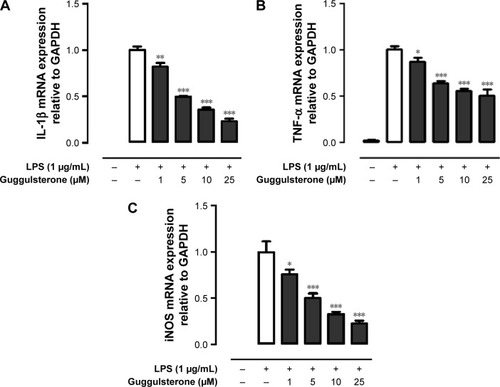
GS inhibited LPS-induced mRNA expression of IL-1β, TNF-α, and iNOS in Raw264.7 cells
We next investigated whether treatment with GS alters the expression of proinflammatory cytokines in LPS-stimulated macrophage. mRNA expressions of IL-1β, TNF-α, and iNOS in LPS-treated Raw264.7 cells were determined using real-time PCR. Results of real-time PCR analysis revealed that mRNA expressions of IL-1β, TNF-α, and iNOS in Raw264.7 cells were significantly enhanced in the presence of LPS, and the elevated mRNA expressions were suppressed in response to GS pretreatment in a dose-dependent manner ().
Figure 1 GS suppressed mRNA expression of proinflammatory cytokines IL-1β, TNF-α, and iNOS in LPS-stimulated Raw264.7 cells.
Abbreviations: GS, guggulsterone; IL-1β, interleukin 1β; TNF-α, tumor necrosis factor-alpha; iNOS, inducible nitric oxide synthase; LPS, lipopolysaccharide; PCR, polymerase chain reaction; GAPDH, glyceraldehyde 3-phosphate dehydrogenase; SD, standard deviation.
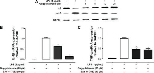
GS inhibited LPS-induced NF-κB pathway in Raw264.7 cells
It is evident that GS exerts its pharmacological effects through manipulating NF-κB pathway. Phosphorylation of IκB, NF-κB inhibitor, is concomitantly required with nuclear translocation of NF-κB. We hence investigated the effect of GS on LPS-induced phosphorylation of IκB in Raw264.7 macrophages. The results showed that Raw264.7 cells exhibited an increased phosphorylation of IκB in response to LPS stimulation (). The elevated IκB phosphorylation was inhibited by GS in a dose-dependent manner. Inhibition of NF-κB using BAY 11-7082 (10 μM), a commercially available inhibitor of NF-κB, led to a significant decrease in the LPS-induced IL-1β expression, whereas GS showed comparatively less suppression effects (). GS treatment significantly decreased the expression of TNF-α in LPS-stimulated macrophages, while BAY 11-7082 also exerted similar inhibitory activity ().
Figure 2 GS inhibited LPS-induced NF-κB pathway in Raw264.7 cells.
Abbreviations: GS, guggulsterone; NF-κB, nuclear factor-kappaB; LPS, lipopolysaccharide; GAPDH, glyceraldehyde 3-phosphate dehydrogenase; IL-1β, interleukin 1β; TNF-α, tumor necrosis factor-alpha; SD, standard deviation.
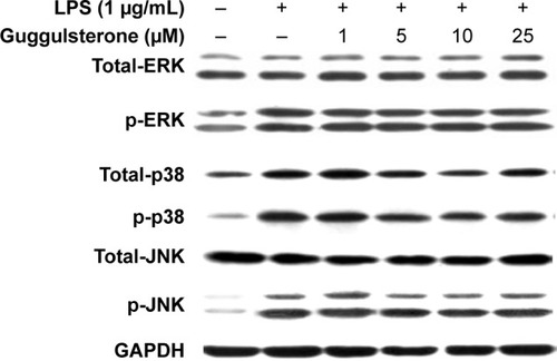
Effects of GS on phosphorylation of ERK1/2, JNK, and p38 in LPS-stimulated Raw264.7 cells
Activation of ERK1/2, JNK, and p38 is suggested to be involved in excessive production of proinflammatory cytokines in presence of LPS. We examined the effects of GS on phosphorylation of ERK1/2, JNK, and p38 in LPS-stimulated Raw264.7 cells. As shown in , exposure to LPS led to significantly increased phosphorylation of ERK1/2, JNK, and p38 in Raw264.7 cells. Treatment of LPS-stimulated Raw264.7 cells with GS at 1, 2.5, 5, 10, 12.5, and 25 μM concentrations resulted in no change in the levels of p-ERK1/2, p-JNK, and p-p38 in comparison to LPS alone.
Figure 3 Effect of GS on phosphorylation of ERK1/2, JNK, and p38 in LPS-stimulated Raw264.7 cells.
Abbreviations: GS, guggulsterone; ERK, extracellular signal-related kinase; JNK, c-Jun N-terminal kinase; p38, p38 mitogen-activated protein kinase; LPS, lipopolysaccharide; GAPDH, glyceraldehyde 3-phosphate dehydrogenase.
Discussion
In the present study, we demonstrated that GS significantly suppressed elevated production of proinflammatory mediators in LPS-stimulated Raw264.7 cells. The GS-induced downregulation of chemokine expression was associated with inhibition of NF-κB activation induced by LPS in macrophages. Moreover, GS had no effects on the LPS-induced phosphorylation of MAP kinase family members, including ERK1/2, JNK, and p38.
Macrophages are known to play a fundamentally critical role in the inflammatory response through producing a variety of mediators and proinflammatory cytokines depending on stimuli. A prolonged activation of macrophages results in a dysregulated inflammatory mediator production, leading to a vicious cycle of chronic inflammation.Citation19 LPS is recognized by TLR4 in association with MD2 and CD14 in macrophages. LPS-induced activation of TLR4 signaling is known to trigger the release and mRNA accumulation of critical proinflammatory cytokines, including IL-1β, TNF-α, and iNOS.Citation20–Citation22 We showed that the upregulated mRNA expression of IL-1β, TNF-α, and iNOS in LPS-stimulated Raw264.7 cells was abolished in the presence of GS. The finding is consistent with previous research in which GS suppresses the activation of transcription factor IRF3 induced by LPS.Citation23 The LPS-induced activation of TLR4 leads to both early and late activation of NF-κB through MyD88- and TRIF-dependent signaling pathways, respectively.Citation24,Citation25 Our data showed that GS was unable to fully exert its inhibitory effects on TNF-α expression in LPS-stimulated Raw264.7 cells. It is indicated that the inhibitory effect of GS in LPS-stimulated macrophages might be attributed to a later step in the NF-κB activation cascade.
NF-κB is a ubiquitous transcription factor, which has a central role in LPS-induced inflammatory responses. In resting macrophages, NF-κB is inactive and sequestered in the cytoplasm through binding with IκB. Interaction of LPS with TLR4 leads to the activation of NF-κB through phosphorylation and degradation of IκB, followed by nuclear translocation of NF-κB.Citation26 Phosphorylation of IκB is catalyzed by IκB kinase (IKK) complex. We found that LPS induced a significantly increased level of p-IκB in the Raw264.7 cells, which was restored by GS treatment. Corresponding to the changes in IκB phosphorylation, LPS-induced elevation of IL-1β and TNF-α mRNA expression was reduced in response to GS treatment. These data suggest that GS exerts anti-inflammatory activity through inhibition of NF-κB activation. Use of IκB inhibitor, BAY 11-7082, which blocks phosphorylation of IκB, resulted in a relatively low expression of IL-1β compared with that of GS-treated cells, whereas GS and BAY 11-7082 shared similar suppression effects on TNF-α expression. It is known that LPS induces biphasic activation of NF-κB.Citation26 LPS-induced early NF-κB activity initiates production of proinflammatory cytokines such as TNF-α and IL-1β, which in turn induce the late NF-κB activation.Citation26,Citation27 Our results suggest that GS suppressed LPS-induced inflammatory response through interfering with late NF-κB activation.
MAPK signaling pathway has been reported to be involved in the regulation of proinflammatory cytokine expression in activated macrophages. In the presence of LPS, signal transduction is initiated with the formation of LPS/TLR/MD2 complex, leading to activation of MAPK pathway cascade.Citation7,Citation28 Researches have shown that p38 MAPK pathway is associated with expression of TNF-α, IL-1β, IL-6, and IL-8.Citation29–Citation31 In our study, ERK1/2, JNK, and p38 were activated in LPS-stimulated Raw264.7 cells in parallel with elevated expression of proinflammatory cytokines. GS treatment showed no alteration in levels of p-ERK, p-JNK, or p-p38, but increased expression of cytokines was abolished. It is suggested that anti-inflammatory property of GS is not mediated by ERK, JNK, or p38 MAPK pathways in part.
Conclusion
In conclusion, we provide evidence highlighting the immunomodulatory activity of GS via the suppression of NF-κB activation but not ERK, JNK, or p38 MAPK pathways in LPS-treated Raw264.7 cells. Our results are expected to contribute to the understanding of mechanism of regulating chronic inflammation, such as sepsis, by using a natural plant component.
Acknowledgments
This study was supported by grants from the Xiamen Medical College, Young Foundation of Fujian Provincial Health Department (2010-2-68), Young Talent Foundation of Fujian Provincial Health Department (2013-ZQN-ZD-31), and Promotion Program for Young and Middle-aged Teacher in Science and Technology Research of Huaqiao University (ZQN-YX205).
Disclosure
The authors report no conflicts of interest in this work.
References
- ParkerLCPrinceLRSabroeITranslational mini-review series on Toll-like receptors: networks regulated by Toll-like receptors mediate innate and adaptive immunityClin Exp Immunol200714719920717223959
- MoonisMAhmadIBachhawatBKMacrophages in host defence – an overviewIndian J Biochem Biophys1992291151221328034
- SchwartzYSvistelnikAVFunctional phenotypes of macrophages and the M1-M2 polarization concept. Part I. Proinflammatory phenotypeBiochemistry (Mosc)20127724626022803942
- CastranovaVRole of nitric oxide in the progression of pneumoconiosisBiochemistry (Mosc)200469323714972015
- MacMickingJXieQWNathanCNitric oxide and macrophage functionAnnu Rev Immunol1997153233509143691
- AnHXuHYuYUp-regulation of TLR9 gene expression by LPS in mouse macrophages via activation of NF-kappaB, ERK and p38 MAPK signal pathwaysImmunol Lett20028116516911947920
- BodeJGEhltingCHaussingerDThe macrophage response towards LPS and its control through the p38(MAPK)-STAT3 axisCell Signal2012241185119422330073
- UrizarNLLivermanABDoddsDTA natural product that lowers cholesterol as an antagonist ligand for FXRScience20022961703170611988537
- SamudioIKonoplevaMSafeSGuggulsterones induce apoptosis and differentiation in acute myeloid leukemia: identification of isomer-specific antileukemic activities of the pregnadienedione structureMol Cancer Ther200541982199216373713
- WangXGreilbergerJLedinskiGThe hypolipidemic natural product Commiphora mukul and its component guggulsterone inhibit oxidative modification of LDLAtherosclerosis200417223924615019533
- WangWCUenYHChangMLProtective effect of guggulsterone against cardiomyocyte injury induced by doxorubicin in vitroBMC Complement Altern Med20121213822920231
- ChanderRRizviFKhannaAKCardioprotective activity of synthetic guggulsterone (E and Z-isomers) in isoproterenol induced myocardial ischemia in rats: a comparative studyIndian J Clin Biochem200318717923105395
- SharmaBSalunkeRSrivastavaSEffects of guggulsterone isolated from Commiphora mukul in high fat diet induced diabetic ratsFood Chem Toxicol2009472631263919635521
- KimJMKimSHKoSHThe guggulsterone derivative GG-52 inhibits NF-kappaB signaling in gastric epithelial cells and ameliorates ethanol-induced gastric mucosal lesions in miceAm J Physiol Gastrointest Liver Physiol2013304G193G20223125156
- MencarelliARengaBPalladinoGThe plant sterol guggulsterone attenuates inflammation and immune dysfunction in murine models of inflammatory bowel diseaseBiochem Pharmacol2009781214122319555671
- NohEMChungEYYounHJCis-guggulsterone inhibits the IKK/NF-kappaB pathway, whereas trans-guggulsterone inhibits MAPK/AP-1 in MCF7 breast cancer cells: guggulsterone regulates MMP9 expression in an isomer-specific mannerInt J Mol Med20133139339923242121
- YangMHLeeKTYangSGuggulsterone enhances antitumor activity of gemcitabine in gallbladder cancer cells through suppression of NF-kappaBJ Cancer Res Clin Oncol20121381743175122699931
- MachaMAMattaAChauhanSSGuggulsterone (GS) inhibits smokeless tobacco and nicotine-induced NF-kappaB and STAT3 pathways in head and neck cancer cellsCarcinogenesis20113236838021177768
- MorimotoYIzumiHKurodaESignificance of persistent inflammation in respiratory disorders induced by nanoparticlesJ Immunol Res2014201496287125097864
- RossolMHeineHMeuschULPS-induced cytokine production in human monocytes and macrophagesCrit Rev Immunol20113137944622142165
- FerwerdaBMcCallMBVerheijenKFunctional consequences of toll-like receptor 4 polymorphismsMol Med20081434635218231573
- GuhaMMackmanNLPS induction of gene expression in human monocytesCell Signal200113859411257452
- YounHSAhnSILeeBYGuggulsterone suppresses the activation of transcription factor IRF3 induced by TLR3 or TLR4 agonistsInt Immunopharmacol2009910811219000789
- KizakiTShiratoKSakuraiTBeta2-adrenergic receptor regulate Toll-like receptor 4-induced late-phase NF-kappaB activationMol Immunol2009461195120319167076
- KangYJKimSOShimadaSCell surface 4-1BBL mediates sequential signaling pathways ‘downstream’ of TLR and is required for sustained TNF production in macrophagesNat Immunol2007860160917496895
- HanSJKoHMChoiJHMolecular mechanisms for lipopolysaccharide-induced biphasic activation of nuclear factor-kappa B (NF-kappa B)J Biol Chem2002277447154472112235132
- HanYBrasierARMechanism for biphasic rel A. NF-kappaB1 nuclear translocation in tumor necrosis factor alpha-stimulated hepatocytesJ Biol Chem1997272982598329092517
- RaoKMMAP kinase activation in macrophagesJ Leukoc Biol20016931011200064
- YougbareIBoireGRoyMNCS 613 exhibits anti-inflammatory effects on PBMCs from lupus patients by inhibiting p38 MAPK and NF-kappaB signalling pathways while reducing proinflammatory cytokine productionCan J Physiol Pharmacol20139135336123656347
- ArmstrongJHarbronCLeaSSynergistic effects of p38 mitogen-activated protein kinase inhibition with a corticosteroid in alveolar macrophages from patients with chronic obstructive pulmonary diseaseJ Pharmacol Exp Ther201133873274021610141
- NeuderLEKeenerJMEckertRERole of p38 MAPK in LPS induced pro-inflammatory cytokine and chemokine gene expression in equine leukocytesVet Immunol Immunopathol200912919219919070370