
 ?Mathematical formulae have been encoded as MathML and are displayed in this HTML version using MathJax in order to improve their display. Uncheck the box to turn MathJax off. This feature requires Javascript. Click on a formula to zoom.
Abstract
The aim of this study was to determine the influence of different combinations of immunosuppressive drugs on the morphology, ultrastructure, and expression of proliferating cell nuclear antigen and cytoskeleton proteins in the rat dorsolateral prostate. The studies were conducted on 48 male Wistar rats. The animals were divided into eight groups: a control group and seven experimental groups. For 6 months, the animals in the experimental groups were administered a combination of drugs including rapamycin (Rapa), cyclosporin A, tacrolimus (Tac), mycophenolate mofetil, and prednisone (Pred), according to the standard three-drug regimens for immunosuppressive therapy used in clinical practice. An evaluation of the morphology and ultrastructure was conducted, and a quantitative evaluation of the expression of proliferating cell nuclear antigen and desmin- and cytokeratin-positive cells with weak, moderate, and strong expression was performed. The combination of Rapa, Tac, and Pred caused the smallest morphological and ultrastructural changes in the rat prostate cells. In the case of rats whose treatment was switched to Rapa monotherapy, a decreased percentage of proliferating cells of both the glandular epithelium and the stroma was found. Decreases in body weight and changes in the expression of cytokeratin and desmin were observed in all the experimental rats. The combination of Rapa, Tac, and Pred would seem to be the most beneficial for patients who do not suffer from prostate diseases. Our results justify the use of inhibitors of the mammalian target of Rapa in the treatment of patients with prostate cancer. The changes in the expression of cytoskeleton proteins may be the result of direct adverse effects of the immunosuppressive drugs, which are studied in this article, on the structure and organization of intermediate filament proteins.
Introduction
Immunosuppressive drugs are used to improve the survival of a patient after organ transplantation by minimizing the reaction of the rejection of vascularized transplanted organs.Citation1,Citation2 The acute or chronic rejection of transplanted organs is a very complex process determined by several immunological and nonimmunological factors.Citation3 Moreover, the choice of an appropriate immunosuppressive treatment protocol is of great importance.Citation4–Citation6 Consideration should be given to protocols with low levels of adverse effects, so that approaches would largely translate into the quality of life of patients.
Many substances with immunosuppressive properties can be used together in different combinations on account of their multidirectional effects.Citation2 For this reason, in clinical practice, immunosuppressive therapy in humans is based on multidrug regimens.Citation7,Citation8 However, the bulk of the published studies focus on the impact of individual drugs on organs and tissues, despite the fact that these drugs may affect each other’s metabolism.Citation1,Citation9–Citation11
Various systemic side effects are known to be caused by immunosuppressive drugs.Citation1,Citation2,Citation12 As a result of the use of such drugs, a considerable decrease in the number of rejected transplants is seen, but this is often proportional to the increased incidence of infections and malignancies. Moreover, studies have shown that renal transplant recipients are at risk of the early occurrence of prostate cancer.Citation2,Citation13,Citation14
Proliferation processes are an important factor in maintaining normal graft function. Analogously, the appropriate structure of the glandular epithelium and the stroma of the prostate, as well as its function, depend on these processes.Citation15 An important proliferation marker is proliferating cell nuclear antigen (PCNA). On the basis of expression studies of PCNA, this protein was determined to be a potential prognostic indicator that contributes to the development of predictive models for cancers and hyperplastic changes.Citation16–Citation19
Another factor that could be used to evaluate tissues in renal transplant recipients is the expression of intermediate filaments (IFs). Cytokeratin and desmin filaments play an important role in tissue regeneration and in proliferation processes.Citation20–Citation23 It has been shown that cytokeratins participate in the resistance of cells against apoptosis, which plays a key role during the response to inflammation.Citation24,Citation25 Research shows that desmin is a marker of late-stage differentiation of smooth muscles, and the changes in its expression are an important prognostic factor for recurrences of prostate cancer.Citation26 The expression patterns of IFs can serve as valuable phenotypic markers in both normal and malignant tissues. Moreover, the knowledge concerning the differentiated expression of IFs (especially cytokeratins) found a practical application in the pathomorphological diagnostics of the prostate.Citation26–Citation28 Expression disorders of cytoskeleton proteins caused by immunosuppressive drugs may contribute to the occurrence of pathological changes in transplanted organs and tissues. Studies addressing this problem may shed some light on the changes caused by these drugs on the cellular level.
One sign of good health among experimental animals is body weight gain.Citation29,Citation30 The measurement of body weight is a simple but very effective way to assess the impact of treatment on important aspects such as appetite. It is worth mentioning that determination of body mass is important in the experimental model, which is proposed in this article, because immunosuppressive drugs have a narrow therapeutic window. Therefore, applied doses are calculated per unit of body weight to avoid the toxic effects of individual drugs associated with high doses.
There are many studies on the effects of immunosuppressants on the male reproductive system, especially on the testes,Citation31–Citation34 but very few articles have dealt with the prostate.Citation35–Citation37 Due to the absence of such information in the literature and given the increased risk of complications in transplant recipients, we have undertaken this research to determine the influence of different combinations of immunosuppressive drugs used in clinical practice on the morphology, ultrastructure, and expression of PCNA and cytoskeleton proteins in the rat dorsolateral prostate. It is worth mentioning that the results presented in this article are the continuation of our earlier studies.Citation37
Materials and methods
Animals
The studies were conducted on 48 sexually mature 14-week-old male Wistar rats. The animals were obtained from a licensed breeding facility from the Institute of Occupational Medicine in Łódź, Poland. Prior to testing, all animals survived the 2-week adaptation period. Before starting the experiment, all animals were weighed, showing a mean weight of 305 g. The animals were housed in cages (six rats per cage) with a 12-hour light/12-hour dark cycle and divided into eight groups: a control group (I) and seven experimental groups (II–VIII). The rats in group I received a placebo. For the period of 6 months, the animals in the experimental groups were administered drugs in their pharmaceutical oral form (in bread pellets) including: rapamycin (Rapa), cyclosporin A (CsA), tacrolimus (Tac), mycophenolate mofetil (MMF), and prednisone (Pred), according to the standard three-drug regimens of immunosuppressive therapy used most frequently in clinical practice. The drug doses administered were based on the literature.Citation9,Citation10,Citation38,Citation39 The study scheme is presented in .
Table 1 Study scheme
Local ethical commission statement
The protocol for this study was in accordance with National Research Council guidelines for the care and use of laboratory animals, and approved by the Local Commission of Ethics for the Care and Use of Laboratory Animals in Szczecin, Poland (permit number 26/2011). The animals had health certificates issued by a veterinarian. Each step of the study protocol was designed in such a way as to minimize the pain and distress of animals.
Collection of material for research
A total of 46 rats completed the study (two rats in group III died in the fourth month of the experiment). After 6 months, the animals were anesthetized with ketamine hydrochloride (50 mg/kg) and their body weights were measured. During the section, the dorsolateral prostates were obtained. We employed rat dorsolateral prostates in this research as they are homologous to the human prostate.Citation40,Citation41
Morphological and ultrastructural studies
Dorsal lobes of the rat prostates of the control and experimental groups were routinely fixed in 4% buffered paraformaldehyde and embedded in paraffin. The paraffin blocks were sliced into 3 μm sections. Next, the sections were stained using standard methods: hematoxylin and eosin, as well as periodic acid Schiff.Citation42
To evaluate the ultrastructure in the transmission electron microscope, the lobes of the rat prostates were cut into pieces of volume of ~1 mm3 and fixed in 2.5% glutaraldehyde solution (Sigma-Aldrich Co., St Louis, MO, USA) in 0.1 M cacodylate buffer (Sigma-Aldrich Co.) at pH 7.4 for 2 hours at 4°C. The material was then fixed in a 1% solution of osmium tetroxide (OsO4; Sigma-Aldrich Co.). The tissue was dehydrated in a series of ethanol and acetone (100%) and gradually supersaturated with Spurr epoxy resin (Polysciences, Inc., Warrington, PA, USA). Subsequently, the material was immersed in capsules filled with epoxy resin. The blocks were cut into ultrathin scraps of 70–80 nm thick. The collected sections were counterstained sequentially with 9% uranyl acetate (Sigma-Aldrich Co.) and 2.66% lead citrate (Sigma-Aldrich Co.).
The histological preparations were examined under a light microscope (BX41; Olympus Corporation, Tokyo, Japan), while the ultrastructure was evaluated with a transmission electron microscope JEM-100CX (JEOL, Tokyo, Japan). The study was blinded, and one experienced pathologist and two histologists independently evaluated the ultrastructure and the histological preparations stained with hematoxylin and eosin.
Immunohistochemistry
For the immunohistochemical reactions, the following antibodies were used: monoclonal mouse antibody IgG against PCNA (clone PC10; Dako Denmark A/S, Glostrup, Denmark) diluted in the ratio of 1:200; monoclonal mouse antibody IgG against cytokeratin (clone MNF116; Dako Denmark A/S), which labels cytokeratins 5, 6, 8, 17, and probably 19, diluted in the ratio of 1:50; and monoclonal mouse antibody IgG against desmin (clone D33; Dako Denmark A/S) diluted in the ratio of 1:50.
The sections of the rat dorsolateral prostates were deparaffinized, hydrated, and boiled in target retrieval solution (Dako Denmark A/S) for 30 minutes at pH 6.0 (for desmin) and pH 9.0 (for PCNA and cytokeratin) to restore the antigens. The activity of endogenous peroxidase was blocked by using peroxidase blocking solution (Dako Denmark A/S) for 10 minutes. The sections were incubated at room temperature with mouse monoclonal primary antibodies in a humid chamber for 30 minutes. Primary antibodies were diluted in diluent (Dako Denmark A/S). Afterward, the sections were incubated with a complex containing secondary antibody conjugated with horseradish peroxidase (Dako REAL EnVision Detection System Peroxidase/diaminobenzidine+, rabbit/mouse; Dako Denmark A/S). After washing out the secondary antibody, diaminobenzidine (Dako Denmark A/S) was applied. As the final step, the slides were counterstained with Mayer’s hematoxylin, dehydrated, and coverslipped. Each step of the procedure was preceded by washing the slides with phosphate-buffered saline. All reactions were performed simultaneously under the same conditions. The slides were examined under a light microscope (BX 41).
Quantitative computer image analysis of immunohistochemistry
First, all the preparations were subjected to a scanning procedure (200× magnification, 0.25 μm resolution) with a ScanScope AT2 scanner (Leica Microsystems, Wetzlar, Germany). Next, the digital images of the slides thus obtained were analyzed on a computer screen using an ImageScope viewer (Version 11.2.0.780; Aperio Technologies, Inc., Vista, CA, USA). For the automatic computer analysis of the nuclear immunohistochemical PCNA reaction, nuclear v9 algorithm (Version 11.2.0.780; Aperio Technologies, Inc.) was used, whereas for cytoplasmic immunohistochemical reactions of cytokeratin and desmin, cytoplasmic v2 algorithm (Version 11.2.0.780; Aperio Technologies, Inc.) was employed. Other parameters were set in such a way as to achieve compliance with the visual evaluation of color intensity, taking into account the threshold for a positive result, namely a brown color of the nucleus or cytoplasm of the cells. Using each algorithm, the percentage of PCNA-positive cells was determined quantitatively and the number of desmin-and cytokeratin-positive cells with weak, moderate, and strong expression was calculated quantitatively. For cells with PCNA expression, the limit of the intensity threshold of the positive nuclei was 162–210 units; for the negative nuclei, this was <162 units. For the cells with cytokeratin and desmin expression, the limit of the intensity threshold was 181–210 units for positive weak (+) cells, 151–180 units for positive moderate (++) cells, 120–150 units for positive strong (+++) cells, and <120 units for negative cells. To obtain reliable and repeatable values, the total number of PCNA-, desmin-, and cytokeratin-positive cells was counted in 60 random fields (ten fields in six slides for each group) with an average area of 0.32 mm2 (for PCNA in the glandular part), 0.14 mm2 (for PCNA in the stroma), 0.24 mm2 (for cytokeratin), and 0.08 mm2 (for desmin).
Statistical analysis
Statistical analysis was conducted using the Statistica 8.0 program for Windows (StatSoft, Krakow, Poland). For quantitative variables of PCNA-positive cells as well as for desmin- and cytokeratin-positive cells with weak, moderate, and strong expression in each group, the minimum and maximum values, arithmetic mean, and standard deviation were calculated. To assess the significance of the differences between the values obtained for each grade of the expression in each group, the Kruskal–Wallis test was performed. The variance analysis of Kruskal–Wallis was used due to the large deviation from the normal distribution. To assess the significance of the differences between the values obtained for PCNA-positive cells in the stroma and the glandular part in each group, a Mann–Whitney U-test was carried out. Differences were considered statistically significant when P-values were <0.05.
Results
An evaluation of the morphology and ultrastructure and the quantitative assessment of PCNA-positive cells were carried out in the glandular epithelium and stroma of rat dorsolateral prostates. Furthermore, a quantitative evaluation of desmin- and cytokeratin-positive cells with weak, moderate, and strong expression was conducted. The prostates of the animals from the experimental groups (II–VIII) and from the control group (I) were compared. Next, the groups in which the conversion of the treatment to Rapa took place were compared with the groups in which the treatment schemes did not include Rapa during the study.
Body mass
Lower body masses were found in all the treated groups II–VIII (). The rats in the control group had the highest body mass, whereas the animals in group II (P<0.01), in which Rapa, Tac, and Pred were administered, had the lowest. Moreover, in group VI, in which the conversion of the treatment with CsA, MMF, and Pred on Rapa occurred, lower body mass was noted than in group V, in which CsA, MMF, and Pred were administered throughout the experiment. Moreover, in group VIII (in which the conversion of the treatment with Tac, MMF, and Pred on Rapa occurred), greater body mass was observed than in group VII, in which Tac, MMF, and Pred were administered throughout the experiment.
Table 2 Body mass of rats
Morphology and ultrastructure
In the glandular part of the rat dorsolateral prostate, the secretory and lead-out sections were lined with single-layer cubical or cylindrical epithelium. Periodic-acid-Schiff-positive eosinophilic secretion was observed in the lumen of the acini. In all the studied groups (II–VIII), changes were observed in the glandular epithelium, mainly in the apical and perinuclear part of the cell. Furthermore, two types of cells were observed – with electron light and dark cytoplasm. Microvilli were seen on the cell surfaces.
Group I: control
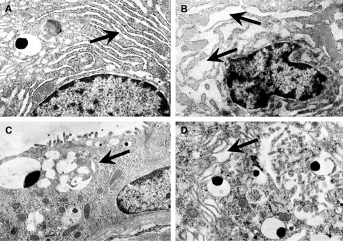
In the control group, acini in the rat dorsolateral prostate were lined with cubical or cylindrical epithelium (), in which no significant changes were observed. Proper cisterns of the rough endoplasmic reticulum in the perinuclear region () and proper Golgi cisterns located in the apical part of the cells were observed. In many cells, different types of acini with transported secretion were present, which took on different forms (condensed, fluffy, and star shaped). In addition, in the perinuclear part of the cells, normal narrow parallel cisterns of rough endoplasmic reticulum were observed.
Figure 1 Morphology of the rat dorsolateral prostate.
Abbreviation: H&E, hematoxylin and eosin.
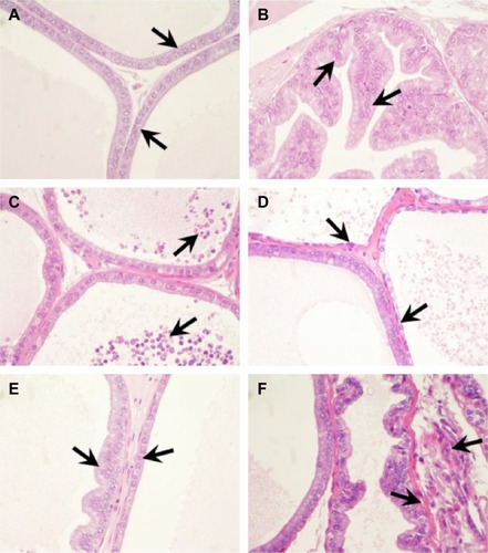
Figure 2 Ultrastructure of the rat dorsolateral prostate.
Group II: Rapa, Tac, and Pred
In this group, the smallest changes were observed in the glandular epithelium, compared to other treatment groups. The glandular epithelium was characterized by small infoldings into the lumen of the acini. During the analysis of the ultrastructure, the epithelium showed the features of atrophy in a small area.
Group III: Rapa, CsA, and Pred
In the acini, numerous infoldings of the glandular epithelium into the lumen of the acini were observed (). Moreover, in most of the cells, bloated cisterns were seen in the Golgi apparatus and in the rough endoplasmic reticulum ().
Group IV: Rapa, MMF, and Pred
Similar to group III, multiple infoldings of the glandular epithelium into the lumen of the acini were observed in the rat dorsolateral prostates of group IV. In the lumen of some acini, inflammatory cells were also seen (). Bloated dictyosomes of Golgi apparatus and rough endoplasmic reticulum (RER) cisterns were also noticed. Moreover, some cells were characterized by disorder in the organization of organelles and by shrunken nucleus.
Group V: CsA, MMF, and Pred
Highly atrophic epithelium was observed in many acini (). In several acini, hyperplasia of the glandular epithelium was also found. The presence of bloated Golgi apparatus cisterns in the apical part of the cells and of rough endoplasmic reticulum in the perinuclear region was noted. Condensed secretion was rarely observed in the secretory acini. On a large surface, the epithelium showed the features of atrophy with clear movement of the secretion apparatus ().
Group VI: CsA, MMF, and Pred/Rapa
Many infoldings of the glandular epithelium into the lumen of acini were found. The epithelium did not show any characteristics of atrophy. In addition, numerous inflammatory cells in the lumen of the acini were observed, along with a disorder in the normal structure of the glandular epithelium (). These changes were accompanied by the foci of the hyperplasia of epithelial cells. Very large bloated cisterns of Golgi apparatus and rough endoplasmic reticulum were also noted. In this group, more severe morphological changes were observed than in group V.
Group VII: Tac, MMF, and Pred
As in the case of group VI, numerous disorders of the normal structure of the epithelium, and focal hyperplasia of the glandular epithelium, were observed, though the changes were clearly smaller. Bloated RER was also seen in the perinuclear region of the cells. The Golgi apparatus was also characterized by bloated cisterns, and condensed secretion was present in the secretory acini.
Group VIII: Tac, MMF, and Pred/Rapa
Focal hyperplasia of the glandular epithelium and infoldings of the epithelium into the lumen of the acini were observed. Moreover, in comparison to group VII, the changes identified in this group were clearly greater. In some areas, the epithelium showed features of atrophy. In addition, in the stroma, edema was observed within the walls of the acini (). As in the case of group VI, very large bloated cisterns of the secretion apparatus were found ().
Immunohistochemistry
PCNA
In the rat dorsolateral prostate, PCNA-positive cells were characterized by brown-stained nuclei (), in both the glandular epithelium and the stroma. The percentage of PCNA-positive cells in the control and experimental groups, in the glandular and stromal part, is presented in and .
Figure 3 Immunolocalization and immunoexpression of PCNA (A, B), cytokeratin (C, D), and desmin (E, F) in the rat dorsolateral prostate.
Abbreviation: PCNA, proliferating cell nuclear antigen.
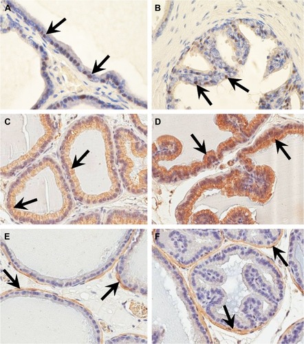
Figure 4 Percentage of PCNA-positive cells in epithelium and stroma in the rat dorsolateral prostate in control (I) and experimental groups (II–VIII).
Abbreviations: CsA, cyclosporin A; MMF, mycophenolate mofetil; Rapa, rapamycin; PCNA, proliferating cell nuclear antigen; Pred, prednisone; Tac, tacrolimus.
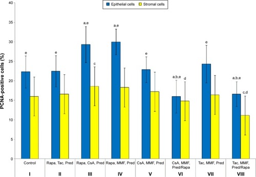
The percentage values obtained for PCNA-positive cells in the glandular epithelium of the control group differed statistically significantly from those of groups III, IV, VI, and VIII (P<0.001). The highest percentage of PCNA-positive cells in the glandular epithelium was noted in group IV, in which Rapa, MMF, and Pred were administered. The lowest percentage of PCNA-positive cells in the epithelium was observed in group VI, in which CsA, MMF, and Pred were administered in the first 3 months, but only Rapa in the next 3 months. The percentage values obtained for PCNA-positive cells in the stroma of the control group differed statistically significantly from those of groups III and VIII (P=0.034 and P<0.001, respectively). The highest percentage of PCNA-positive cells in the stroma was found in group III (P=0.034), in which Rapa, CsA, and Pred were administered. The lowest percentage of PCNA-positive cells in the stroma was noted in group VIII (P<0.001), in which Tac, MMF, and Pred were administered during the first 3 months and only Rapa in the next 3 months. The percentages of PCNA-positive cells in the glandular epithelium of all groups, apart from group VI, in comparison to the values for the stroma in particular groups, demonstrated statistically significant differences (P<0.001). The percentage values obtained in group VI (in which CsA, MMF, and Pred were administered during the first 3 months and Rapa alone in the remaining period) were significantly lower than in group V, in which throughout the experiment CsA, MMF, and Pred were administered. The situation was similar when groups VII and VIII were compared. In group VIII (in which Tac, MMF, and Pred were administered for the first 3 months and Rapa alone during the next 3 months), the percentage of PCNA-positive cells was significantly lower than in group VII, in which Tac, MMF, and Pred were administered throughout the experiment.
Cytokeratin
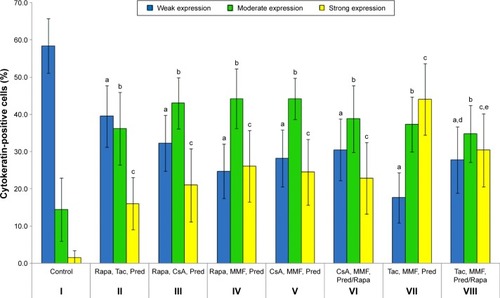
Cytokeratin-positive cells were found in the glandular epithelium and vascular endothelium of the stroma of the rat prostate. These cells were characterized by brown-stained cytoplasm (). The percentages of cytokeratin-positive cells with weak, moderate, and strong expression in individual groups (I–VIII) are presented in and . The percentage of cytokeratin-positive cells with weak, moderate, and strong expression in group I differed statistically from all the other groups: II, III, IV, V, VI, VII, and VIII (P=0.001 and P<0.001, respectively). The highest percentage of cytokeratin-positive cells with weak expression was found in the control group (I), as well as in group II (P=0.001 vs control), in which Rapa, Tac, and Pred were administered. The lowest percentage of cytokeratin-positive cells with weak expression was found in group VII (P<0.001 vs control), in which Tac, MMF, and Pred were administered. The highest percentage of cytokeratin-positive cells with moderate expression was found in group IV (P<0.001 vs control), in which Rapa, MMF, and Pred were administered, and in group V (P<0.001 vs control), in which CsA, MMF, and Pred were administered. The lowest percentage of cytokeratin-positive cells with moderate expression was found in the control group, as well as in group VIII (P<0.001 vs control), in which Tac, MMF, and Pred were administered in the first 3 months and Rapa alone in the following 3 months. The highest percentage of cytokeratin-positive cells with strong expression was found in group VII (P<0.001 vs control), in which Tac, MMF, and Pred were administered. The lowest percentage of cytokeratin-positive cells with strong expression was found in the control group, as well as in group II (P<0.001 vs control), in which Rapa, Tac, and Pred were administered. In group VI (in which CsA, MMF, and Pred were administered in the first 3 months and Rapa along during the remaining period), the percentage of the cells showing moderate and strong expression of cytokeratin was lower (statistically insignificant) than in group V (in which CsA, MMF, and Pred were administered during the whole duration of the experiment). The comparison of groups VII and VIII gave similar outcomes.
Figure 5 Percentage of cytokeratin-positive cells in epithelium in the rat dorsolateral prostate in control (I) and experimental groups (II–VIII).
Abbreviations: CsA, cyclosporin A; MMF, mycophenolate mofetil; Rapa, rapamycin; Pred, prednisone; Tac, tacrolimus.
Desmin
Desmin-positive cells were localized in the stroma. The expression of desmin was observed in the smooth myocytes around the acini and in the smooth myocytes of the vascular walls; they were characterized by brown-stained cytoplasm (). The percentage of desmin-positive cells with weak, moderate, and strong expression in each of the groups (I–VIII) is presented in and .
Figure 6 Percentage of desmin-positive cells in epithelium in the rat dorsolateral prostate in control (I) and experimental groups (II–VIII).
Abbreviations: CsA, cyclosporin A; MMF, mycophenolate mofetil; Rapa, rapamycin; Pred, prednisone; Tac, tacrolimus.
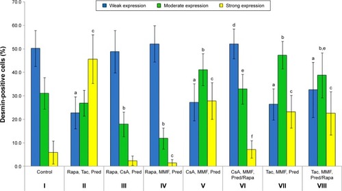
The percentage of desmin-positive cells with weak expression of group I differed statistically significantly from that of groups II, V, VII, and VIII (P<0.001). The highest percentage of desmin-positive cells with weak expression was found in group IV (statistically insignificant vs control), in which Rapa, MMF, and Pred were administered, and in group VI (statistically insignificant vs control), in which CsA, MMF, and Pred were administered in the first 3 months and Rapa alone in the following 3 months. The lowest percentage of desmin-positive cells with weak expression was found in group II (P<0.001 vs control), in which Rapa, Tac, and Pred were administered. The percentage of desmin-positive cells with moderate expression for group I differed statistically significantly from that for groups III, IV, V, VII, and VIII (P<0.001 and P=0.025, respectively). The highest percentage of desmin-positive cells with moderate expression was found in group VII (P<0.001 vs control), in which Tac, MMF, and Pred were administered. The lowest percentage of desmin-positive cells with moderate expression was found in group IV (P<0.001 vs control), in which Rapa, MMF, and Pred were administered. The percentage of desmin-positive cells with strong expression for group I differed statistically significantly from that for groups II, IV, V, VII, and VIII (P<0.001 and P=0.002, respectively). The highest percentage of desmin-positive cells with strong expression was found in group II (P<0.001 vs control), in which Rapa, Tac, and Pred were administered. The lowest percentage of desmin-positive cells with strong expression was found in group IV (P=0.002 vs control), in which Rapa, MMF, and Pred were administered. In group VI (in which CsA, MMF, and Pred were administered in the first 3 months and Rapa alone during the remaining period) the percentage of the cells showing moderate and strong expression of desmin was significantly lower than in group V (in which CsA, MMF, and Pred were administered throughout the experiment). Comparison of groups VII and VIII gave similar results. In group VIII (in which Tac, MMF, and Pred were administered in the first 3 months and Rapa alone during the next 3 months), the percentage of the cells showing moderate and strong desmin expression was significantly lower in comparison to group VII (in which Tac, MMF, and Pred were administered during the whole duration of the experiment).
Discussion
In the available literature, there are few publications on the effects of different combinations of immunosuppressive drugs on the prostate. Only our earlier studies relate to the effects of various combinations of immunosuppressants on the morphology and the expression of PCNA and cytokeratin in the prostate.Citation37,Citation43 So far, no research has been found concerning the impact of the standard three-drug combinations of immunosuppressive drugs on the ultrastructure and the expression of desmin in the prostate. Moreover, a quantitative evaluation, with the division of the prostate into glandular and stromal parts, was performed. Our study can thus be regarded as innovative.
In the experiment conducted, it was attempted to develop a model of the action of the various immunosuppressive drugs that might be comparable to long-term immunosuppressive therapy in humans following organ transplantation. The experiment was conducted for 6 months owing to the fact that the rat’s average life span varies from 2 years to 3 years, which refects ~12–15 years of a human life.Citation44,Citation45 As a result, the presented findings relate to the long-term effects of the particular combinations of immunosuppressive drugs. To compare, in many studies on the effects of immunosuppressive drugs on various organs, the time of the drug administration was relatively short.Citation10,Citation32,Citation35,Citation46 The fact that a physiological enteral route of the drug administration was chosen, rather than an intraperitoneal or intravenous route, as in some research on animal models, speaks to the advantage of the presented experimental model.Citation47–Citation49
In this experiment, a loss of body weight was observed in all the experimental rats. The highest (statistically significant) decrease in body mass was found in the animals in group II, in which Rapa, Tac, and Pred were administered. Other researchers have also observed a weight loss in both animals and humans undergoing immunosuppressive therapy.Citation31,Citation35,Citation47,Citation50–Citation52 The effect of particular immunosuppressive drugs on the body mass was also examined. Rovira et alCitation52 observed that rats receiving CsA had higher body weight than animals on the CsA-free treatment scheme. In our experimental model, the positive effect exerted by CsA in CsA-based schemes on the weight of the rats was confirmed, while a considerable body weight loss was also found in rats treated with Rapa. Moreover, a decrease in the body weight was also found in the case of the conversion treatment from CsA-based regimens to Rapa. Other authors have also confirmed these observations.Citation50 The weight loss caused by the administration of various combinations of immunosuppressive drugs may be related to their influence on cell growth processes and metabolism.
Both morphology and ultrastructure studies have shown that, in rat dorsolateral prostates in the group in which CsA, MMF, and Pred were administered, the epithelium was markedly atrophic. In our earlier studies conducted on the rat ventral prostate, a highly atrophied epithelium was observed in the group in which Rapa, CsA, and Pred were administered.Citation37 This can be explained by the fact that the ventral lobe differs structurally and functionally from the dorsolateral unit.Citation41 Unfortunately, other researchers have focused on identifying the impact of individual immunosuppressive drugs on the prostate.Citation35,Citation36 In our study, in all the groups, in which the atrophic epithelium was observed, calcineurin inhibitors (CsA or Tac) were administered. Some authors have observed that CsA causes a significant reduction in the absolute volume of all prostate components, including the epithelium in the ventral lobe of the rat prostate.Citation35,Citation36 These authors observed that, as a result of the administration of CsA, the glandular epithelial cells were clearly lower. Moreover, the ultrastructural analysis confirmed the epithelial atrophy. The morphological changes observed in groups with the conversion of the treatment to Rapa were more severe than in groups in which animals did not receive Rapa. Literature data confirm that, in rats, the conversion of the treatment from CsA to Rapa ultimately leads to larger structural damages and testicle dysfunctions than does the continuation of treatment with CsA.Citation50
In the glandular epithelium and stroma of the rat dorsolateral prostate, the lowest percentage of PCNA-positive cells was observed in groups with the conversion of the treatment to monotherapy with Rapa. Interestingly, these values were lower than in groups in which animals did not receive Rapa at all during the experiment. In our earlier studies on rat ventral prostates, the results had a similar distribution.Citation37 Our results show that Rapa has antiproliferative activity. Other researchers have confirmed that Rapa exerts antiproliferative effects.Citation38,Citation46 In groups III and IV, in which rats received Rapa in combination with other drugs throughout the experiment, there were no fewer PCNA-positive cells than in the groups in which animals did not receive Rapa. The results were analogous in our earlier studies. In this experimental model, Rapa in combination with other drugs had less influence on the proliferation of cells in the dorsolateral lobe of the rat prostate. Unfortunately, we could not find any studies on the impact of immunosuppressive drugs on the expression of PCNA in the prostate. However, studies carried out on other models confirm the results obtained. Zhuang et alCitation53 showed that the prolonged supply of Rapa inhibits the proliferation of PC-3 cells of prostate cancer. Other researchersCitation54 have confirmed the antiproliferative properties of Rapa, observing that Rapa inhibits the proliferation of human keratinocyte stem cells by blocking the cell progression cycle in the G1 phase.
Cytokeratins constitute the most durable part of the cell cytoskeleton; for that reason, they are primarily responsible for protecting epithelial cells from mechanical tensions. They also play an important role in the regulation of cell growth.Citation20,Citation21 The expression of individual cytokeratins depends on the type of epithelium, the period of embryonic development, the level of cell differentiation, and the condition of the organism.Citation55 Disturbances in the expression of cytokeratin may have a negative impact on processes in which regulation IFs are involved. In the dorsolateral lobe, in all the treatment groups (II–VIII), more cells exhibiting moderate and strong expression of cytokeratin were observed than in the control group. Unfortunately, we could not find any publications on the impact of even single immunosuppressant drugs on the organization of cytokeratin filaments in the prostate. The available literature indicates the negative impact of only certain immunosuppressive drugs on cytokeratins. Such studies have shown that CsA has a selectively toxic effect on cytokeratin filaments through a plurality of modifications in their structure. Research conducted on cell cultures subjected to 24 hours of exposure to CsA has shown that the fibers of the cytokeratin filaments are characterized by thickening and weakening adhesion to the cell membrane.Citation56 Klawitter et alCitation57 found that immunosuppressive drugs may increase the tissue concentration of selected cytoskeletal proteins. Other researchers confirm that CsA affects the organization of the cellular cytoskeleton.Citation58 These reports may explain the increase in the expression of cytokeratin in the groups in which the treatment schemes contained CsA (III, V, VI). The results also show that Rapa causes a decrease in the expression of cytokeratin, in comparison to groups without the conversion to Rapa.
Research has shown that cytokeratin filaments are involved in the process of resistance against apoptosis.Citation24,Citation25 The resistance of epithelial cells to apoptosis is an important factor in the response to inflammation and is closely associated with the presence of cytokeratin filaments. As a result, it may be stated that mechanisms acting against inflammation could contribute to an increase in the expression of cytokeratins.
The smooth muscle cells (SMCs) of the prostate synthesize the regulatory and structural components of the extracellular matrix to create a microenvironment that regulates the growth and function of other cell types.Citation59,Citation60 Desmin is an integral part of the SMC and is necessary for maintaining both structural integrity and muscle function.Citation22,Citation23 It is also believed that desmin may be involved in the regulation processes of gene expression.Citation61 Disorders in the expression of desmin may have a negative impact on processes in which this protein is involved.
The percentage of cells showing moderate and strong expression of desmin in groups with the conversion of treatment to monotherapy with Rapa was lower than in groups in which animals did not receive Rapa at all. Unfortunately, we could not find any publications on the impact of even single immunosuppressant drugs on the desmin in the prostate. Experiments on other experimental models can merely indirectly explain the decrease in the expression of desmin through the inhibition of muscle cell proliferation. Poon et alCitation62 showed that Rapa inhibits the ability of vascular SMCs to migrate. Moreover, the inhibitory effect on the migration of SMC was maintained for up to 2 weeks after the administration of Rapa was halted. Li et alCitation22 confirmed the antiproliferative effects of Rapa on SMCs.
Studies suggest that many patients who have undergone kidney transplantation continue to report sexual dysfunctions despite normal graft function.Citation63,Citation64 Many factors influence the proper function of the male reproductive systems in these individuals, including age, comorbid diseases, and the received immunosuppressive drugs as an integral part of therapy. It has been shown that, in patients who have received transplants, immunosuppressive drugs are the main cause of reduction in testosterone and of the occurrence of histopathological changes in the gonads.Citation31,Citation50,Citation65 However, it is known that not only the testes but also other organs are responsible for fertility and reproductive capacity. Therefore, research into the influence of immunosuppressive drugs on other organs of the male reproductive system, including the prostate, seems to be justified.
Conclusion and perspectives
It can be stated that, in the case of rats whose treatment was switched to monotherapy with Rapa during the study treatment, a decreased percentage of proliferating cells of both the glandular epithelium and stroma was found. The presented results provide further evidence for and justify the use of inhibitors of the mammalian target of Rapa in the treatment of patients with cancers, including men suffering from prostate cancer following kidney transplantation.
Based on these results, it may be also stated that immunosuppressive drugs in combination with Rapa, Tac, and Pred caused the smallest morphological and ultrastructural changes in rat prostate cells. Another advantage of this combination is the percentage of proliferating cells, which was similar to that in the control group. If the prostate is considered in isolation from the other organs of the body, this treatment scheme would seem to be the most beneficial for patients who do not suffer from prostate diseases. However, it must be remembered that an immunosuppressive therapy must always be tailored individually to the patient and must take into account any comorbid diseases.
Conducted experiment has also shown abnormalities in the ultrastructure of the prostate cells of the rats. The numerous distended cisterns of the Golgi apparatus and of the rough endoplasmic reticulum may explain the irregularities in the synthesis and secretion processes. The observed changes indicate the adverse effects of the immunosuppressive drugs on processes occurring on the intracellular level.
It is difficult to clearly interpret the observed changes in the expression of cytokeratin and desmin in the rat dorsolateral prostate. The changes in the expression profiles of the cytoskeleton proteins in the experimental groups may be the result of the direct adverse effects of the immunosuppressive drugs on the structure and organization of the IF proteins. Moreover, a number of defense mechanisms against inflammation could be activated in the cells, which, in result, could contribute to an increase in the expression of cytokeratins. The increase in the expression of these proteins may also indicate an increase in cell resistance to the apoptosis process.
Hopefully, our results not only contribute to the deeper understanding of the processes occurring in the prostate in patients undergoing immunosuppressive therapy but will also help in the individual selection of the most favorable treatment regimens that best minimize the future risk of complications, especially in men planning fatherhood.
Acknowledgments
This work was supported by the fund of Pomeranian Medical University in Szczecin for the young scientists, no MB-322-96/13, and the funds were allocated for the maintenance of the research potential.
Supplementary materials
Table S1 Percentage of PCNA-positive epithelial and stromal cells in the rat dorsolateral prostate in control (I) and experimental (II–VIII) groups
Table S2 Percentage of cytokeratin-positive cells in the rat dorsolateral prostate in control (I) and experimental (II–VIII) groups
Table S3 Percentage of desmin-positive cells in the rat dorsolateral prostate in control (I) and experimental (II–VIII) groups
Disclosure
The authors report no conflicts of interest in this work.
References
- CruzadoJMBestardOGrinyóJMNew immunosuppressive protocols with the advent of novel biological drugsTransplantation2009883 supplS20S2319667957
- GummertJFIkonenTMorrisRENewer immunosuppressive drugs: a reviewJ Am Soc Nephrol19991061366138010361877
- NankivellBJAlexanderSIRejection of the kidney allograftN Engl J Med2010363151451146220925547
- MahmudNKlipaDAhsanNAntibody immunosuppressive therapy in solid-organ transplantMAbs20102214815620150766
- ThomsonAWForresterJVTherapeutic advances in immunosuppressionClin Exp Immunol19949833513577994898
- LiuYYLiCPHuaiMSComprehensive comparison of three different immunosuppressive regimens for liver transplant patients with hepatocellular carcinoma: steroid-free immunosuppression, induction immunosuppression and standard immunosuppressionPLoS One2015103e012093925816221
- CherikhWSMyronKHMaghirangJBleyerAJJohnsonCPA comparison of discharge immunosuppressive drug regimens in primary cadaveric kidney transplantationTransplantation200376346347012923430
- KnoopCHaverichAFischerSImmunosuppressive therapy after human lung transplantationEur Respir J200423115917114738248
- OgawaTTokudaMTomizawaKOsteoblastic differentiation is enhanced by rapamycin in rat osteoblast-like osteosarcoma (ROS 17/2.8) cellsBiochem Biophys Res Commun199824912262309705862
- WestrhenenRAtenJHajjiNCyclosporin A induces peritoneal fibrosis and angiogenesis during chronic peritoneal exposure to a glucose-based, lactate-buffered dialysis solution in the ratBlood Purif2007255–646647218087149
- XiaoZLiCShanJMechanisms of renal cell apoptosis induced by cyclosporine A: a systematic review of in vitro studiesAm J Nephrol201133655856621613783
- MarinariRFleischmajerRSchraggerAHRosenthalALMycophenolic acid in the treatment of psoriasis: long-term administrationArch Dermatol19771137930932879814
- KleinclaussFGiganteMNeuzilletYProstate cancer in renal transplant recipientsNephrol Dial Transplant2008232374238018283085
- VasudevBHariharanSCancer after renal transplantationCurr Opin Nephrol Hypertens200716652352818089965
- HarrissDRSavillJApoptosis and the prostateBr J Urol199575127337613855
- NemotoRKawamuraHMiyakawaIImmunohistochemical detection of proliferating cell nuclear antigen (PCNA)/cyclin in human prostate adenocarcinomaJ Urol199314911651698093265
- McNealJEHaillotOYemotoCCell proliferation in dysplasia of the prostate: analysis by PCNA immunostainingProstate19952752582687479393
- BierhoffEVogelJBenzMGieferTWernertNPfeiferUStromal nodules in benign prostatic hyperplasiaEur Urol19962933453548740022
- ZhongWPengJHeHKi-67 and PCNA expression in prostate cancer and benign prostatic hyperplasiaClin Invest Med2008311E8E1518312749
- FuchsEWeberKIntermediate filaments: structure, dynamics, function and diseaseAnnu Rev Biochem1994633453827979242
- MaginTMVijayarajPLeubeREStructural and regulatory functions of keratinsExp Cell Res2007313102021203217434482
- LiWLiQQinLRapamycin inhibits smooth muscle cell proliferation and obstructive arteriopathy attributable to elastin deficiencyArterioscler Thromb Vasc Biol20133351028103523493289
- PaulinDLiZDesmin: a major intermediate filament protein essential for the structural integrity and function of muscleExp Cell Res200430111715501438
- CaulinCWareCFMaginTMOshimaRGKeratin-dependent, epithelial resistance to tumor necrosis factor-induced apoptosisJ Cell Biol20001491172210747083
- InadaHIzawaINishizawaMKeratin attenuates tumor necrosis factor-induced cytotoxicity through association with TRADDJ Cell Biol2001155341542611684708
- AyalaGTuxhornJAWheelerTMReactive stroma as a predictor of biochemical-free recurrence in prostate cancerClin Cancer Res20039134792480114581350
- OkadaHTsuburaAOkamuraAMorii S: keratin profiles in normal/hyperplastic prostates and prostate carcinomaVirchows Arch A Pathol Anat Histopathol199242121571611381129
- Van LeendersGJAaldersTWHulsbergen-van de KaaCARuiterDJSchalkenJAExpression of basal cell keratins in human prostate cancer metastases and cell linesJ Pathol2001195556357011745692
- Abou-IsmailUAMahboubHDThe effects of enriching laboratory cages using various physical structures on multiple measures of welfare in singly-housed ratsLab Anim201145314515321498639
- SpangenbergEMAugustssonHDahlbornKEssén-GustavssonBCvekKHousing-related activity in rats: effects on body weight, urinary corticosterone levels, muscle properties and performanceLab Anim2005391455715703124
- ChenYZhangZLinYLong-term impact of immunosuppressants at therapeutic doses on male reproductive system in unilateral nephrectomized rats: a comparative studyBiomed Res Int2013201369038223936832
- MasudaHFujihiraSUenoHKagawaMKatsuokaYMoriHUltrastructural study on cytotoxic effects of cyclosporine A in spermiogenesis in ratsMed Electron Microsc200336318319114505063
- SeethalakshmiLDiamondDAMalhotraRKMazanitisSGKumarSMenonMCyclosporine-induced testicular dysfunction: a separation of the nephrotoxic component and an assessment of a 60-day recovery periodTransplant Proc1988203100510103133851
- SrinivasMAgarwalaSDatta GuptaSEffect of cyclosporine on fertility in male ratsPediatr Surg Int1998135–63883919639624
- FreitasKMMonteiroJCGomesMLTabogaSRDolderHCyclosporin A causes impairment of the ventral prostate tissue structure of Wistar ratsHum Exp Toxicol201231121262127022549095
- FreitasKMMonteiroJCGomesMLTabogaSRDolderHHeteropterys tomentosa (A. Juss.) infusion counteracts Cyclosporin a side effects on the ventral prostateBMC Complement Altern Med20131330303923406403
- GrabowskaMSłuczanowska-GłąbowskaSKramAThe influence of immunosuppressants on the morphology, proliferating cell nuclear antigen (PCNA) and apoptosis in the rat ventral prostateHistol Histopathol20153091089110025772584
- JolicoeurEMQiSXuDDumontLDalozePChenHCombination therapy of mycophenolate mofetil an rapamycin in prevention of chronic renal allograft rejection in the ratTransplantation2003751545912544871
- KatzIATakizawaMJaffeIISteinBFallonMDEpsteinSComparision of the effects of FK506 and cyclosporine on bone mineral metabolism in the rat. A pilot studyTransplantation19915235715741716801
- AumüllerGEnderle-SchmittUSeitzJMuntzingJChandlerJAUltrastructure and immunohistochemistry of the lateral prostate in aged ratsProstate19871032452562884651
- SikorskiAComparative light microscopic study of the lobes of the rat prostateFolia Morphol19814016372
- BancroftJDGambleMStandard hematoxylin and eosin stain for paraffin sectionsBancroftJDGambleMTheory and Practice of Histological TechniquesLondonChurchill Livingstone2002135136
- GrabowskaMSłuczanowska-GłąbowskaSRyłAPiaseckaMLaszczyńskaMThe impact of selected immunosuppressive drugs: rapamycin, cyclosporine A and prednisone on immunolocalization and immunoexpression of cytokeration in prostate gland – experimental modelJ Public Health Nurs Med Rescue201447782
- RomijnHJHofmanMAGramsbergenAAt what age is the developing cerebral cortex of the rat comparable to that of the full-term newborn human baby?Early Hum Dev199126161671914989
- QuinnRComparing rat’s to human’s age: how old is my rat in people years?Nutrition200521677577715925305
- MunivenkatappaRHaririanAPapadimitriouJCDrachenbergCBDinits-PensyMKlassenDKTubular epithelial cell and podocyte apoptosis with de novo sirolimus based immunosuppression in renal allograft recipients with DGFHistol Histopathol201025218919620017105
- CaneguimBHCerriPSSpolidórioLCMiragliaSMSasso-CerriEStructural alterations in the seminiferous tubules of rats treated with immunosuppressor tacrolimusReprod Biol Endocrinol200971919243597
- DamoiseauxJGDefresneMPReutelingspergerCPVan Breda VriesmanPJCyclosporin-A differentially affects apoptosis during in vivo rat thymocyte maturationScand J Immunol200256435336012234256
- YsebaertDKDe GreefKEVercauterenSREffect of immunosuppression on damage, leukocyte infltration, and regeneration after severe warm ischemia/reperfusion renal injuryKidney Int200364386487312911536
- HeZQiuJLiJZhaoDChenGChenLLong-term effects of conversion from cyclosporine to rapamycin on testicular function and morphology in a rat transplantation modelTransplant Proc201345276376923498818
- RoviraJDiekmannFRamírez-BajoMJBañón-ManeusEMoya-RullDCampistolJMSirolimus-associated testicular toxicity: detrimental but reversibleTransplantation201293987487922357177
- RoviraJMarceloAEBurkeJTEffect of mTOR inhibitor on body weight: from an experimental rat model to human transplant patientsTranspl Int2008211099299818657090
- ZhuangQYChenXGDongZQLiuJHYeZQEffects of rapamycin on prostate cancer PC-3 cellsAi Zheng200928885185519664332
- JavierAFBata-CsorgoZEllisCNKangSVoorheesJJCooperKDRapamycin (sirolimus) inhibits proliferating cell nuclear antigen expression and blocks cell cycle in the G1 phase in human keratinocyte stem cellsJ Clin Invest1997999209420999151781
- SteinertPIntermediate filaments in health and diseaseExp Mol Med1996285563
- VernettiLAGandolfAJNagleRBSelective alteration of cytokeratin intermediate filament by cyclosporine A is a lethal toxicity in PTK2 cell culturesAdv Exp Med Biol19912838478511712536
- KlawitterJKlawitterJKushnerEAssociation of immunosuppressant-induced protein changes in the rat kidney with changes in urine metabolite patterns: a proteo-metabonomic studyJ Proteome Res20109286587519994912
- PrignanoFDomenici-LombardoLGerliniGPimpinelliNRomagnoliPCyclosporin-A affects the organization of cytoskeleton of normal human keratinocytes in cultureHistol Histopathol19961148898948930631
- AntonioliEDella-ColletaHHCarvalhoHFSmooth muscle cell behavior in the ventral prostate of castrated ratsJ Androl2004251505614662786
- ThomsonAATimmsBGBartonLCunhaGRGraceOCThe role of smooth muscle in regulating prostatic inductionDevelopment200212981905191211934856
- CostaMLEscaleiraRCataldoAOliveiraFMermelsteinCSDesmin: molecular interactions and putative functions of the muscle intermediate filament proteinBraz J Med Biol Res200437121819183015558188
- PoonMMarxSOGalloRBadimonJJTaubmanMBMarksARRapamycin inhibits vascular smooth muscle cell migrationJ Clin Invest19969810227722838941644
- HricikDEHalbertRJBarrMLLife satisfaction in renal transplant recipients: preliminary results from the transplant learning centerAm J Kidney Dis200138358058711532692
- MalavaudBRostaingLRischmannPSarramonJPDurandDHigh prevalence of erectile dysfunction after renal transplantationTransplantation200069102121212410852609
- BererhiLFlamantMMartinezFKarrasAThervetELegendreCRapamycin-induced oligospermiaTransplantation200376588588614501876