
Abstract
Environmental exposure to arsenic represents a serious challenge to humans and other animals. The aim of the present study was to test the protective effect of antioxidant N-acetylcysteine (NAC) either individually or in combination with a chelating agent, meso-2,3-dimercaptosuccinic acid (DMSA), against sodium arsenite oral toxicity in male rats. Five groups were used: control; arsenic group (orally administrated in a concentration of 2 mg/kg body weight [b.w.]); the other three groups were orally administrated sodium arsenite in a concentration of 2 mg/kg b.w. followed by either NAC (10 mg/kg b.w., intraperitoneally [i.p.]), DMSA (50 mg/kg b.w., i.p.) or NAC plus DMSA. Arsenic toxicity caused significant rise in serum aspartate aminotransferase, alanine aminotransferase and total bilirubin, and a significant decrease in total protein (TP) and albumin levels after 3 weeks of experimental period. In addition, arsenic-treated rats showed significantly higher arsenic content in liver and significant rise in hepatic malondialdehyde level. By contrast, sharp decreases in glutathione content and catalase and glutathione reductase activities were discernible. NAC and/or DMSA counteracted most of these physiologic and biochemical defects. NAC monotherapy was more effective than DMSA in increasing TP, while DMSA was more effective in decreasing alanine aminotransferase. The combined treatment was superior over monotherapies in recovery of TP and glutathione. Biochemical data were well supported by histopathological and ultrastructural findings. In conclusion, the combination therapy of NAC and DMSA may be an ideal choice against oxidative insult induced by arsenic poisoning.
Introduction
Arsenic contamination of drinking water and human diet poses serious health problems across the world. This element is abundant in some rocks of earth’s crust, and the excessive applications of agricultural insecticides and rodenticides increase arsenic contamination in ground water.Citation1,Citation2 Exposure to arsenic results in cardiovascular abnormalities, hepatic and renal diseases as well as neurological deficits.Citation3 In fact, arsenic has been implicated in the occurrence of various tumors and it is classified as a group I carcinogen.Citation4–Citation6 Liver is one of the target organs of arsenic toxicity and carcinogenesis.Citation7,Citation8 Epidemiological studies have indicated that chronic arsenic exposure causes abnormal liver function and hepatotoxicity.Citation9 Previous studies have revealed that arsenic induces hepatic oxidative injury and overproduction of reactive oxygen species (ROS), which could be detrimental to biological membranes.Citation10,Citation11 Thus, increasing the cellular antioxidant capacity has become an appropriate approach to abolish arsenic toxicity.Citation12,Citation13
N-acetylcysteine (NAC) is an organosulfur antioxidant derived from Allium plants. It is reported to exert a hepatoprotective activity.Citation14 NAC has both mucolytic and anticarcinogenic properties.Citation15 As a source of sulfhydryl groups, NAC is able to restore endogenous antioxidant potential, promote detoxification and act as a strong scavenger of toxic radicals such as OH• and H2O2.Citation16 NAC was also shown to have anti-inflammatory and immunomodulatory effects, leading to increase of liver repair.Citation17 It is a standard chemoprotective drug against the toxicity of carcinogenic metals.Citation18,Citation19 Additionally, NAC has cytoprotective effect caused by inorganic arsenic.Citation20 On the other hand, chelating agents such as meso-2,3-dimercaptosuccinic acid (DMSA) were used as antidotes for acute and chronic metal poisoning in laboratory animals.Citation21 DMSA is hydrophilic and belongs to the mercapto family, which has vicinal dithiol moiety for the binding of metals. Clinical trials and research establish this compound as the major metal chelator, based on renal metal excretion and its safety attributes compared to other types of chelating legends.Citation22 Interestingly, several authors highlighted the usefulness of combination therapy instead of monotherapy with chelators/antioxidants against toxic effects of metals.Citation23–Citation28 In this regard, FloraCitation29 and Kannan and FloraCitation30 reported the reduction of tissue oxidative stress by DMSA (or its analogs) and NAC in rats exposed to chronic arsenic via drinking water. In the present study, we demonstrated the pharmaceutical effects of DMSA/NAC on liver injury induced by short-term (subacute) toxicity of arsenic in male rats. Specifically, biochemical parameters indicative of hepatocellular damage and ROS production were analyzed. Liver histological and ultrastructural anomalies were also monitored at the same time.
Materials and methods
Chemicals
Sodium arsenite (NaAsO2, molecular weight 129.9), NAC and DMSA were purchased from Sigma Chemicals, St Louis, MO, USA. All other chemicals and reagent used in this work were of analytical grade. NAC was dissolved and diluted in sterile normal saline (pH 7.2). DMSA was freshly prepared in 10% NaHCO3 solution. The arsenic compound, NaAsO2, was dissolved in distilled water instantly prior to use.
Animals
Adult male albino rats (10–13 weeks, 150–200 g) were obtained from an animal facility at Alexandria University, Egypt. Animals were housed in plastic cages in a well-ventilated room; they had free access to standard lab chow and water for 1 week before and during the experiment. Rats were also maintained at controlled temperature of 25°C–27°C in 12-hour light/12-hour dark photoperiod schedule.
Treatments and tissue collection
Male albino rats were divided randomly into five groups (eight rats per group); animals were treated for 3 weeks as follows:
Group 1: injected (i.p.) with saline (control).
Group 2: received 2 mg NaAsO2/kg b.w. orally once daily.
Group 3: received NaAsO2 as described in Group 2 followed by 10 mg NAC/kg b.w. (i.p.) once daily.
Group 4: received NaAsO2 as described in Group 2 followed by 50 mg DMSA/kg b.w. (i.p.) once daily.
Group 5: received NaAsO2 followed by NAC and DMSA (as described in Groups 2, 3 and 4).
Doses of arsenic, NAC, and DMSA were selected based on previous studies.Citation27,Citation31 Animals were fasted overnight after the end of treatment, and then they were sacrificed under diethyl ether anesthesia. Blood was taken from abdominal aorta, then serum was separated via centrifugation and stored at −20°C until assay. Half portion of liver was removed, rinsed in cold saline (0.9% NaCl), blotted, weighted and processed immediately for biochemical estimation, and a small part of tissue was stored at −20°C for wet acid digestion and analysis of arsenic content. The remaining intact liver tissues were taken for light and electron microscopy. The experimental design was in accordance with the National Institute of Health (NIH) guidelines for the care and use of laboratory animals and the treatment protocols were approved by a local committee review at Alexandria University.
Biochemical analyses
Liver function tests
Aspartate aminotransferase (AST), alanine aminotransferase (ALT), and total bilirubin (TB) were assayed in serum according to the standard spectrophotometric procedures described by the manufacturers (Sigma Diagnostics (I) Pvt. Ltd., Baroda, India). Serum albumin was estimated using a specific kit (Thermo Trace-BECGMAN, Germany). Total protein (TP) content was determined by the method of Lowry et alCitation32 using bovine serum as standard.
Assay for redox status in tissue homogenates
An amount of 200 mg of liver sample was taken and homogenized in 50 mM potassium phosphate buffer containing 1 mM ethylenediaminetetraacetic acid (EDTA) (pH 7.4). The homogenate (10%) was centrifuged at 10,000× g for 20 minutes at 4°C; the resultant supernatant was used for oxidative stress-related parameters. Lipid peroxidation (LPO) was determined by thiobarbituric acid method, which estimates the malondialdehyde (MDA) formation.Citation33 Levels of reduced glutathione (GSH) were detected by the method of Ellman.Citation34 The activity of glutathione reductase (GR) was measured by monitoring the oxidation of nicotinamide adenine dinucleotide phosphate (NADPH) at 340 nm in the presence of oxidized GSH according to Beutler.Citation35 Catalase (CAT) activity was evaluated as previously described by the method of Sinha.Citation36
Estimation of arsenic residues in liver
Liver samples were wet acid digestedCitation37 and the concentration of arsenic in the digested samples were measured according to Nandi et alCitation38 using atomic absorption spectrophotometer (ECIL-4141; ECIL, Hyderabad, India) at 193.7 nm wavelength and 10 mA current. Samples were analyzed against standards within the linear range of the calibration. The values were expressed in microgram per gram of tissue. The detection limit for arsenic was 0.02 ppm. Analytical accuracy was ensured by repeated analysis of test samples; eight freshly prepared standards and reagent blanks were run with each analytical series.
Histological study
Livers were fixed in 10% neutral buffered formalin. From each tissue sample, 4 μm sections were prepared and stained with routine hematoxylin–eosin. The micrographs of the relevant stained sections were subsequently taken with the aid of Olympus light microscope.
Electron microscopic examination
Small pieces of liver (1–3 mm) were fixed in 2.5% glutaraldehyde (buffered) for 3 hours at 4°C. Subsequently, samples were postfixed in 1% osmium tetroxide (cold) in 0.1 M phosphate or cacodylate buffer (pH 7.2) for 1 hour. Samples were then flushed in phosphate buffer, dehydrated in increasing series of ethanol and embedded in Araldite. Ultrathin sections (80–100 nm) were obtained with an ultramicrotome (Nova; LKB Inc., Bromma, Sweden). Sections were mounted on 200 mesh Cu grids, double stained with 4% uranyl acetate (15 minutes) followed by 1% lead citrate (2 minutes) and materials were viewed under a Jeol-CX-100 transmission electron microscope at 80 kV.
Statistics
Experimental data were analyzed using analysis of variance followed by post hoc comparisons for mean values. Results were presented as mean ± standard error of mean, and P-values less than 0.05 were considered significant.
Results
Biochemical findings
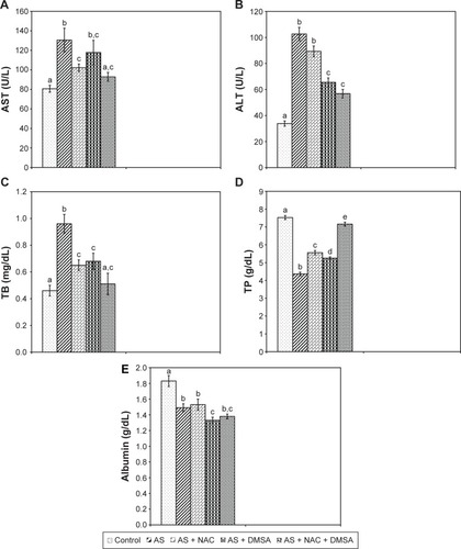
Oral administration of arsenic caused a significant increase in the activities of serum AST (+61.8%), ALT (+203.4%), and bilirubin level (+108.7%), but statistically significant reduction was found in TP concentration (−42%) and albumin level (−18.5%) when compared with control group (). Monotherapy with NAC and the combined treatment of NAC + DMSA resulted in a significant decrease in serum AST activity compared to the arsenic group; AST was restored to normal values only after combined treatment (). Meanwhile, NAC-alone did not significantly reduce serum ALT activity, which responded significantly to both DMSA-alone and NAC + DMSA. However, only NAC + DMSA combined treatment revealed a maximum reduction in the enzyme activity without attaining control values (). However, treatment with NAC, DMSA and their combination after arsenic exposure significantly reduced the increased serum bilirubin level compared to the arsenic-treated group. Three weeks of co-treatment with NAC + DMSA provoked a more intensive lowering in arsenic-induced elevation of serum bilirubin level compared to individual treatments and restored its level near to the control (). The inhibited level of serum TP after treatment with arsenic was significantly elevated in rats treated with NAC, DMSA, and NAC + DMSA compared to arsenic-intoxicated group. Remarkably, the combination treatment with NAC + DMSA produced a recovery in serum protein level over that by either of them alone, although it was still significantly lower than those of healthy controls (). Anywise, NAC or DMSA monotherapy and the combined treatment of NAC + DMSA failed to protect against arsenic-induced reduction of serum albumin ().
Figure 1 Effect of NAC and/or DMSA on arsenic induced changes in serum levels of AST (A), ALT (B), TB (C), TP (D), and albumin (E). Each bar represents mean ± SE of eight animals in each group. Bar superscripts with no common letters (a–e) are significantly different (P<0.05).
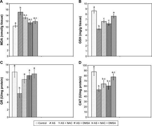
Compared to the control group, LPO level was markedly increased in the arsenic-exposed rats as exhibited by a significant increase in the level of hepatic MDA (+46.2%). In addition, the level of GSH (−40%) and activities of GR (−47.6%) and CAT (−39.6%) were significantly decreased (). DMSA-alone treatment or in combination with NAC showed a more pronounced reduction and retrieved MDA level toward normalization, while treatment with NAC-alone was unable to reduce the increase in the hepatic MDA level (). The level of GSH and the activity of GR were significantly elevated after treatment with NAC, DMSA, and NAC + DMSA compared to the arsenic-treated group. Strikingly, only the combination of NAC + DMSA efficiently restored the inhibited GSH level to near normal (), whereas treatment with NAC and/or DMSA enhanced GR activity toward normalization (). After arsenic administration, the activity of CAT in rats given separate treatments of NAC or DMSA was the same as those treated with arsenic alone. Notably, only the combination of NAC + DMSA efficiently restored the inhibited CAT activity to near normal ().
Figure 2 Effect of NAC and/or DMSA on arsenic induced changes in hepatic MDA (A), GSH (B), GR (C), and CAT (D). Each bar represents mean ± SE of eight animals in each group. Bar superscripts with no common letters (a–c) are significantly different (P<0.05).
Changes of arsenic accumulation
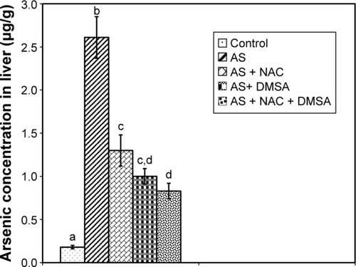
In arsenic group, there was a significant increase in arsenic accumulation in hepatic tissue (+1,350%) compared to controls (). A notable reduction of arsenic levels in liver tissue was recorded in arsenic-exposed rats given NAC-alone (−50.2%) and DMSA-alone (−61.6%) as compared to the arsenic group. However, a better recovery in arsenic accumulation was observed in hepatic tissue in rats treated with combined NAC + DMSA (−68.2%) as compared to the arsenic-intoxicated group. The level of arsenic remained significantly higher than the control in all exposed groups.
Figure 3 Effect of NAC and/or DMSA on the concentration of arsenic in liver. Each bar represents mean ± SE of eight animals in each group. Bar superscripts with no common letters (a–d) are significantly different (P<0.05).
Histopathological observations
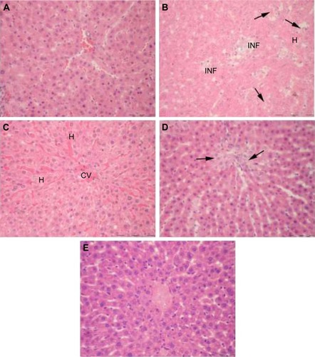
As shown in , the livers of control rats showed normal histology. The hepatic tissue from arsenic-treated rats displayed focal hepatocyte degeneration, hemorrhagic lesions, and massive inflammatory cell infiltration (). The damage was clearly less severe after treatment with NAC () and DMSA (), but discrete changes were still observed. The liver architecture was normal in most areas after combined NAC/DMSA treatment regimen with no evidence of leukocyte infiltrates ().
Figure 4 Microphotographs of H and E-stained liver sections of rats. (A) Control depicts normal liver parenchyma. (B) Arsenic: note degeneration of hepatocytes (arrows), vascular congestion/hemorrhage (H), and multiple inflammatory sites (INF). (C) Arsenic + NAC: hepatocytes are intact but congestion and widening of sinusoidal spaces are still found. (D) Arsenic + DMSA: foci of parenchymal degeneration and necrosis are obvious (arrows). (E) Arsenic + NAC + DMSA: liver architecture is comparable to control without inflammatory characteristics. Original magnification 400× (A–E).
Liver ultrastructural changes
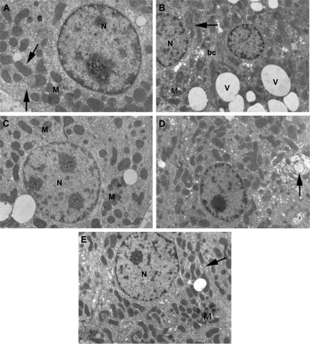
Liver of control rats had normal cytoarchitecture (). Fine structure of liver of the arsenic-treated group revealed dramatic modifications in cell structure, including cytoplasmic vacuolization, diminished rough endoplasmic reticulum (RER) cisternae, few number of disrupted mitochondria, and condensation of nuclear chromatin (ie, pyknosis) (). Decreased degenerative areas and vacuolization, increased RER membranes in hepatocytes, and rarely pyknotic nuclei were seen after NAC treatment (). Also, DMSA treatment after arsenic showed an ameliorative appearance of RER, mitochondria, and nucleus (). Hepatocytes regained their normal appearance after combined treatment of NAC + DMSA ().
Figure 5 Electron micrographs of liver sections of rats. (A) Control shows hepatocyte with normal cellular organelles and intact nuclear chromatin; N indicates nucleus, M indicates mitochondria, and arrows indicate endoplasmic reticulum. (B) Arsenic observe increased peripheral chromatin in the nucleus (N), mitochondria (M) with ill-defined cristae, few endoplasmic reticulum (arrow) and numerous obvious vacuoles all over the cytoplasm (V); bc indicates bile canaliculus. (C) Arsenic + NAC: note that less condensed chromatin in the nucleus (N), scattered mitochondria (M), and in-between short strands of endoplasmic reticulum are visible. (D) Arsenic + DMSA: the cytoplasm is heavily populated with mitochondria and less fragmented endoplasmic reticulum (compared to B). Notable increase in organelle degeneration is still evident (arrow). (E) Arsenic + NAC + DMSA: hepatocyte is preserved without vacuolization, nucleus (N) without abnormal chromatin, normal mitochondria (M) with intact cristae, normally shaped rough endoplasmic reticulum (arrow). Original magnification 7,500× (A) and 5,000× (B–E).
Discussion
The present study aimed at comparing the effectiveness of NAC and DMSA on subacute hepatotoxicity of arsenic in rats. Exposure to arsenic increased serum levels of AST, ALT, TB, and TP, and decreased albumin level, reflecting a broad hepatocellular damage. These results are in good accordance with those in previous studies.Citation39–Citation43 Administration of NAC counteracted arsenic-induced hepatotoxicity as shown by decreased levels of AST and TB, and elevated TP content. In support to the latter results, NAC was reported to have a tendency to avert liver damage via membrane stabilization, thereby suppressing the leakage of hepatic enzymes through membranes.Citation30 Also in this work, DMSA monotherapy was even better than NAC in relieving ALT, probably due to relatively efficient elimination of arsenic via chelation (). Of special interest is the observation that the combined therapy was more beneficial than individual treatments in reversing TP.
There is a growing evidence indicating that arsenic toxicity involves oxidative damage that plays an imperative role in biochemical alterations. Various studies have reported that arsenic exposure generates ROS-like superoxide anion (O2•−), hydroxyl radical (•OH), hydrogen peroxide (H2O2), singlet oxygen (1O2), and peroxyl radicals.Citation44 A primarily measure of arsenic-induced oxidative stress is the increase in MDA level (a marker of LPO), which is a hallmark of the excess generation of intracellular ROS and hepatotoxicity.Citation29 In the present study, arsenic-treated animals showed significantly increased levels of free radical-mediated LPO and decreased levels of GSH, CAT, and GR compared to control. These changes agreed by other researchers such as Flora et al,Citation24,Citation25 Messarah et al,Citation41 El-Demerdash et al,Citation45 Jain et al,Citation46 Manimaran et al,Citation47 Kotyzová et al,Citation48 and Mohanta et al.Citation49 The increase in O2•− by arsenic decreases CAT activity.Citation50,Citation51 The decreased CAT activity may relate to their effective functioning for elimination of hepatic ROS. Moreover, the inhibition of GR leads to accumulation of the pro-oxidant glutathione disulfide by preventing reduction of GSSG to GSH, suggesting that GR may not compensate the consumption of GSH. Some of the ultrastructural changes observed in arsenic-treated rats including fragmentation of RER, breakdown of mitochondrial cristae and disturbed nuclear chromatin are typical for increased ROS and impairment of protein synthesis. By contrast, the ultrastructural picture of liver following antioxidant and/or chelator therapies revealed a healthy appearance. The improvement was more obvious with NAC + DMSA, which assisted to an overall enhancement of the antioxidant defense system. In full broad agreement with these structural observations, the biochemical data showed that the combined NAC + DMSA treatment after arsenic yielded more GSH levels compared with NAC and DMSA alone. NAC interacts most strongly with •OH radicals;Citation52 it exerts an indirect effect on antioxidant status, since it restores CAT and is a precursor for synthesis of GSH, thus providing enhanced protection against toxin-induced oxidative insult.Citation53 DMSA has also an important role in decreasing the oxidative stress, either by eliminating the toxic metal from the target organ and/or by directly scavenging ROS via its sulfhydryl groups.Citation25 DMSA chelator effectively interacts with arsenic in the cell, forming a saturated five-member heterocyclic ring namely 1,3-dithiol-2-arsolan. Following this, mobilization of arsenic is increased via the kidneys, which might be one of the reasons for the increase in the activities of enzymatic antioxidants.Citation54
Polyunsaturated fatty acids in cell membrane are very susceptible to free radical attack, which initiates the self-propagating LPO reactionsCitation55 resulting in the loss of membrane fluidity, decreased membrane potential, increased permeability of protons and Ca2+, and inactivation of a several membrane-bound enzymes.Citation56 Thus, arsenic-induced LPO leads to the degradation of phospholipids and finally prompts cellular deterioration in liver. In this research, DMSA or NAC + DMSA therapeutic regimens achieved the best effect and were able to normalize LPO levels.
Conclusion
The present study confirmed the valuable effect of NAC and DMSA chelator against oxidant/antioxidant imbalance induced by short-term arsenic. The action of NAC against arsenic burden may be complementary to that of DMSA, but the underlying molecular mechanisms need further investigations.
Disclosure
The authors report no conflicts of interest in this work.
References
- JiangJ-QAshekuzzamanSMJiangAArsenic contaminated groundwater and its treatment options in BangladeshInt J Environ Res Public Health20131011846
- DwivediNFloraGKushwahaPFloraSJAlpha-lipoic acid protects oxidative stress, changes in cholinergic system and tissue histopathology during co-exposure to arsenic-dichlorvos in ratsEnviron Toxicol Pharmacol201437172324291368
- AnetorJIWanibuchiHFukushimaSArsenic exposure and its health effects and risk of cancer in developing countries: micronutrients as host defenceAsian Pac J Cancer Prevention200781323
- FloraSJSPachauriVChelation in metal intoxicationInt J Environ Res Public Health2010772745278820717537
- NaujokasMFAndersonBAhsanHThe broad scope of health effects from chronic arsenic exposure: update on a worldwide public health problemEnviron Health Perspect2013121329530223458756
- IARCInternational agency for research on cancer IARC monographs on the evaluation of carcinogenic risks to humansIARC Monogr Eval Carcinog Risks Hum2002961390
- PatraPHBandyopadhyaySKumarRQuantitative imaging of arsenic and its species in goat following long term oral exposureFood Chem Toxicol20125061946195022497900
- DixitGSinghAPKumarASulfur mediated reduction of arsenic toxicity involves efficient thiol metabolism and the antioxidant defense system in riceJ Hazard Mater201529824125126073379
- Guha MazumderDNChronic arsenic toxicity & human healthIndian J Med Res2008128443644719106439
- SantraAMaitiAChowdhuryAMazumderDNOxidative stress in liver of mice exposed to arsenic-contaminated waterIndian J Gastroenterol200019311211510918716
- GhatakSBiswasADhaliGKChowdhuryABoyerJLSantraAOxidative stress and hepatic stellate cell activation are key events in arsenic induced liver fibrosis in miceToxicol Appl Pharmacol20112511596921134390
- DasKBagSSahuRProtective effect of Corchorus olitorius leaves on sodium arsenite-induced toxicity in experimental ratsFood Chem Toxicol201048132633519852998
- RanaTBeraAKDasSEffect of ascorbic acid on blood oxidative stress in experimental chronic arsenicosis in rodentsFood Chem Toxicol20104841072107720122981
- NissarAUFarrukhMRKaiserPJEffect of N-acetyl cysteine (NAC), an organosulfur compound from Allium plants, on experimentally induced hepatic prefibrogenic events in Wistar ratPhytomedicine2013201082883323578993
- FloraSJSArsenic-induced oxidative stress and its reversibilityFree Radic Biol Med201151225728121554949
- BaumgardnerJNShankarKHenningsLAlbanoEBadgerTMRonisMJJN-acetylcysteine attenuates progression of liver pathology in a rat model of nonalcoholic steatohepatitisJ Nutr2008138101872187918806095
- LasramMMLamineAJDhouibIBAntioxidant and anti-inflammatory effects of N-acetylcysteine against malathion-induced liver damages and immunotoxicity in ratsLife Sci20141071–2505824810974
- SmithSSReyesJRArbonKSHarveyWAHuntLMHegglandSJCadmium-induced decrease in RUNX2 mRNA expression and recovery by the antioxidant N-acetylcysteine (NAC) in the human osteoblast-like cell line, Saos-2Toxicol In Vitro2009231606619017541
- LuczakMWZhitkovichARole of direct reactivity with metals in chemoprotection by N-acetylcysteine against chromium(VI), cadmium(II), and cobalt(II)Free Radic Biol Med20136526226923792775
- ChenGWangKYangBYSynergistic antitumor activity of oridonin and arsenic trioxide on hepatocellular carcinoma cellsInt J Oncol201240113914721947421
- El-SayedMFAbdel-GhafarSKAdlyMASalimAAAbdel-SameiWMThe ameliorative effects of DMSA and some vitamins against toxicity induced by lead in the testes of albino rats. IIJ Basic Appl Zool2015716065
- MillerALDimercaptosuccinic acid (DMSA), a non-toxic, water-soluble treatment for heavy metal toxicityAltern Med Rev1998331992079630737
- FloraSJSPandeMMehtaABeneficial effect of combined administration of some naturally occurring antioxidants (vitamins) and thiol chelators in the treatment of chronic lead intoxicationChem Biol Interact2003145326728012732454
- FloraSJSBhadauriaSDhakedRKPantSCArsenic induced blood and brain oxidative stress and its response to some thiol chelators in ratsLife Sci200577182324233715964026
- FloraSJSChouhanSKannanGMMittalMSwarnkarHCombined administration of taurine and monoisoamyl DMSA protects arsenic induced oxidative injury in ratsOxid Med Cell Longev200811394519794907
- KokilavaniVDeviMASivarajanKPanneerselvamCCombined efficacies of DL-α-lipoic acid and meso 2,3 dimercaptosuccinic acid against arsenic induced toxicity in antioxidant systems of ratsToxicol Lett200516011715998567
- ModiMKaulRKKannanGMFloraSJCo-administration of zinc and n-acetylcysteine prevents arsenic-induced tissue oxidative stress in male ratsJ Trace Elem Med Biol200620319720416959597
- GuptaRDubeyDKKannanGMFloraSJConcomitant administration of Moringa oleifera seed powder in the remediation of arsenic-induced oxidative stress in mouseCell Biol Int2007311445617055307
- FloraSJSArsenic-induced oxidative stress and its reversibility following combined administration of N-acetylcysteine and meso 2, 3-dimercaptosuccinic acid in ratsClin Exp Pharmacol Physiol1999261186586910561806
- KannanGMFloraSJSCombined administration of N-acetylcysteine and monoisoamyl DMSA on tissue oxidative stress during arsenic chelation therapyBiol Trace Elem Res20061101435916679547
- KaliaKNarulaGDKannanGMFloraSJSEffects of combined administration of captopril and DMSA on arsenite induced oxidative stress and blood and tissue arsenic concentration in ratsComp Biochem Physiol Pharmacol Toxicol20071444372379
- LowryOHRosebroughNJFarrALRandallRJProtein measurement with the Folin phenol reagentJ Biol Chem1951193126527514907713
- NairVTurnerGEThe thiobarbituric acid test for lipid peroxidation structure of the adduct with malondialdehydeLipids198419108495
- EllmanGLTissue sulfhydryl groupsArch Biochem Biophys1959821707713650640
- BeutlerEEffect of flavin compounds on glutathione reductase activity: in vivo and in vitro studiesJ Clin Invest19694810195719665822598
- SinhaAKColorimetric assay of catalaseAnal Biochem19724723893944556490
- HersheyJWOostdykTSKeliherPNDetermination of arsenic and selenium in environmental and agricultural samples by hydride generation atomic absorption spectrometryJ Assoc Off Anal Chem1988716109010933240958
- NandiDPatraRCSwarupDEffect of cysteine, methionine, ascorbic acid and thiamine on arsenic-induced oxidative stress and biochemical alterations in ratsToxicology20052111–2263515863245
- YousefMIEl-DemerdashFMRadwanFMSodium arsenite induced biochemical perturbations in rats: ameliorating effect of curcuminFood Chem Toxicol200846113506351118809455
- ChattopadhyaySMaitiSMajiGDebBPanBGhoshDProtective role of Moringa oleifera (Sajina) seed on arsenic-induced hepatocellular degeneration in female albino ratsBiol Trace Elem Res2011142220021220661662
- MessarahMKlibetFBoumendjelAHepatoprotective role and antioxidant capacity of selenium on arsenic-induced liver injury in ratsExp Toxicol Pathol201264316717420851583
- JoshiDMittalDKShuklaSSrivastavAKSrivastavSKN-acetyl cysteine and selenium protects mercuric chloride-induced oxidative stress and antioxidant defense system in liver and kidney of rats: A histopathological approachJ Trace Elem Med Biol201428221822624485406
- MuthumaniMMiltonprabuSAmeliorative efficacy of tetrahydrocurcumin against arsenic induced oxidative damage, dyslipidemia and hepatic mitochondrial toxicity in ratsChem Biol Interact20152359510525869292
- FloraSJSArsenic and dichlorvos: possible interaction between two environmental contaminantsJ Trace Elem Med Biol201635436027049126
- El-DemerdashFMYousefMIRadwanFMEAmeliorating effect of curcumin on sodium arsenite-induced oxidative damage and lipid peroxidation in different rat organsFood Chem Toxicol200947124925419049818
- JainAYadavABozhkovAIPadalkoVIFloraSJTherapeutic efficacy of silymarin and naringenin in reducing arsenic-induced hepatic damage in young ratsEcotoxicol Environ Saf201174460761420719385
- ManimaranASarkarSNSankarPInfluence of repeated preexposure to arsenic on acetaminophen-induced oxidative stress in liver of male ratsFood Chem Toxicol201048260561019932728
- KotyzováDBludovskáMEyblVDifferential influences of various arsenic compounds on antioxidant defense system in liver and kidney of ratsEnviron Toxicol Pharmacol20133631015102124095718
- MohantaRKGargAKDassRSEffect of vitamin E supplementation on arsenic induced alteration in blood biochemical profile, oxidant/antioxidant status, serum cortisol level and retention of arsenic and selenium in goatsJ Trace Elem Med Biol20152918819425240912
- KonoYFridovichISuperoxide radical inhibits catalaseJ Biol Chem198225710575157546279612
- KirkmanHNGaetaniGFCatalase: a tetrameric enzyme with four tightly bound molecules of NADPHProc Natl Acad Sci U S A198481434343476589599
- ArakawaMItoYN-acetylcysteine and neurodegenerative diseases: basic and clinical pharmacologyCerebellum20076430831417853088
- Abu El-SaadAMElgerbedMSDimethoate induced hepatotoxicity in rats and the protective roles of vitamin E and N-acetylcysteineEgypt J Exp Biol201062219230
- PalaniappanPRVijayasundaramVThe effect of arsenic exposure and the efficacy of DMSA on the proteins and lipids of the gill tissues of Labeo rohitaFood Chem Toxicol20094781752175919394394
- PepicelliOFedeleEBerardiMMinghettiLCyclo-oxygenase-1 and -2 differently contribute to prostaglandin E 2 synthesis and lipid peroxidation after in vivo activation of N-methyl-D-aspartate receptors in rat hippocampusJ Neurochem20059361561156715935072
- HalliwellBGutteridgeJMCFree Radicals in Biology and Medicine3rd editionOxford, UKClarendon Press1999