
Abstract
Leukemia is a clonal malignant hematopoietic stem cell disease. It is the sixth most lethal cancer and accounts for 4% of all cancers. The main form of treatment for leukemia is chemotherapy. While some cancer types with a higher incidence than leukemia, such as lung and gastric cancer, have shown a sharp decline in mortality rates in recent years, leukemia has not followed this trend. Drug resistance is often regarded as the main clinical obstacle to effective chemotherapy in patients diagnosed with leukemia. Many resistance mechanisms have now been identified, and multidrug resistance (MDR) is considered the most important and prevalent mechanism involved in the failure of chemotherapy in leukemia. In order to reverse MDR and improve leukemia prognosis, effective detection methods are needed to identify drug resistance genes at initial diagnosis. This article provides a comprehensive overview of published approaches for the detection of MDR in leukemia. Identification of relevant MDR genes and methods for early detection of these genes will be needed in order to treat leukemia more effectively.
Introduction
The treatment of cancers which have developed multidrug resistance (MDR) is a major challenge, and rates of morbidity and mortality are extremely high.Citation1 Also, in clinical practice we have found that the emergence of MDR has caused considerable levels of pain in many leukemia patients. MDR is defined as when cancer cells become resistant to a chemotherapeutic drug, after one or more treatment cycles, and this may lead to resistance to other chemotherapeutic drugs that have different structures and mechanisms.Citation2
Numerous reports have focused on the different mechanisms of MDR in leukemia. These resistance mechanisms include: a) increased drug metabolism due to altered molecular targets; b) defective apoptotic machinery; c) over-expression of efflux pumps, such as P-glycoprotein (P-gp), multidrug resistance-associated proteins (MRP1, MRP2), lung resistance protein/major vault protein, and breast cancer resistance protein (BCRP); d) enzyme-mediated drug resistance mechanisms such as overexpression of glutathione S-transferase; e) microenvironmental resistance; and f) enhanced repair of drug-induced DNA damage.Citation3,Citation4 It is well known that the appearance of MDR has made effective treatment of leukemia very challenging. Therefore, the early, accurate, and sensitive detection of MDR genes is vital, and it is also beneficial to search for more effective chemotherapeutic approaches for use in the clinical setting. Notably, MDR efflux pumps are usually localized in epithelial cells, and their location is polarized. Moreover, they are often expressed in combination with other membrane proteins. All of these can serve as potential targets for convenient testing. MDR genes can usually be potentially detected by the RNA and protein levels by various approaches as described in this article.
Multidrug resistance
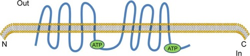
The occurrence of MDR is mainly due to the extracellular efflux of chemotherapy agents which involves mechanisms mediated by over expression of ATP-binding cassette (ABC), and MDR proteins.Citation5 Both P-gp and MRP1 are thought to play pivotal roles in the MDR process.Citation6 P-gp is a membrane-associated drug efflux pump which belongs to the ABC protein family and is encoded by the MDR1 gene (). P-gp represents a 170 kDa glycosylated integral plasma membrane protein,Citation7,Citation8 which acts as a drug efflux pump to decrease intracellular drug concentrations.Citation9 Multidrug resistance and chemotherapy failure are produced by intracellular anticancer drugs that increasingly flow from cells through the efflux pump.Citation10,Citation11 The protein includes two halves. Each part of the protein contains six hydrophobic trans-membrane domains, and one ATP binding domain.Citation12 Powered by the hydrolysis of ATP, they efflux structurally diverse compounds. Hydrolysis of ATP is believed to reset the protein to the inward-facing form to begin a new cycle of drug binding and release.Citation13 P-gp, which is located on chromosome 7q21, was the first MDR gene to be identified.Citation14 Two isoforms of P-gp are expressed in humans, class I and III isoforms are drug transporters (MDR1/ABCB1), while the function of class II isoforms (MDR2/3/ABCB4) is to export phosphatidylcholine into bile.Citation15 In addition to exporting chemotherapeutic agents, P-gp is able to transport a broad range of substrates, including amino acids, sugars, peptides, organic ions, metabolites, and numerous hydrophobic compounds.Citation16,Citation17 Several reports suggest that P-gp may regulate apoptosis, chloride channel activity, cholesterol metabolism, differentiation, proliferation, adhesion, and immune cell function.Citation18 Significantly, elevated P-gp expression was found in chronic B-cell leukemia patients. Alterations in P-gp expression may also occur because of single nucleotide polymorphisms, which have been found in MDR1, and which affect drug-metabolism and result in altered pharmacokinetics of chemotherapeutics.Citation19 Some investigations have proposed a model in which expression of ABC transporters prevent drug accumulation in resistant cells by increasing efflux, at the same time, the models also consider the other side of the equation in which drug influx is suppressed by the active reduction of endocytosis.Citation20
Figure 1 Schematics of protein ABCB1.
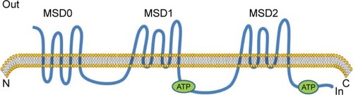
MRP1 was one of the first human ABC proteins identified and is encoded by the ABCC1 gene (), located on chromosome 16p13.Citation12 MRP1 is a glycosylated protein with molecular weight ~190 kDa and functions in the transport of sulfate, glutathione or glucuronate and anionic substances, in an ATP-dependent manner.Citation16,Citation21,Citation22 Structurally, MRP1 has three membrane spanning domains (MSD0, MSD1, MSD2). MSD1 and MSD2 each has six transmembrane (TM) helixes, MSD0 has five TM segments with approximately 200 amino acids. MRP1 is also made up of two cytosolic nucleotide binding domains (NBDs). The cytosolic NBD is responsible for binding and hydrolysis of ATP to provide energy for substrate transport.Citation16,Citation23 A variety of anticancer drugs transported by MRP1, are bulky hydrophobic molecules that acquire entry to cells by simple diffusion across the lipid bilayer of the cell’s outer membrane.Citation23,Citation24 The mechanism of MRP1 involved multidrug resistance has not been exactly understood. GSH possibly plays a role in the occurrence of multidrug resistance.Citation25 MRP1 also possibly serves a sequestration function to prevent drugs from reaching their intracellular targets, because it has been found in other subcellular organelles such as the endoplasmic reticulum and endocytic vesicles.Citation26,Citation27
Figure 2 Schematics of protein ABCC1.
Detection approaches
The mechanisms underlying MDR are complex. Accurate and sensitive detection of these mechanisms is thought to be vital to improved treatment of leukemia. Since MDR1 gene amplification is almost non-existent in human tumors, detection of alterations in DNA copy number is not appropriate. Therefore, at present we usually measure MDR1 mRNA expression levels. Commonly used methods include polymerase chain reaction (PCR), in situ hybridization (ISH), and RNase protection assays (RPAs). Western blotting and immunohistochemistry (IHC) may also be used for protein detection.
Nucleic acid-based detection methods
PCR
PCR is a technique for the selective amplification of DNA or RNA segments of up to 2 kb or more in length.Citation28 The three stages of PCR are exponential amplification, followed by leveling off, and the plateau stage. DNA polymerase and specific oligonucleotide primers are currently used for amplification of P-gp cDNAs.Citation29 Specifically, PCR amplification relies on thermal cycling, consisting of cycles of repeated heating and cooling for DNA melting and enzymatic replication of the DNA.Citation30 Primers for the PCR reactions are based on conserved sequences of the catalytic domain that are shared among all known drug efflux pumps protein tyrosine phosphatases.Citation31 Primers could produce optimal amplicon size for quantitative PCR when amplifying regions for the partial P-gp sequences. For each reaction, for optimization, it was implemented to detect each primer pair and undertake amplification of a single PCR product only.Citation29 Repeated cycles of three different reaction temperatures are needed: the first high temperature step for heat-denaturation of the DNA, the second for annealing, and the third for extension of the primers.Citation32 As PCR is sensitive to contamination, care must be taken to include appropriate negative controls. In order to ensure that any contaminating material would not act as a template for PCR, buffers should be incubated with restriction enzymes that cut within the amplified fragment before amplification.Citation33 This approach has been greatly simplified by automated procedures that use a thermostable enzyme DNA polymerase.Citation34 This allows a single sample to be simultaneously amplified for several different markers, and has important economic implications.Citation35 PCR is now routinely and universally applied in human genetics, basic molecular biology, and clinical investigations with the goal of monitoring the causes of disease.Citation28 At the end of the 20th century, Vogelstein described a digital PCR method. Digital PCR is both a qualitative and quantitative method, and can sensitively and accurately detect genetic alterations.Citation36
Fluorescence in situ hybridization (FISH)
FISH is a method to detect specific nucleic acids in fixed but otherwise intact cells, that has been shown to be both sensitive and specific.Citation37,Citation38 FISH for visualization of nucleic acids was developed as an alternative to older methods that used radio-labeled probes.Citation39 The basic elements of the FISH procedure include selection of probe(s) for a sequence complementary to the target of interest, probe labeling, slide preparation, slide pretreatment, denaturation of probe and target, hybridization, washing, analysis, and interpretation.Citation40 Probes are labeled either directly, by incorporation of fluorescent nucleotides, or indirectly, by incorporation of reporter molecules that are subsequently detected by fluorescent antibodies or other high affinity molecules (eg, streptavidin/biotin).Citation41 Hybridization to the target loci is visualized by the detection of fluorescent signals on metaphase chromosomes or interphase nuclei.Citation42 Many new technologies have been invented to overcome the shortcomings of FISH. For example, a high resolution multi-color banding method was developed to overcome the disadvantage of lack of detection of perientric and paracentric DNA inversions, and therefore to precisely identify chromosome breakpoints.Citation43 Quantitative FISH can measure the length of telomere repeats in preparations of metaphase chromosomes.Citation44 Many applications based on FISH have been proposed in different fields of investigation, including evolutionary studies, interphase nucleus architecture analyses, DNA sequence mapping, toxicology, microbial ecology, cancer diagnostics, and forensics.Citation38,Citation45 This technology provides a wide range of choices for us to target any desired tumor biomarkers or any of the chromosomes to be examined.Citation46
RPA
The RPA is a sensitive means of quantitating mRNA transcripts initiated at a specific nucleotide, which was developed in the early 1980s.Citation47 The procedure is based on the hybridization of the analyzed RNA to a radioactively labeled RNA probe, and the hybridization reactions are treated with ribonuclease to remove free probe. This leaves intact fragments of probe annealed to homologous sequences in the sample RNA.Citation48,Citation49 Notably, the position of radioactively labeled probe fragments can be detected by autoradiography. Alternatively, samples can be transferred to a membrane for secondary visualization if non-isotopically labeled probes are used.Citation50 The hybridized RNA is then digested with ribonucleases, followed by precipitation and resuspension of the protected RNAs. Ultimately, electrophoresis and autoradiography of the protected RNAs is done on a denaturing polyacrylamide gel.Citation51 The assay of mRNA by RPA is achievable because double-stranded RNA is not susceptible to degradation by ribonuclease.Citation52 The advantage of the assay is that multiple mRNA species can be measured simultaneously in a single total RNA sample. Also, there are no amplification steps involved in ribonuclease protection, and therefore quantitative data are more reliable than reverse transcription (RT)-PCR. However, RNase protection is more time-consuming than RT-PCR and takes several days to complete, even by an experienced technician.Citation53 For further technological development of this approach, microchip electrophoresis was considered in the analytical stages due to its characteristics: high speed, high throughput, low consumption of samples and reagents, miniaturization, and automation.Citation54
Protein-based detection methods
Western blot
Western blotting is a technique that was first developed between 1977 and 1979 by Towbin and Staehelin, that has since become a common technique applied in research laboratories globally for the immune detection and quantitation of specific proteins.Citation55,Citation56 The principle of this method is based on specific antibody-antigen interaction. It could achieve the qualitative or semiquantitative identification of specific proteins. What is more important, the molecular weight of the proteins also be measured from a complex mixture. Gel electrophoresis of a protein sample, transfer of protein from a gel to a membrane support, and immune detection of a target antigen are the three necessary steps.Citation57 Pelleting the cells by low speed centrifugation in order to drain the cell pellet well is usually performed when in the protein extraction. Also, it should be done at 4°C with protease inhibitors to prevent denaturing of the proteins, and incubated at room temperature for 5 min.Citation58,Citation59 Polyacrylamide gel electrophoresis is used to separate proteins in Western blotting.Citation57 Some researchers indicated that the sensitivity of Western blotting was dependent on the quality of both the antigen and the antibody detection reagents.Citation60 Similarly, increasing the volume of serum for low-titer specimens is critical.Citation61 The critical steps in generating high-quality, quantitative Western blots are the following: 1) measuring protein concentrations; 2) sample preparation; 3) blocking the membrane; and 4) obtaining good quality and caliber of the primary antibody.Citation62 A Western blotting minimal reporting standard and stain-free technology are recommended to improve the reproducibility of Western blot analysis.Citation63 These are novel and unique quality control tools for data normalization in Western blotting workflows.Citation55 The advantages of Western blotting are: simplicity, speed, and sensitivity. Furthermore, immunoblotting can distinguish different molecular forms of an antigen.Citation64 Western blotting has been used to detect changes in MRP1 levels.Citation65 Expression of MDR-1/P-gp levels in P388, P388/ADR, and HCT-15 cell lines have also been tested by Western blot analysis.Citation7
IHC
IHC is a powerful method for detecting specific proteins in formalin-fixed, paraffin-embedded tissues based on antigen–antibody interactions. The process involves tissue processing, sectioning and epitope retrieval, antigen–antibody interaction for protein detection, and visualization through various approaches.Citation66 Successful immunostaining depends on the source, specificity, and quality of the primary antibody. The condition of the specimen is also very important, as is the fixation procedure and antigen-detection strategy.Citation67 Excellent morphology can be obtained if the cells are fixed while they are still in growth-culture medium dish/flask before removing them.Citation68 IHC is a highly standardized, sensitive, and simple technique.Citation66 In addition, it possesses the ability to delineate admixed normal cells bearing P-gp. All of these are beneficial when investigating the development and emergence of multidrug resistance in leukemia. Subjectivity is the primary disadvantage.Citation69 Antigen localization, cell and tissue morphology can be observed simultaneously because IHC utilizes a light microscope for visualization.Citation67 However, IHC is subject to qualitative and subjective assessment, and has been criticized due to a lack of stringency compared with PCR.Citation70 As the enhanced diagnostic utility of IHC was realized, the demand for this technique has also increased. It will continue to be a rapidly evolving field with the ever-increasing numbers of new antibodies available.Citation71 The development of automation has progressed to eliminate many of the manual steps in the process.Citation72 The combination of mass spectroscopy with IHC has allowed multiplexed, direct quantitative imaging of tissue samples for basic and clinical research.Citation73 Multiplexed IHC methods permit identification of at least three, and up to 30 discrete antigens.Citation74 Some studies suggest that IHC may be sufficiently sensitive and specific to predict response to targeted therapy.Citation75
Conclusion
The outcome of leukemia is usually fatal. Recent surveys indicate that leukemia is the most common cause of cancer-related death in men under the age of 40. Meanwhile, about half of new cancer-related deaths among females were attributed to leukemia. Equally alarming is the fact that leukemia is a major cause of death in females under the age of 20. For children under the age of 14, acute lymphocytic leukemia is the most common form of cancer.Citation76 Chronic myeloid leukemia (CML) has an incidence of 1–1.5 per 100,000,Citation77 with relapse occurring in 25% of patients being associated with a poor prognosis.Citation78 As a global problem of increasing concern, MDR leads to inadequate treatment and poor prognosis in leukemia patients. In recent years, some progress has been made in understanding the mechanisms of MDR.Citation79 Despite this poor prognosis, there has been some progress in the treatment of leukemia in recent years. Tyrosine kinase inhibitors (TKIs) have changed the clinical course of CML, improving the 5-year survival from 35% to more than 90% with imatinib mesylate.Citation80 Some new therapies aimed at reducing drug resistance are being studied, such as targeting JAK/STAT signaling, and the combination of ATRA with TKIs.Citation14,Citation81 However, both P-gp and MRP1 have been shown to interact with imatinib.Citation82 The challenges are sure to remain difficult in the future. Detection of MDR genes has been described using the methodology outlined in this article. For example, 56 cell membrane glycoproteins in leukemia samples could be successfully quantitated by antibody analysis of non-glycopeptides.Citation83 Detection technology is constantly developing. At the end of the 20th century, DNA microarrays were starting to be developed, allowing an opportunity to test the expression of hundreds to thousands of genes in a single assay.Citation84 Nowadays, high-throughput sequencing has risen to prominence and can also be used to detect MDR genes, diagnose cancer, and so on. The technology makes the direct application of RNA sequencing, while not through DNA synthesis, possible.Citation85 DNA microarray and next generation sequencing are products of the implementation of high-throughput techniques. Microarrays have greater advantages in terms of parallelism, automation, and miniaturization. Without PCR-based signal amplification, next-generation DNA sequencing technologies also have the ability to read signals of a single fluorescent molecule.Citation86 In comparison to traditional Sanger sequencing, next-generation sequencing allows multiplexing of samples and gene targets in one experimental setup.Citation87 At the same time, it can be combined with microarrays, DNA co-immunoprecipitation, and so on. However, they still have some shortcomings and technical limitations. It is of critical importance to establish the most convenient and efficient method to detect MDR genes in the future. The ultimate goal is to detect resistance genes and reverse their activities so as to reduce the risk of relapse and improve patient prognosis.
Acknowledgments
This work was supported by the National Key Basic Research Program 973 of the People’s Republic of China (number 2010CB732404), the National Nature Science Foundation of the People’s Republic of China (numbers 81170492, 81370673), and the National High Technology Research and Development Program 863 Projects of the People’s Republic of China (number 2012AA022703).
Disclosure
The authors report no conflicts of interest in this work.
References
- KandiVKandiSAntimicrobial properties of nanomolecules: potential candidates as antibiotics in the era of multi-drug resistanceEpidemiol Health201537 e201502025968114
- WuQYangZNieYShiYFanDMulti-drug resistance in cancer chemotherapeutics: mechanisms and lab approachesCancer Lett20143472 159 16624657660
- PatelNRPattniBSAbouzeidAHTorchilinVPNanopreparations to overcome multidrug resistance in cancerAdv Drug Deliv Rev20136513–14 1748 176223973912
- UllahMFCancer multidrug resistance (MDR): a major impediment to effective chemotherapyAsian Pac J Cancer Prev200891 1 618439063
- YuPDuYYangLFanSWuJZhengSSignificance of multidrug resistance gene-related proteins in the postoperative chemotherapy of gastric cancerInt J Clin Exp Pathol2014711 7945 795025550836
- MaHChengLHaoKReversal effect of ST6GAL 1 on multidrug resistance in human leukemia by regulating the PI3K/Akt pathway and the expression of P-gp and MRP1PLoS One201491 e8511324454800
- MaPDongXSwadleyCLDevelopment of idarubicin and doxorubicin solid lipid nanoparticles to overcome Pgp-mediated multiple drug resistance in leukemiaJ Biomed Nanotechnol200952 151 16120055093
- ZhangLXiaoRXiongJActivated ERM protein plays a critical role in drug resistance of MOLT4 cells induced by CCL25PLoS One201381 e5238423326330
- XuYOhmsSJLiZChanges in the expression of miR-381 and miR-495 are inversely associated with the expression of the MDR1 gene and development of multi-drug resistancePLoS One2013811 e8206224303078
- HuTToKKWangLReversal of P-glycoprotein (P-gp) mediated multidrug resistance in colon cancer cells by cryptotanshinone and dihydrotanshinone of Salvia miltiorrhizaPhytomedicine20142111 1264 127225172788
- JingXZhangHHuJβ-arrestin 2 is associated with multidrug resistance in breast cancer cells through regulating MDR1 gene expressionInt J Clin Exp Pathol201582 1354 136325973019
- MilojkovicMMilacicNRadovicJLjubisavljevicSMDR1 gene polymorphisms and P-glycoprotein expression in respiratory diseasesBiomed Pap Med Fac Univ Palacky Olomouc Czech Repub20151593 341 34624993742
- ChufanEESimHMAmbudkarSVMolecular basis of the polyspecificity of P-glycoprotein (ABCB1): recent biochemical and structural studiesAdv Cancer Res2015125 71 9625640267
- CarmoCRLyons-LewisJSecklMJCosta-PereiraAPA novel requirement for janus kinases as mediators of drug resistance induced by fibroblast growth factor-2 in human cancer cellsPLoS One201165 e1986121625473
- Kapse-MistrySGovenderTSrivastavaRYergeriMNanodrug delivery in reversing multidrug resistance in cancer cellsFront Pharmacol20145 15925071577
- de MoraesACMaranhoCKRauberGSSantos-SilvaMCImportance of detecting multidrug resistance proteins in acute leukemia prognosis and therapyJ Clin Lab Anal2013271 62 7123292860
- Carrett-DiasMAlmeidaLKPereiraJLCell differentiation and the multiple drug resistance phenotype in human erythroleukemic cellsLeuk Res201642 13 2026852002
- CerezoDLencinaMRuiz-AlcarazAJAcquisition of MDR phenotype by leukemic cells is associated with increased caspase-3 activity and a collateral sensitivity to cold stressJ Cell Biochem20121134 1416 142522173742
- PennaGAllegraAAlonciAMDR-1 polymorphisms (G2677T and C3435T) in B-chronic lymphocytic leukemia: an impact on susceptibility and prognosisMed Oncol2011284 1549 155420496015
- PiscoAOJacksonDAHuangSReduced intracellular drug accumulation in drug-resistant leukemia cells is not only solely due to MDR-mediated efflux but also to decreased uptakeFront Oncol20144 30625401091
- ColeSPTargeting multidrug resistance protein 1 (MRP1, ABCC1): past, present, and futureAnn Rev Pharmacol Toxicol201454 95 11724050699
- MahjoubiFGolalipourMGhavamzadehAAlimoghaddamKExpression of MRP1 gene in acute leukemiaSao Paulo Med J20081263 172 17918711657
- YinJZhangJMultidrug resistance-associated protein 1 (MRP1/ABCC1) polymorphism: from discovery to clinical applicationZhong Nan Da Xue Xue Bao Yi Xue Ban20113610 927 93822086004
- FazlinaNMahaAJamalRExpression of multidrug resistance (MDR) proteins and in vitro drug resistance in acute leukemiasHematology2007121 33 3717364990
- AkanIAkanSAkcaHSavasBOzbenTMultidrug resistance-associated protein 1 (MRP1) mediated vincristine resistance: effects of N-acetylcysteine and Buthionine sulfoximineCancer Cell Int200551 2216042792
- ColeSPMultidrug resistance protein 1 (MRP1, ABCC1), a “multitasking” ATP-binding cassette (ABC) transporterJ Biol Chem201428945 30880 3088825281745
- LuJFPokharelDBebawyMMRP1 and its role in anticancer drug resistanceDrug Metab Rev2015474 406 41926541366
- VosbergHPThe polymerase chain reaction: an improved method for the analysis of nucleic acidsHum Genet1989831 1 152475423
- WilliamsonSMWolstenholmeAJP-glycoproteins of Haemonchus contortus: development of real-time PCR assays for gene expression studiesJ Helminthol2012862 202 20821729384
- ErlichHAPolymerase chain reactionJ Clin Immunol198996 437 4472698397
- YiTClevelandJLIhleJNIdentification of novel protein tyrosine phosphatases of hematopoietic cells by polymerase chain reaction amplificationBlood1991789 2222 22281932742
- CollasiusMFalkHCieslerCValetGHow to build an inexpensive cyclotherm instrument for automated polymerase chain reactionAnal Biochem19891811 163 1662817375
- LawlerMHumphriesPMcCannSREvaluation of mixed chimerism by in vitro amplification of dinucleotide repeat sequences using the polymerase chain reactionBlood19917711 2504 25142039832
- SaitoIServeniusBComptonTFoxRIDetection of Epstein-Barr virus DNA by polymerase chain reaction in blood and tissue biopsies from patients with Sjogren’s syndromeJ Exp Med19891696 2191 21982543732
- BellJThe polymerase chain reactionImmunol Today19891010 351 3552679632
- VogelsteinBKinzlerKWDigital PCRProc Natl Acad Sci U S A19999616 9236 924110430926
- KlingerKWFISH: sensitivity and specificity on sorted and unsorted cellsAnn N Y Acad Sci1994731 48 567944134
- VautrotVAigueperseCBranlantCBehm-AnsmantIFluorescence In situ hybridization of small non-coding RNAsMethods Mol Biol20151296 73 8325791592
- LevskyJMSingerRHFluorescence in situ hybridization: past, present and futureJ Cell Sci2003116Pt 14 2833 283812808017
- TsuchiyaKDFluorescence in situ hybridizationClin Lab Med2011314 525 54222118735
- VolpiEVBridgerJMFISH glossary: an overview of the fluorescence in situ hybridization techniqueBiotechniques2008454 385 38618855767
- MurthySKDemetrickDJNew approaches to fluorescence in situ hybridizationMethods Mol Biol2006319 237 25916719359
- ChudobaIPleschALörchTLemkeJClaussenUSengerGHigh resolution multicolor-banding: a new technique for refined FISH analysis of human chromosomesCytogenet Cell Genet1999843–4 156 16010393418
- PoonSSLansdorpPMQuantitative fluorescence in situ hybridization (Q-FISH)Curr Protoc Cell Biol2001 Chapter 18: Unit 18.4
- NathJJohnsonKLA Review of fluorescence in situ hybridization (FISH): current status and future prospectsBiotech Histochem2000752 54 7810941509
- LinPPIntegrated EpCAM-independent subtraction enrichment and iFISH strategies to detect and classify disseminated and circulating tumors cellsClin Transl Med201541 3826718583
- CareyMFPetersonCLSmaleSTThe RNase protection assayCold Spring Harb Protoc201320133
- MironovVNVan MontaguMInzéDHigh throughput RNase protection assayNucleic Acids Res19952316 3359 33607545289
- GilmanMRibonuclease protection assayCurr Protoc Mol Biol2001 Chapter 4: Unit 7
- EylerEExplanatory chapter: nuclease protection assaysMethods Enzymol2013530 89 9724034316
- YoungHASubleskiJJKrebsSMMultiprobe ribonuclease protection assay for simultaneous measurement of mRNA expressionCurr Protoc Immunol2003 Chapter 10: Unit 10.29
- MitchellAFidgeNDetermination of apolipoprotein mRNA levels by ribonuclease protection assayMethods Enzymol1996263 351 3638749022
- RottmanJBThe ribonuclease protection assay: a powerful tool for the veterinary pathologistVet Pathol2002391 2 912102215
- YamaguchiYYatsushiroSYamamuraSRibonuclease protection assay on microchip electrophoresisAnalyst201113611 2247 225121509398
- TaylorSCPoschAThe design of a quantitative western blot experimentBiomed Res Int20142014 36159024738055
- TowbinHStaehelinTElectrophoretic transfer of proteins from polyacrylamide gels to nitrocellulose sheets: procedure and some applicationsProc Natl Acad Sci U S A1979769 4350 4354388439
- HnaskoTSHnaskoRMThe Western blotMethods Mol Biol20151318 87 9626160567
- EslamiALujanJWestern blotting: sample preparation to detectionJ Vis Exp201044
- MahmoodTYangPCWestern blot: technique, theory, and trouble shootingN Am J Med Sci201249 429 43423050259
- GalloDDiggsJLShellGRDaileyPJHoffmanMNRiggsJLComparison of detection of antibody to the acquired immune deficiency syndrome virus by enzyme immunoassay, immunofluorescence, and Western blot methodsJ Clin Microbiol1986236 1049 10513011854
- AshleyRLMilitoniJLeeFNahmiasACoreyLComparison of Western blot (immunoblot) and glycoprotein G-specific immunodot enzyme assay for detecting antibodies to herpes simplex virus types 1 and 2 in human seraJ Clin Microbiol1988264 662 6672835389
- SilvaJMMcMahonMThe fastest Western in town: a contemporary twist on the classic Western blot analysisJ Vis Exp201484 e5114924561642
- GildaJEGhoshRCheahJXWestTMBodineSCGomesAVWestern blotting inaccuracies with unverified antibodies: need for a Western blotting minimal reporting standard (WBMRS)PloS One2015108 e013539226287535
- HoweJGHersheyJWA sensitive immunoblotting method for measuring protein synthesis initiation factor levels in lysates of Escherichia coliJ Biol Chem198125624 12836 128397031052
- MaSLHuYPWangFLapatinib antagonizes multidrug resistance-associated protein 1-mediated multidrug resistance by inhibiting its transport functionMol Med201420 390 39925105301
- SchachtVKernJSBasics of immunohistochemistryJ Invest Dermatol20151353 e30
- HofmanFMTaylorCRImmunohistochemistryCurr Protoc Immunol2013103 Unit 21 4
- NuovoGJThe Basics of ImmunohistochemistryIn Situ Molecular Pathology and Co-Expression AnalysesLondon, UKAcademic Press2013 133 165
- BeckWTGroganTMWillmanCLMethods to detect P-glycoprotein-associated multidrug resistance in patients’ tumors: consensus recommendationsCancer Res19965613 3010 30208674056
- ElliottKMcQuaidSSalto-TellezMMaxwellPImmunohistochemistry should undergo robust validation equivalent to that of molecular diagnosticsJ Clin Pathol20156810 766 77026280782
- FerringerTImmunohistochemistry in dermatopathologyArch Pathol Lab Med20151381 83 105
- PrichardJWOverview of automated immunohistochemistryArch Pathol Lab Med201413812 1578 158225427039
- RimmDLNext-gen immunohistochemistryNat Methods2014114 381 38324681723
- DixonARBathanyCTsueiMWhiteJBaraldKFTakayamaSRecent developments in multiplexing techniques for immunohistochemistryExpert Rev Mol Diagn2015159 1171 118626289603
- SwansonPEImmunohistochemistry as a surrogate for molecular testing: a reviewAppl Immunohistochem Mol Morphol2015232 81 9625675083
- ChenAHTsauYWLinCHNovel methods to identify biologically relevant genes for leukemia and prostate cancer from gene expression profilesBMC Genomics201011 27420433712
- RoundhillEABurchillSADetection and characterisation of multi-drug resistance protein 1 (MRP-1) in human mitochondriaBr J Cancer20121066 1224 123322353810
- KourtiMVavatsiNGombakisNExpression of multidrug resistance 1 (MDR1), multidrug resistance-related protein 1 (MRP1), lung resistance protein (LRP), and breast cancer resistance protein (BCRP) genes and clinical outcome in childhood acute lymphoblastic leukemiaInt J Hematol2007862 166 17317875533
- ZhouHMaHWeiWB4GALT family mediates the multidrug resistance of human leukemia cells by regulating the hedgehog pathway and the expression of p-glycoprotein and multidrug resistance-associated protein 1Cell Death Dis20134 e65423744354
- IchimCVKinase-independent mechanisms of resistance of leukemia stem cells to tyrosine kinase inhibitorsStem Cells Transl Med201434 405 41524598782
- WangZLiuZWuXATRA-induced cellular differentiation and CD38 expression inhibits acquisition of BCR-ABL mutations for CML acquired resistancePLoS Genet2014106 e100441424967705
- PengXXTiwariAKWuHCChenZSOverexpression of P-glycoprotein induces acquired resistance to imatinib in chronic myelogenous leukemia cellsChin J Cancer2012312 110 11822098951
- LiKSunZZhengJIn-depth research of multidrug resistance related cell surface glycoproteome in gastric cancerJ Proteomics201382 130 14023470797
- MartinezMASoto-Del Rio MdeLGutierrezRMDNA microarray for detection of gastrointestinal virusesJ Clin Microbiol2015531 136 14525355758
- ParkSJSaito-AdachiMKomiyamaYNakaiKAdvances, practice, and clinical perspectives in high-throughput sequencingOral Dis2016225 353 36426602181
- TengXXiaoHPerspectives of DNA microarray and next-generation DNA sequencing technologiesSci China C Life Sci2009521 7 1619152079
- VollbrechtCMairingerFDKoitzschUComprehensive analysis of disease-related genes in chronic lymphocytic leukemia by multiplex PCR-based next generation sequencingPLoS One2015106 e012954426053404