
Abstract
Dexamethasone (Dex) is an effective glucocorticoid in treating inflammation and preventing rejection reaction. However, the side effects limit its clinical application. To improve its druggable profile, the conjugates of RGD-peptide-modified Dex were presented and their enhanced anti-inflammation activity, minimized osteoporotic action, and nanoscaled assembly were explored. (RGD stands for Arg-Gly-Asp. Standard single letter biochemical abbreviations for amino acids have been used throughout this paper.) In respect of the rejection reaction, the survival time of the implanted myocardium of the mice treated with 1.43 µmol/kg/d of the conjugates for 15 consecutive days was significantly longer than that of the mice treated with 2.5 µmol/kg/d of Dex, and the conjugates, but not Dex, exhibited no toxic action. At a single dose of 14.3 µmol/kg (100 times minimal effective dose, 0.143 µmol/kg), the conjugates induced no liver, kidney, or systemic toxicity. At the dose of 1.43 µmol/kg, the conjugates, but not Dex, prolonged the bleeding time of the mice, and inhibited the thrombosis of the rats. In water and rat plasma, the conjugates formed nanoparticles of 14–250 and 101–166 nm in diameter, respectively. Since the nanoparticles of ~100 nm in size cannot be entrapped by macrophages in the circulation, RGDF-Dex would particularly be worthy of development, since its nanoparticle diameter is 101 nm.
Introduction
As one of most effective glucocorticoids, dexamethasone (Dex) is clinically used to treat inflammatory diseases,Citation1,Citation2 rheumatoid arthritis,Citation3–Citation5 postoperative induced nausea and vomiting, and low-level emetogenic chemotherapy/radiotherapy.Citation6–Citation8 It has also been used to treat castration-resistant prostate cancer,Citation9 postoperative pain,Citation10 diabetic macular edema, as well as asthma exacerbation.Citation11,Citation12 In addition, Dex is an effective therapeutic for treating primary immune thrombocytopenia and perioperative immunosuppression of cardiac transplantation, preventing rejection of the transplanted organ, and preserving graft function.Citation13–Citation16
However, the clinical use of Dex is limited by its side effects. In cardiac surgery, intraoperative high-dose of Dex is associated with rethoracotomy risk.Citation17 A single dose of Dex was associated with myocardial hypertrophy of laryngeal edema in a preterm infant.Citation18 For patients in valvular cardiac surgery, high-dose Dex has no protective effect on the occurrence of postpericardiotomy syndrome,Citation19 while low-dose Dex may worsen the survival of surgery patients with rectal cancer.Citation20 The psychiatric and cognitive symptoms resembling major depression could be observed in patients’ elevated levels of Dex.Citation21 Preterm labor risk could result from Dex use.Citation22 The treatment with Dex (postnatally) is associated with adverse motor outcome, such as abnormal fine motor, ball skills and balance,Citation23 and ubiquitous distribution is correlated with systemic toxicity.Citation24 Dex administration is also the most common cause of secondary osteoporosis.Citation25 Therefore, developing safe and effective Dex derivatives is of great clinical importance.
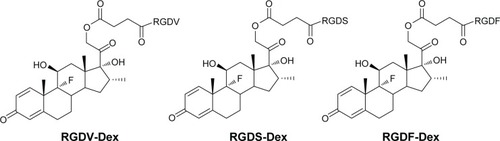
Arg-Gly-Asp-Val (RGDV), Arg-Gly-Asp-Ser (RGDS) and Arg-Gly-Asp-Phe (RGDF) are well-known antiadhesive peptides, which are the motifs of integrins in recognizing collagen, fibronectin, vitronectin, laminin, immunoglobulin superfamily, and plasma proteins. Recently, these peptides were used to modify Dex, yielding three nanoscale conjugates RGDV-Dex, RGDS-Dex, and RGDF-Dex (), which are capable of targeting integrins. In a mouse model, the conjugates are superior to Dex in inhibiting inflammatory and osteoporotic actions.Citation26 However, whether they are worthy of development remains unclear. In this context, the nanostructures, immunosuppressive activities, thrombosis risks, and toxicities of these three conjugates were investigated.
Figure 1 Structure of RGDV-Dex, RGDS-Dex, and RGDF-Dex.
Materials and methods
Animals
Male Wistar rats (male, weighing 250–300 g), BALB/C mice (male, 14 weeks in age), and ICR mice (male, 22±2 g) were purchased from Laboratory Animal Center of Capital Medical University. The animals were housed in polypropylene cages at 22°C±2°C and relative humidity of 60%–70%, with automatically controlled 12/12 hours light/dark cycle, and provided with the standard pellet diet and drinking water in plastic bottles. All assays were conducted between 9 am and 4 pm. Overnight fast was imposed before assays.
Work performed was based on a protocol reviewed and approved by the ethics committee of Capital Medical University. The committee assures that the welfare of the animals was maintained in accordance to the requirements of the Animal Welfare Act and in accordance to the NIH Guide for Care and Use of Laboratory Animals. Statistical analyses of all the biological data were carried out by use of analysis of variance (ANOVA). P-values <0.05 were considered statistically significant.
RGDV-Dex, RGDS-Dex, and RGDF-Dex
Dex was modified with RGD-peptides. In the 9-step modification procedure, the strategy with butane diacid bridging the hydroxy group of Dex at 21-position and the N-terminal amino group of the RGD-peptides was used, and three novel conjugates RGDV-Dex, RGDS-Dex, and RGDF-Dex were obtained. The high-performance liquid chromatography (HPLC) purity (CHIRALPAK AH-H column, 4.6×250 mm, Daicel Chemical Industries Ltd., Osaka, Japan) of these conjugates was 97%–98%. The chromatograms and the optical spectra are shown in –.
Transmission electron microscopy
The examinations of the nanostructures of RGDV-Dex, RGDS-Dex, and RGDF-Dex were performed on a transmission electron microscope (TEM, JSM-6360 LV, JEOL, Tokyo, Japan). The solutions of 10−8, 10−10, and 10−12 M of RGDV-Dex, RGDS-Dex, or RGDF-Dex in water (pH 7.0) were dripped onto a formvar coated copper grid, and then a drop of anhydrous ethanol was added to promote the removal of water. At first, the grid was allowed to thoroughly dry in air and then was heated at 37°C for 24 hours. The copper grids were viewed under the TEM. The nanostructures were determined from counting over 100 species in randomly selected regions on the copper grids. All determinations were carried out in triplicate grids. The TEM was operated at 80 kV, electron beam accelerating voltage. Images were recorded on an imaging plate (Gatan Bioscan Camera model 1792, Gatan, Inc., Pleasanton, CA, USA) with 20 eV energy windows at 6,000–400,000× and were digitally enlarged.
Atomic force microscopy
To show that our solution was transparent, but not colloidal, we used 10−8 M solution, the highest concentration among the three concentrations, to record the atomic force microscopy (AFM) images. AFM tests were performed by using the contact mode on Nanoscope 3D AFM (Veeco Instruments, Ltd, Plainview, NY, USA) under ambient conditions. Samples of rat plasma alone or ultrapure water alone and rat plasma or ultrapure water plus RGDV-Dex, RGDS-Dex, and RGDF-Dex (10−8 M) were used for recording the images, and all samples were imaged in triplicate.
Faraday–Tyndall effect test
The self-assembly property of RGDV-Dex, RGDS-Dex, and RGDF-Dex in aqueous solution was visualized with laser (650 nm) induced Faraday–Tyndall effect.Citation27 For this purpose, pH 7.0 ultrapure water and pH 2.0 ultrapure water were irradiated with laser beam of 650 nm, and Faraday–Tyndall effect was recorded. Similarly, the 10−8 M solution of RGDV-Dex or RGDS-Dex or RGDF-Dex in ultra-pure water (pH 7.0 and pH 2.0) was irradiated with laser beam of 650 nm and Faraday–Tyndall effect was recorded.
Size distribution test
After 7-day storage at room temperature, size distribution test of the 10−8 M solutions of RGDV-Dex or RGDS-Dex or RGDF-Dex in ultrapure water (pH 7.0 and pH 2.0) was carried out on a Malvern Zetasizer (Nano-ZS90; Malvern Instruments, Malvern, UK) with the DTS (Nano) Program.
ζ-Potential test
Seven days after room temperature storage, the ζ-potential of 10−8 M solution of RGDV-Dex or RGDS-Dex or RGDF-Dex in ultrapure water (pH 7.0) was measured using the automatic mode on a Malvern’s Zetasizer (Nano-ZS90; Malvern Instruments) with the DTS (Nano) Program.
Concanavalin A-induced lymphocyte proliferation assay
BALB/C mouse was killed, the spleen was extirpated in asepsis condition, triturated with injector core on stainless steel mesh of 200, washed with PBS (0.5 mL ×2), at 1,500 rpm/min centrifuged for 10 minutes suspended in Roswell Park Memorial Institute-1640 medium to form 5×10⁶ cells/mL suspension. To each well of the 96-well microplate, 100 µL cell suspension and 20 µL concanavalin A (ConA, final concentration, 5 µg/mL) were successively added, the 96-well microplate was cultured at 37°C in a humidified atmosphere of 5% CO2 for 4 hours. ConA induced proliferation of spleen lymphocytes of BALB/C mice was measured. Based on the tested optical density (OD) and % inhibition = [(OD4a–c − ODConA)/ODConA] ×100% (where in OD4a–c represents the OD value derived from the lymphocytes treated with RGDV-Dex, or RGDS-Dex, or RGDF-Dex, ODConA represents the OD value derived from the lymphocytes treated with ConA), the inhibitory rates of RGDV-Dex, or RGDS-Dex, or RGDF-Dex were calculated, of which the inhibitions at various concentrations (1×10⁻⁴, 8×10⁻⁵, 5×10⁻⁵, 2×10⁻⁵, 1×10⁻⁵, 8×10⁻⁶, 5×10⁻⁶, and 1×10⁻⁶ M) were calculated to draw the proliferation curve of mouse spleen cells and to calculate the half-maximal inhibitory concentration (IC50) values.
Myocardium survival time assay
The split hearts (3×3 mm in size) were prepared with the hearts of C57bl/6 mice of near full term fetuses to 48 hours old and transplanted into the base of the “pocket” near the distal edge of the ear of BALB/C mice (male, 14 weeks in age) by following the standard procedure. One day after transplantation, the mice in positive control group were intraperitoneally injected the suspension of cyclosporine A (CsA) in 0.5% carmellose sodium (CMCNa, 2.5 µmol/kg/d), the mice in negative control group were orally given 0.2 mL of 0.5% CMCNa/mouse/d, the mice in RGDV-Dex or RGDS-Dex treated groups were orally given the suspensions of the respective drugs in 0.5% CMCNa (1.43 µmol/kg/d), the mice in RGDF-Dex treated groups were orally given the suspension of the drug in 0.5% carmellose sodium (CMCNa, 1.43, 0.143, and 0.0143 µmol/kg/d), and the treatment was continued for 15 days. From seventh day, the split hearts implanted near the distal edge of the mouse ear received cardiogram examination every day to monitor their survival.
In vivo antithrombotic assay
Wistar rats were used in this assay. CMCNa (negative control) or aspirin (positive control) or RGDV-Dex or RGDS-Dex or RGDF-Dex suspended in CMCNa before administration was kept in an ice bath. Thirty minutes after the oral administration of 3 mL/kg of CMCNa or 3 mL/kg of aspirin (Beijing Pharmaceutical Works, Beijing, People’s Republic of China) in CMCNa (167 µmol/kg, 16.7 µmol/kg), or 3 mL/kg of RGDV-Dex in CMCNa (1.43 µmol/kg) or 3 mL/kg of RGDS-Dex in CMCNa (1.43 µmol/kg), or 3 mL/kg of RGDF-Dex in CMCNa (1.43 µmol/kg) the rats were anesthetized with pentobarbital sodium (Beijing Pharmaceutical Works, 80.0 mg/kg, intraperitoneal injection) and the right carotid artery and left jugular vein were separated. A weighed 6 cm thread was inserted into the middle of a polyethylene tube which was filled with heparin sodium (50 IU/mL in normal saline). One end of the polyethylene tube was inserted into the left jugular vein. From the other end of the polyethylene tube, heparin sodium was injected as anticoagulant, and this end was inserted into the right carotid artery. Blood was allowed to flow from the right carotid artery to the left jugular vein through the polyethylene tube for 15 minutes. The thread was retrieved to obtain the thrombus weight. After operation, the treated rats were euthanized immediately by exsanguination from the right carotid artery under pentobarbital sodium anesthesia.
In vivo tail bleeding time assay
The assay was performed by following a standard procedure. In brief, male ICR mice (18–22 g) were orally administered with CMCNa (0.5%, 0.2 mL/mouse of dose) or the suspension of Dex in CMCNa (dose, 2.5 µmol/kg), and orally administered with the suspension of RGDF-Dex or RGDS-Dex or RGDV-Dex in CMCNa (dose, 1.43 µmol/kg). Thirty minutes after the administration, the mouse was placed in a tube holder, the tail of the mouse was protruded, and 2 mm of tail tine was cut off. The flowing blood was gently wiped away with a filter paper every 30 seconds until blood flow stopped; this helped calculate the bleeding time.
Acute toxicity assay
Male ICR mice were purchased from Laboratory Animal Center of Capital Medical University for determining acute toxicity. The mice were maintained at 21°C with a natural day/night cycle in a conventional animal colony and were 10 weeks old at the beginning of the experiment. Twelve hours after fasting, mice were randomly divided into eight groups (six per group). The mice were orally administered with the suspension of RGDV-Dex or RGDS-Dex or RGDF-Dex in CMCNa (dose: 14.3 µmol/kg, 100 times of 0.143 µmol/kg of the minimal effective dose) or CMCNa (0.5%, 0.2 mL/mouse). Four hours or 7 days after the administration, the mice received ether anesthesia and were sacrificed to sample the blood and organs for toxic analysis.
Determination of serum ALT, AST, and Cr
To estimate the effect of RGDV-Dex, or RGDS-Dex, or RGDF-Dex on liver and kidney function, acute toxicity assay was performed on the blood of ICR mice and the blood was centrifuged to obtain serum for measuring alanine transaminase (ALT), aspartate transaminase (AST), and creatinine (Cr) by using alanine aminotransferase assay kit, aspartate aminotransferase assay kit, and creatinine assay kit, respectively, on a microtiter plate reader within 15 minutes and recording OD value. All kits were purchased from Nanjing Jiancheng Bioengineering Institute. According to the standard curve, the serum concentrations of ALT, AST, and Cr were calculated (n=12). Data were statistically analyzed by Student’s t-test. P-values <0.05 were considered statistically significant.
Results
Concentration and structure-dependent TEM images
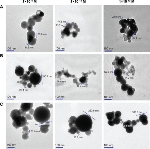
In order to examine the effect of the concentration on the nanofeature, the TEM images of 10−8, 10−10, and 10−12 M of RGDV-Dex, RGDS-Dex, and RGDF-Dex in ultrapure water (pH 7.0) were recorded and are shown in . The nanoparticle diameters of 10−8, 10−10, and 10−12 M of RGDF-Dex are 35–121, 25–75, and 23–85 nm, respectively. The nanoparticle diameters of 10−8, 10−10, and 10−12 M of RGDS-Dex are 23–186, 18–92, and 14–123 nm, respectively. The nanoparticle diameters of 10−8, 10−10, and 10−12 M of RGDV-Dex are 33–250, 11–254, and 26–103 nm, respectively. The diameter of the nanoparticles is decided by both concentration and the sequence of RGD-peptides.
Figure 2 TEM images showing the effects of concentration.
Abbreviations: TEM, transmission electron microscope; RGDF, Arg-Gly-Asp-Phe; RGDS, Arg-Gly-Asp-Ser; RGDV, Arg-Gly-Asp-Val; Dex, dexamethasone.
Rat plasma and water do not affect the AFM image
To examine nanofeature of the conjugates in blood, the AFM images of 10−8 M of RGDV-Dex, RGDS-Dex, and RGDF-Dex in rat plasma were recorded and are shown in . As seen, in rat plasma RGDF-Dex, RGDS-Dex, and RGDV-Dex form the nanoparticles of ~101, ~163, and ~160 nm in size, respectively, while rat plasma alone does not give any comparable nanoparticles. Of three conjugates, RGDF-Dex forms the smallest nanoparticles.
Figure 3 AFM images.
Abbreviations: AFM, atomic force microscopy; RGDF, Arg-Gly-Asp-Phe; RGDS, Arg-Gly-Asp-Ser; RGDV, Arg-Gly-Asp-Val; Dex, dexamethasone.
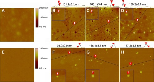
To exclude the effect of TEM and AFM models on the nanoparticle size, the AFM images of RGDV-Dex, RGDS-Dex, and RGDF-Dex in ultrapure water (10−8 M) were also recorded and are shown in . As seen in both rat plasma and ultrapure water, 10−8 M of RGDV-Dex, RGDS-Dex, and RGDF-Dex form similar nanoparticles.
The aqueous solution of the conjugates show nanoproperty
To show that the aqueous solution of the conjugates has nanoproperty, Faraday–Tyndall effect, size distribution, and ζ-potential tests were carried out in the 10−8 M solution of RGDF-Dex, RGDS-Dex, and RGDV-Dex in ultrapure water. indicate that similar to ultrapure water, without laser beam irradiation the solutions of RGDF-Dex, RGDS-Dex, and RGDV-Dex in ultrapure water are clean, while the irradiation of 650 nm laser beam induces the solutions of the conjugates in pH 7.0 and pH 2.0 ultrapure water to show Faraday–Tyndall effect.
Figure 4 Nanoproperties of 10−8 M solution of RGDF-Dex, RGDS-Dex, and RGDV-Dex in water.
Abbreviations: St dev, standard deviation; RGDF, Arg-Gly-Asp-Phe; RGDS, Arg-Gly-Asp-Ser; RGDV, Arg-Gly-Asp-Val; Dex, dexamethasone.
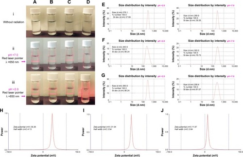
The 10−8 M of conjugate aqueous solutions were also tested on a Malvern’s Zetasizer (Nano-ZS90; Malvern Instruments) with the DTS (Nano) Program to characterize the size distribution. indicate that the sizes of RGDF-Dex, RGDS-Dex, and RGDV-Dex in pH 2.0 and 7.0 ultrapure water are 276.1±37.7 and 268.9±39.3, 263.9±86.2, and 302.0±76.7 nm as well as 263.9±86.2 and 258.4±81.6 nm, respectively. The size distribution of the conjugates in pH 7.0 ultrapure water is less than that in pH 2.0 ultrapure water.
ζ-Potential tests were measured in the 10−8 M conjugate aqueous solutions. indicate that the ζ-potentials of RGDF-Dex, RGDS-Dex, and RGDV-Dex in pH 7.0 ultrapure water are 39.3, 31.5, and 11.9 mV, respectively; RGDF-Dex possesses the highest value.
The conjugates inhibit spleen lymphocytes’ proliferation
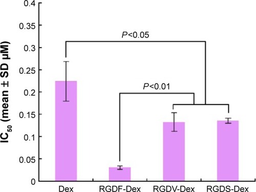
The immunosuppressive activities of the conjugates were evaluated by ConA-induced proliferation of BALB/c mouse spleen lymphocytes. indicates that the IC50 values of Dex, RGDV-Dex, RGDS-Dex, and RGDF-Dex against spleen lymphocyte proliferation are 0.223, 0.133, 0.136, and 0.031 µM, respectively. The IC50 values of RGDV-Dex and RGDS-Dex are significantly lower than that of Dex, but significantly higher than that of RGDF-Dex. Thus, RGDF-Dex possesses the highest inhibition on the spleen lymphocyte proliferation of BALB/C mice in vitro.
Figure 5 The effect of RGDV-Dex, RGDS-Dex, and RGDF-Dex on ConA-induced proliferation of the spleen lymphocytes of BALB/C mouse, n=3.
The conjugates prolong survival time of the implanted myocardium
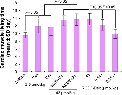
The immunosuppressive activities of the conjugates were also evaluated by survival time of the myocardium implanted in BALB/C. The mice were treated with oral CMCNa (0.5%, negative control), or intraperitoneal CsA (positive control, 2.5 µmol/kg/d), or oral Dex (reference control, 2.5 µmol/kg/d), or oral conjugate (1.43 µmol/kg/d) for 15 days. indicates that compared to the survival time of the myocardium implanted in the mice treated with CMCNa (9.6 days), those treated with CsA (12.0 days) and Dex (11.7 days) led to the increase of survival time by 2.4 and 2.1 days, respectively. Comparing the survival time of the myocardium implanted in the mice treated with Dex, treating with RGDV-Dex (13.4 days), RGDS-Dex (13.7 days), and RGDF-Dex (13.8 days) led to the increase of survival time by 1.7, 2.0, and 2.1 days, respectively. The myocardium implanted in the mice treated with RGDF-Dex exhibits the longest survival time. Thus, the dose-dependent action of RGDF-Dex was tested. indicates that the survival time of the myocardium implanted in the mice treated with 1.43, 0.143, and 0.0143 µmol/kg/d of RGDF-Dex is 13.8, 12.3, and 9.9 days, respectively, suggesting that RGDF-Dex dose-dependently increases the survival time.
Figure 6 The survival time of the implanted myocardium in the “pocket” near the distal edge of the ear of BALB/C mice, n=12.
The conjugates induce no coagulation risk
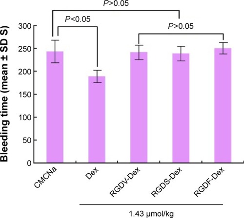
The tail bleeding time assay was used to evaluate the coagulation risk of the conjugates. indicates that the tail bleeding time of the mice treated with Dex (1.43 µmol/kg of dose) is significantly shorter than that of the mice treated with CMCNa. In contrast, 1.43 µmol/kg of RGDF-Dex, RGDS-Dex, and RGDV-Dex do not alter the tail bleeding time of CMCNa treated mice.
Figure 7 Tail bleeding time of RGDV-Dex-, RGDS-Dex-, and RGDF-Dex-treated mice, n=12.
The conjugates have antithrombotic action
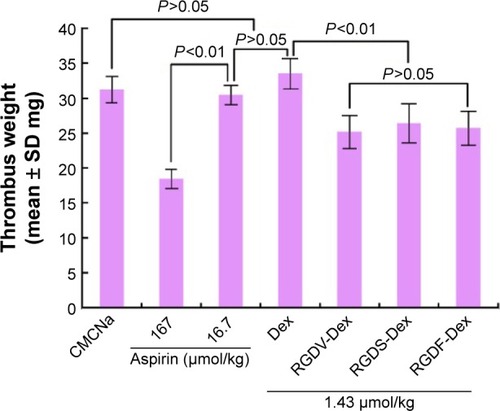
The antithrombotic activities of the conjugates were evaluated with rat thrombosis assay and represented as thrombus weight. In the assay, the rats were orally given CMCNa (0.5%, negative control) or the suspension of aspirin in CMCNa (positive control, 167 and 16.7 µmol/kg), or the suspension of Dex in CMCNa (1.43 µmol/kg dose), or the suspension of the conjugate in CMCNa (1.43 µmol/kg dose). indicates that the thrombus weight of Dex treated rats is equal to that of CMCNa- or 16.7 µmol/kg aspirin-treated rats. The thrombus weights of RGDV-Dex-, RGDS-Dex-, and RGDF-Dex-treated rats are significantly lower than that of CMCNa treated rats.
Figure 8 Anti-thrombotic activities of Dex, RGDV-Dex, RGDS-Dex, and RGDF-Dex, n=12.
The conjugates do not slow mouse growth
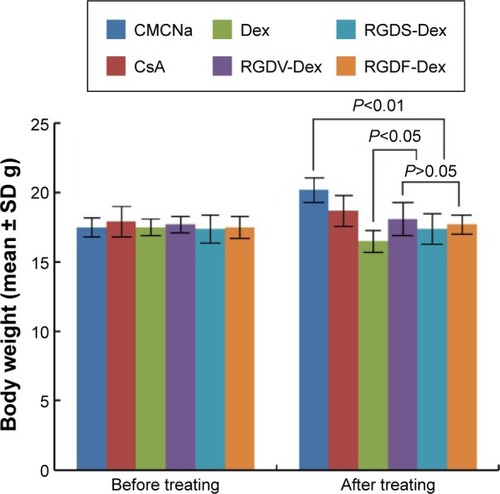
The toxic reaction of the conjugates was mirrored by the body weight of the treated BALB/C mice. indicates that 15 days after the treatment, the body weight of the mice treated with 2.5 µmol/kg/d of Dex is significantly lower than that of the mice treated with 1.43 µmol/kg/d of RGDV-Dex, RGDS-Dex, or RGDF-Dex.
Figure 9 Body weight of the treated BALB/C mice, n=12.
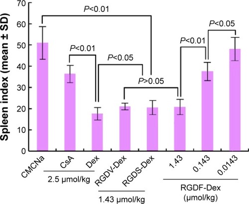
The toxic reaction of the conjugates was also mirrored with the spleen index (spleen weight/body weight) of the BALB/C mice. indicates that 15 days after the treatment, the spleen index of the mice treated with 2.5 µmol/kg/d of Dex is significantly lower than that of the mice treated with 1.43 µmol/kg/d of RGDV-Dex, RGDS-Dex, or RGDF-Dex.
Figure 10 Spleen index of the treated BALB/C mice, n=12.
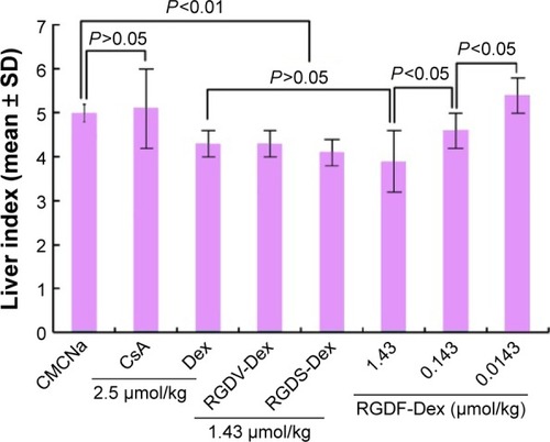
The toxic reaction of the conjugates was further mirrored with the liver index (liver weight/body weight) of the BALB/C mice. shows that 15 days after the treatment, the liver index of the mice treated with 2.5 µmol/kg/d of Dex is significantly lower than that of the mice treated with 1.43 µmol/kg/d of RGDV-Dex, RGDS-Dex, and RGDF-Dex.
Figure 11 Liver index of the treated BALB/C mice, n=12.
The conjugates do not induce acute toxicity
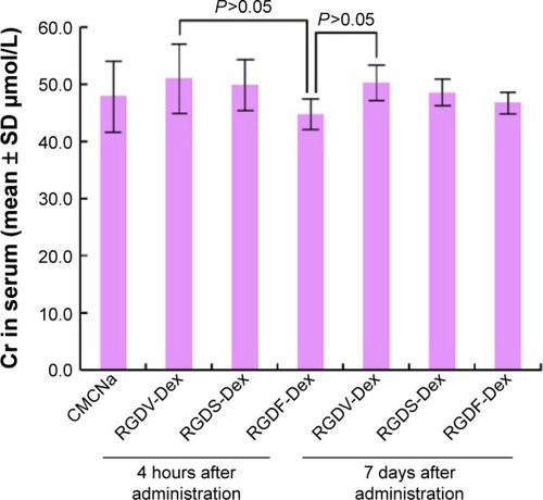
Acute toxicity assay was carried out in the healthy ICR mice by orally treating with the conjugates at a single dose of 14.3 µmol/kg, a dose of 100 times of the minimal effective dose 0.143 µmol/kg, and the mice were monitored for 7 days. It was found that during 7 days, the mice had no abnormal behavior, while the autopsy showed no morphological changes in the organs. Serum ALT, AST, and Cr of the mice were determined by the kits. – show that the serum ALT, AST, and Cr of the mice treated with 14.3 µmol/kg RGDV-Dex or RGDS-Dex or RGDF-Dex have normal values.
Figure 12 Serum ALT levels of the mice on which acute toxicity assay was performed, n=12.
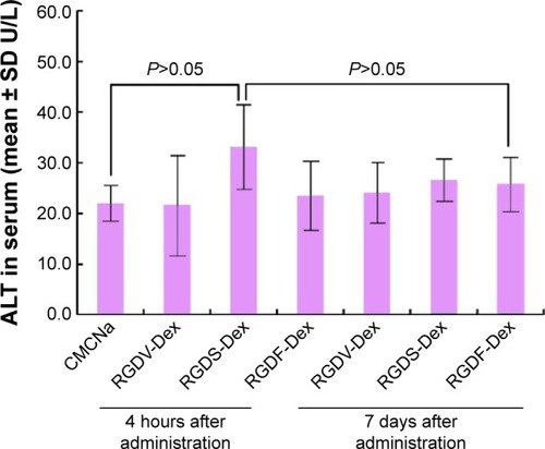
Figure 13 AST levels of the mice on which acute toxicity assay was performed, n=12.
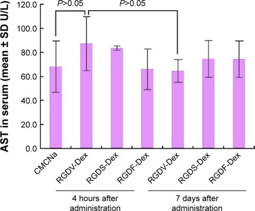
Figure 14 Serum Cr levels of the mice on which acute toxicity assay was performed, n=12.
Discussion
Dex is the most effective anti-inflammatory glucocorticoid in treating several chronic and acute inflammatory conditions,Citation1–Citation5 preventing rejection of the transplanted organ, and preserving graft function.Citation15,Citation16 However, the coagulation and osteoporosis risks restrict its clinical application.Citation25,Citation28,Citation29 Developing safe and effective novel derivatives of Dex is of clinical importance. RGDF, RGDV, and RGDS, the so called RGD-peptides, are antiadhesive peptides and have been widely used to modify various drugs, of which motif-like action with integrins can potentially enhance the therapeutic efficacy and reduce the coagulation and osteoporosis risks of drugs.Citation26,Citation30 Aimed at this, the present study examines the development profile of covalent conjugates of RGD-peptides and Dex (RGDV-Dex, RGDS-Dex, and RGDF-Dex) as new drugs. In vitro and in vivo, the activities of the conjugates are significantly higher than Dex itself and induce no coagulation risk. shows that of three conjugates against ConA-induced proliferation of spleen lymphocytes, RGDF-Dex exhibits the smallest IC50 value. indicates that of three conjugates affecting the implanted myocardium RGDF-Dex gives the longest survival time. Of three conjugates, in respect of the efficacies, RGDF-Dex is more worthy of developing as new drug. Besides, the in vitro and in vivo observations that the covalent introduction of RGDF benefit the efficacy are consistent with our previous work.Citation31,Citation32 shows that RGDF-Dex, RGDS-Dex, and RGDV-Dex, but not Dex itself, prolong the tail bleeding time of the mice. Taking RGDF-Dex, RGDS-Dex, and RGDV-Dex, but not Dex itself, inducing no osteoporosis into account,Citation26 RGD-peptide modification leads to the complete relief of side effects of Dex. The safety of RGDF-Dex, RGDS-Dex, and RGDV-Dex was further evaluated with acute toxicity assay. – indicate that even if the dose is 100-fold higher than the minimal effective dose, three conjugates are still unable to induce toxic reaction. Furthermore, RGD-peptide modification leads to Dex having an additional antithrombotic activity.
The effect of nanofeature and size on bioactivity is an interesting profile. Showing the relationships between the bioactivities of small molecules with their TEM images, between the concentrations of small molecules with their TEM images, and between the structures of small molecules with their TEM images has been our interests.Citation33–Citation36 To elucidate the contribution of the nanoproperty to the advantages of three conjugates, this study systematically characterizes their nanofeatures. The Faraday–Tyndall effect, size distribution, and ζ-potential of three conjugates in water ensure their properties as a true solution and their use as nanomedicine (). The TEM images of three conjugates in pH 7.0 and 2.0 ultrapure water of three concentrations clearly show the effect of pH, concentration, and RGD-peptide sequence on the nanofeatures and size. As seen in , RGDF sequence, pH 7.0, and lower concentration benefit the formation of smaller nanoparticles. Since the particles of 100 nm in diameter have minimal probability of being entrapped by macrophages in blood circulation,Citation37 the AFM images of small molecules in rat plasma were recorded in particular to simulate the feature of nanoparticles in blood circulation.Citation34–Citation36 The AFM images and the diameters of RGDF-Dex nanoparticles (close to 100 nm) shown in suggest that it can be safely delivered in blood circulation.
Conclusion
The development of novel, effective, and safe derivatives is of practical importance for Dex therapy. Our previous data and the present study demonstrate that covalent modification of Dex with RGD-peptides provides a strategy to enhance the anti-inflammatory activity, to prolong the survival time of the implanted myocardium, to minimize the side effects of osteoporosis and coagulation, to prevent the liver and the kidney from acute and chronic damage, and to provide a beneficial antithrombotic action. With respect to the size of the nanoparticles of three conjugates, RGDF-Dex is the most suitable for delivery in the circulation.
Supplementary materials
Optical spectra
Figure S1 1HNMR of RGDV-Dex.
Figure S2 13C NMR of RGDV-Dex.
Figure S3 1H NMR of RGDS-Dex.
Figure S4 13C NMR of RGDS-Dex.
Figure S5 1H NMR of RGDF-Dex.
Figure S6 13C NMR of RGDF-Dex.
Figure S7 HPLC chromatogram of RGDF-Dex.
Abbreviations: HPLC, high performance liquid chromatography; RGDF, Arg-Gly-Asp-Phe; Dex, dexamethasone.
Figure S8 HPLC chromatogram of RGDV-Dex.
Abbreviations: HPLC, high performance liquid chromatography; RGDV, Arg-Gly-Asp-Val; Dex, dexamethasone.
Figure S9 HPLC chromatogram of RGDS-Dex.
Abbreviations: HPLC, high performance liquid chromatography; RGDS, Arg-Gly-Asp-Ser; Dex, dexamethasone.
Acknowledgments
This work was supported by Beijing Municipal Science & Technology Commission (Z141100002114049), the Project of Construction of Innovative Teams and Teacher Career Development for Universities and Colleges under Beijing Municipality, the NSFC (81172930, 81273379, 81373265, 81202412, 81373264), 863 program (2015AA020902), Beijing Talent Program (2014000026833ZK23) and Joint Research and Development of protective agents for low dose radiation injury (2014DFR30930).
Disclosure.
The authors report no conflicts of interest in this work.
References
- BarnesPJGlucocorticosteroids: current and future directionsBr J Pharmacol20111631294321198556
- BarnesPJMechanisms and resistance in glucocorticoid control of inflammationJ Steroid Biochem Mol Boil201012027685
- Goldbach-ManskyRLipskyPENew concepts in the treatment of rheumatoid arthritisAnnu Rev Med200354119721612359827
- van VollenhovenRFTreatment of rheumatoid arthritis: state of the art 2009Nat Rev Rheumatol200951053154119798027
- ButtgereitFSaagKGCutoloMda SilvaJABijilsmaJWThe molecular basis for the effectiveness, toxicity, and resistance to glucocorticoids: focus on the treatment of rheumatoid arthritisScand J Rheumatol2005341142115903020
- SekhavatLDavarRBehdadSEfficacy of prophylactic dexamethasone in prevention of postoperative nausea and vomitingJ Epidemiol Glob Health20155217517925922327
- LowYWhiteWDHabibASPostoperative hyperglycemia after 4- vs 8–10-mg dexamethasone for postoperative nausea and vomiting prophylaxis in patients with type II diabetes mellitus: a retrospective database analysisJ Clin Anesth201527758959426303274
- ChuC-CHsingC-HShiehJ-PChienCCHoCMWangJJThe cellular mechanisms of the antiemetic action of dexamethasone and related glucocorticoids against vomitingEur J Pharmacol20147225485424184695
- VenkitaramanRLorenteDMurthyVA randomised Phase 2 trial of dexamethasone versus prednisolone in castration-resistant prostate cancerEur Urol201567467367925457497
- BadawyAAEl SakkaAPreoperative gabapentin alone or in combination with dexamethasone on postoperative pain relief after abdominal hysterectomies. A randomized controlled trialEgypt J Anaesth2015312107113
- BoyerDSYoonYHBelfortRJrThree-year, randomized, sham-controlled trial of dexamethasone intravitreal implant in patients with diabetic macular edemaOphthalmology2014121101904191424907062
- ParikhKHallMMittalVComparative effectiveness of dexamethasone versus prednisone in children hospitalized with asthmaJ Pediatr2015167363964426319919
- ZhanYZouSHuaFHigh-dose dexamethasone modulates serum cytokine profile in patients with primary immune thrombocytopeniaImmunol Lett20141601333824657323
- CochraneABHusainANAndersonASKimAYFedsonSEIncreased rejection rates in cardiac transplant associated with dexamethasoneSteroids200873444144818243261
- FuchsKMCoustanDRImmunosuppressant therapy in pregnant organ transplant recipientsSemin Perinatol200731636337118063120
- WeiselKDimopoulosMSongKWPomalidomide and low-dose dexamethasone improves health-related quality of life and prolongs time to worsening in relapsed/refractory patients with multiple myeloma enrolled in the MM-003 randomized phase III trialClin Lymphoma Myeloma Leuk201515951953026149712
- van OschDDielemanJMNathoeHMIntraoperative high-dose dexamethasone in cardiac surgery and the risk of rethoracotomyAnn Thorac Surg201510062237224226319483
- KaleYAydemirOCeylanOBasAYDemirelNHypertrophic cardiomyopathy after a single dose of dexamethasone in a preterm infantPediatr Neonatol201556426827023639746
- BungeJJHvan OschDDielemanJMDexamethasone for the prevention of postpericardiotomy syndrome: a dexamethasone for cardiac surgery substudyAm Heart J2014168112613124952869
- YuHCLuoYXPengHKangLHuangMJWangJPAvoiding perioperative dexamethasone may improve the outcome of patients with rectal cancerEur J Surg Oncol201541566767325744813
- WróbelASerefkoAWlaźPPoleszakEThe depressogenic-like effect of acute and chronic treatment with dexamethasone and its influence on the activity of anti-depressant drugs in the forced swim test in adult miceProg Neuropsychopharmacol Biol Psychiatry2014541524324824984273
- MakrydimaSFPistikiACChreliasCGThe immunomodulatory and anti-apoptotic effect of dexamethasone in imminent preterm labor: an experimental studyEur J Pharmacol20147305313524582761
- HitzertMMVan BraeckelKNde BokMMaathuisCGRozeEBosAFFunctional outcome at school age of preterm-born children treated with high-dose dexamethasoneEarly Hum Dev201490625325824602475
- RenKDusadAYuanFMacromolecular prodrug of dexamethasone prevents particle-induced peri-implant osteolysis with reduced systemic side effectsJ Control Release2014175101924326124
- RenHLiangDJiangXVariance of spinal osteoporosis induced by dexamethasone and methylprednisolone and its associated mechanismSteroids2015102657526216207
- YuHMeiSLiZZhaoMWangYZhuHRGD-peptides modifying dexamethasone: to enhance the efficacy of anti-inflammation and limit the risk of osteoporosisMed Chem Commun2015613451351
- LiawJ-WTsaiS-WLinH-HYenT-CChenB-RWavelength-dependent Faraday-Tyndall effect on laser-induced microbubble in gold colloidJ Quant Spectrosc Radiat Transfer20121131722342242
- ZamagniaEValdrèLPalaretiGCavoMPrevention of VTE in multiple myeloma patientsThromb Res20071206S133S13618023708
- MusallamKMDahdalehFSShamseddineAITaherATIncidence and prophylaxis of venous thromboembolic events in multiple myeloma patients receiving immunomodulatory therapyThromb Res2009123567968618992924
- XiongYZhaoMWangCChangHWPengSImproved antiosteoporosis potency and reduced endometrial membrane hyperplasia during HRT with estrogen-RGD peptide conjugatesJ Med Chem200750143340335317571867
- LiNKangGGuiLNovel Cu(II)-RGD-octapeptides: synthesis, coordination mode, in vitro anti-platelet aggregation/in vivo anti-thrombotic evaluation and correlation of sequence with nano-structureNanomedicine20117440340921272663
- WangYWuJKangGNovel nano-materials, RGD-tetrapeptide-modified 17β-amino-11α-hydroxyandrost-1,4-diene-3-one: synthesis, self-assembly based nano-images and in vivo anti-osteoporosis evaluationJ Mater Chem2012221146524659
- GuiLZhaoMWangYQinYLiuJPengSSynthesis, nano-features, in vitro thrombus lysis activity and in vivo thrombolytic activity of poly-α, β-aspartyl-l-alanineNanomedicine (Lond)20105570371420662642
- WuJWangYWangYCu2+-RGDFRGDS: exploring the mechanism and high efficacy of nanoparticles in anti-thrombotic therapyInt J Nanomedicine2015102925293825931819
- LiSWangYWangFSmall molecule PZL318: forming fluorescent nanoparticles capable of tracing their interactions with cancer cells and activated platelets, slowing tumor growth and inhibiting thrombosisInt J Nanomedicine2015105273529226345234
- JinSWangYZhuHNano-sized aspirin-Arg-Gly-Asp-Val: delivery of aspirin to thrombus by a target carrier Arg-Gly-Asp-Val tetrapeptideACS Nano2013797664767323931063
- FujitaYMieMKobatakeEConstruction of nanoscale protein particle using temperature-sensitive elastin-like peptide and polyaspartic acid chainBiomaterials200930203450345719324406