
Abstract
Background
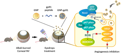
Corneal neovascularization (NV) is a process of abnormal vessel growth into the transparent cornea from the limbus and can disturb the light passing through the cornea, resulting in vision loss or even blindness. The use of nanomedicine as an effective therapeutic formulation in ophthalmology has led to higher drug bioavailability and a slow drug release rate. In this research, we designed and explored the feasibility of a new nanomedicine, gp91 ds-tat (gp91) peptide-encapsulated gelatin nanoparticles (GNP-gp91), for inhibiting corneal angiogenesis.
Methods
GNP-gp91 were prepared by a two-step desolvation method. The characterization and cytocompatibility of GNP-gp91 were analyzed. The inhibition effect of GNP-gp91 on HUVEC cell migration and tube formation was observed by an inverted microscope. The drug retention test in mouse cornea was observed by in vivo imaging system, fluorescence microscope, and DAPI/TAMRA staining. Finally, the therapeutic efficacy and evaluation of neovascularization-related factors were conducted through the in vivo corneal NV mice model via topical delivery.
Results
The prepared GNP-gp91 had a nano-scale diameter (550.6 nm) with positive charge (21.7 mV) slow-release behavior (25%, 240hr). In vitro test revealed that GNP-gp91 enhanced the inhibition of cell migration and tube formation capacity via higher internalization of HUVEC. Topical administration (eyedrops) of the GNP-gp91 significantly prolongs the retention time (46%, 20 min) in the mouse cornea. In chemically burned corneal neovascularization models, corneal vessel area with a significant reduction in GNP-gp91 group (7.89%) was revealed when compared with PBS (33.99%) and gp91 (19.67%) treated groups via every two days dosing. Moreover, GNP-gp91 significantly reduced the concentration of Nox2, VEGF and MMP9 in NV’s cornea.
Conclusion
The nanomedicine, GNP-gp91, was successfully synthesized for ophthalmological application. These data suggest that GNP-gp91 contained eyedrops that not only have a longer retention time on the cornea but also can treat mice corneal NV effectively delivered in a low dosing frequency, GNP-gp91 eyedrops provides an alternative strategy for clinical ocular disease treatment in the culture.
Graphical Abstract
Introduction
Corneal neovascularization (NV) is a pathological condition of the cornea characterized by the invasion of new blood vessels into the avascular corneal regions from the limbus.Citation1 Common causes of corneal neovascularization include chemical burns, trauma, contact lens wear, hypoxia, infection, corneal graft rejection, and immunological disease.Citation1 The increased permeability of these new vessels leads to chronic corneal edema, lipid exudation, inflammation, and scar formation.Citation2–4 The mechanism underlying inflammation-induced NV results from the disruption of angiogenic and antiangiogenic balance in the cornea, which leads to significant corneal transparency change and visual impairment.Citation3,Citation5,Citation6 Several medical and surgical options have been used to treat corneal NV, such as laser ablation, photodynamic therapy, and anti-vascular endothelial growth factor (VEGF) treatment.Citation3,Citation7 Anti-VEGF treatment administered by topical, subconjunctival, and intraocular application is one of the major therapeutics used for ocular neovascularization treatment nowadays.Citation3,Citation7
While anti-VEGF treatment is promising, partial efficacy, delivered in an invasive manner, resistance for longer treatment, and side effects have limited its clinical effect. In addition to VEGF signaling, there are several other angiogenic factors are also known to be involved in ocular neovascularization.Citation8–10 Thus, new compounds/drugs or formulas have also been developed for application in inhibiting vessel formation to effectively treat ocular NV. For instance, blockade of nicotinamide adenine dinucleotide phosphate (NADPH) oxidases, especially NADPH oxidase 2 (Nox2), by gp91ds-tat peptide prevents diabetes-induced premature retinal endothelial cell senescence.Citation11 Previous studies demonstrated that gp91ds-tat peptide (gp91) can mediate oxidative stress and reduce reactive oxygen species (ROS),Citation12,Citation13 which mediates many biological responses, including angiogenesis.Citation14 Therefore, gp91 is considered a candidate for anti-angiogenic therapy.
For drug delivery to the eye, subconjunctival injection and topical therapy are the two feasible approaches.Citation15,Citation16 Compared with subconjunctival injection, topical therapy is the preferred method for patients because of the relatively safe and widely applicable approach. However, a low bioavailability (<5%) of topical delivery due to various anatomical and physiological barriers, such as not allowing therapeutic agents to penetrate the corneal epithelium due to intracellular tight junctions,Citation17,Citation18 and constraints such as blinking and lacrimal draining have limited its applications.Citation18,Citation19 Higher concentrations or repeated dosing is a way to overcome these limitations of drug administration, but these methods are not accurate due to a dramatic variation in the therapeutic concentration in the ocular tissue.Citation19–21 These limitations have prompted researchers to develop new and effective drug delivery systems for eye treatment.
Nanoparticles (NPs) are distinct particulate systems with a size of 10–1000 nm and a specific surface charge.Citation19 With a size lower than 1000 nm, not only the scratching and irritation of the eyes can be avoided when using nanoparticles,Citation22,Citation23 but the corneal permeability of drugs will also be enhanced.Citation24,Citation25 Variant kinds of nanoparticles such as liposome, gold nanoparticles, and polymeric nanoparticles were studied.Citation1,Citation19,Citation24 The biological polymer, gelatin, was chosen in the study as the particle matrix because of its biocompatibility and biodegradation.Citation26,Citation27 Gelatin nanoparticles (GNPs) can be fabricated with different surface charge (manipulated by different types of gelatins), and the drug release behavior of GNP can be controlled by changing the crosslinking agent and time.Citation27 Owing to the cornea/conjunctiva having a negatively charged ocular surface, cationic NPs can be attracted to the ocular surface for topical drug delivery,Citation26,Citation28,Citation29 and the corneal permeability of drug-loaded cationic GNP will also increase due to electrostatic interaction.Citation26,Citation29,Citation30 Furthermore, GNPs were successful in enhancing drug retention on the ocular surface and can be applied as eye drops for treating corneal disease.Citation25,Citation26,Citation29,Citation30
In this study, we used gp91 as the active component for treating pathological angiogenesis in cornea. The nanomedicine, gp91-loaded GNP (GNP-gp91) was designed, synthesized, and proceeded properties’ examination. The therapeutic efficacy of GNP-gp91 for the inhibition of vessel function was evaluated in vitro and in vivo. The schematic drawing of this study is revealed in .
Figure 1 Schematic illustration of gp91ds-tat peptide (gp91)-loaded gelatin nanoparticles (GNP-gp91) as eye drops to treat alkali-burned induced corneal neovascularization (NV) in a mouse model.
Materials and Methods
Materials
Human umbilical vein endothelial cells (HUVECs) were purchased from the Bioresource Collection and Research Center (Hsinchu, Taiwan). Medium 199 and penicillin-streptomycin-neomycin (PSN) antibiotic mixture were purchased from Life Technologies (Camarillo, CA, USA). Fetal bovine serum (FBS) was obtained from HyClone (South Logan, Utah, USA). The endothelial cell growth supplement (ECGS) was bought from Merck Millipore (Billerica, MA, USA). The gp91-ds-tat peptide (gp91) (RKKRRQRRRCSTRIRRQL) and fluorescein isothiocyanate (FITC)-labeled gp91-ds-tat (gp91FITC) were synthesized by MDBio Inc. (Taipei, Taiwan). Gelatin type B (derived from bovine skin, Bloom 225), heparin, TritonTM X-100, 4’,6-diamidino-2-phenylindole (DAPI), cell counting Kit-8 (CCK-8), live/dead cell double staining kit, lipopolysaccharide (LPS), and 2’7’-dichlorofluorescin diacetate (DCFH-DA) were purchased from Sigma-Aldrich (St. Louis, MO, USA). The Vivaspin 500 ultrafiltration device was purchased from Sartorius (Göttingen, Germany). Float-A-LyzerⓇG2 Dialysis Device (MWCO 20 kDa) was acquired from Spectrum Laboratories, Inc. (Rancho Dominguez, CA, USA). Tetramethyl rhodamine succinyl ester (TAMRA-SE) was purchased from Invitrogen (Carlsbad, CA, USA). The MatrigelTM matrix was purchased from Corning (Corning, NY, USA). Rompun solution (2%) was obtained from Bayer Korea, Ltd. (Ansan City, Gyeonggi-do, Korea), and Zoletil 50 was purchased from Virbac Animal Health (Vauvert, Nice, France). Topical anesthesia solution (Alcaine® 0.5% ophthalmic solution) was obtained from Alcon-Couvreur N.V. (Puurs, Belgium). Grafco® silver nitrate applicators were purchased from Medline Industries Inc. (Mun-delein, IL, USA). All other chemicals were purchased from Sigma-Aldrich.
Preparation of GNP and gp91-Loaded GNP (GNP-gp91)
The GNP and GNP-gp91 were prepared by a two-step desolvation method with slightly modified.Citation30,Citation31 First, 5% (w/v) type B gelatin solution was purified using acetone for the first desolvation to remove gelatin with low molecular weight.Citation31,Citation32 The residual gelatin was then redissolved in hot water to get gelatin solution (1% w/v) again and adjusted its pH value to 8 or 9 after cooling. Then, 300 μL of deionized water or gp91 peptide solution (gp91) (10 mg/mL) was added into 1 mL gelatin solution separately for preparation of GNP or peptide loaded GNP (GNP-gp91). After that, 1.6 mL ethanol was then added dropwise to the gelatin solution to form gelatin nanoparticles (GNPs); followed by adding 16.25 μL glutaraldehyde (8% (w/v)) and stirring for 30 minutes for crosslinking reaction.Citation30 Finally, the ethanol was evaporated using a rotary evaporator (EYELA, Tokyo, Japan), and the excess impurities were removed using an ultrafiltration device (Vivaspin 500, MWCO 30 kDa). The GNP and GNP-gp91 were stored at 4 °C in deionized water for further application.
Characterization of GNP and GNP-gp91
The particle size distribution and zeta potential were analyzed using a zetasizer (Nano ZS 90, Malvern Instruments, Malvern, UK, scattering light: 90°, equilibration time: 180s, temperature: 25°C). The particle morphology was observed using a transmission electron microscope (TEM, Hitachi HT-7700, Tokyo, Japan, acceleration voltage: 75.0 kV). Samples were dropped on carbon-coated nickel mesh and dried overnight before analysis. The quantification of gp91 was determined using the Bicinchoninic Acid (BCA) protein assay to measure the unencapsulated gp91 peptide content after GNP-gp91 preparation, and the encapsulation efficiency (EE) was calculated as the final peptide content compared with the initial added peptide content. For the drug release examination, the content of fluorescent dye (FITC) released out the dialysis bag was quantified for dye/drug release. The FITC labeled gp91 (gp91FITC) and gp91FITC-loaded GNP (GNP-gp91FITC) were added into a A-LyzerⓇG2 Dialysis Device, then soaked in PBS and stirred at a condition of 100 rpm and 37°C. Solutions were sampled at the indicated time and examined using a microplate reader (Varioskan Flash, Thermo Fisher Scientific) at excitation wavelength: 494 nm and emission wavelength: 518 nm. We compared the optical density (OD) value of tested samples with FITC standard curve for dye quantification. The total amount of dye re-leased was calculated as the accumulated release rate for the release evaluation.
In vitro Study
Cell Viability
Human umbilical vascular endothelial cells (HUVECs) were used in this study and cultured in M199 medium contained supplemented with 10% fetal bovine serum, 1% penicillin/streptomycin, 30 μg/mL endothelial cell growth supplement, and 25 U/mL heparin. All in vitro experiments were incubated in an incubator at 37°C in a 5% CO2 atmosphere. For cytotoxicity test, HUVECs were seeded in a 96-well plate at a density of 5 × 10³ cells/well and cultured overnight. After removing the cultured medium, HUVECs were co-cultured with various formulations (GNP, gp91, and GNP-gp91) at peptide concentrations of 50–150 μg/mL for 24 hours and 72 hours. After removing the co-cultured medium, 100 μL medium and 10 μL Cell Counting Kit-8 (CCK-8) reagent were mixed and added into each well for 3 hours. Finally, the cell viability was calculated after analyzing the absorbance using a microplate reader (Epoch 2, BioTek, Winooski, VT, USA) at 450 nm. The percentage of viable cells was calculated in comparison to that of the control cells cultured with medium only. For Live&Dead staining, HUVECs were seeded in a 24-well plate at a density of 3.3 × 10⁴ cells/well and cultured overnight. After removing the cultured medium, HUVECs were co-cultured with various formulations (333 μg/mL GNP, 100 μg/mL gp91, and GNP-gp91: 333 μg/mL GNP with 100 μg/mL gp91) for 1 and 3 days. After removing the co-cultured medium, the diluted calcein-AM and propidium iodide (PI) were added into each well for 10 min. Finally, the cell morphology was observed using an inverted fluorescence microscope (DMi8, Leica, Wetzlar, Germany).
Cellular Uptake
HUVECs were seeded in a 24-well plate at a density of 2 × 10⁵ cells/well and cultured overnight. After removing the cultured medium, HUVECs were co-cultured with gp91FITC and GNP-gp91FITC (gp91FITC concentration: 120 μg/mL) for 0.5 hours and 2 hours. After removing the co-cultured medium, HUVECs were collected by adding 0.05% trypsin-EDTA for 5 minutes, centrifuged, and resuspended in PBS. Finally, the cellular uptake results were analyzed using a flow cytometer (Attune™ NxT Acoustic Focusing Cytometer, Thermo Fisher Scientific, Carlsbad, CA). The auto-fluorescent intensity of cells was adjusted to the range of 102–103 as the background value.
Reactive Oxygen Species (ROS) Examination
The ROS content of cells was determined using dichloro-dihydro-fluorescein diacetate (DCFH-DA) assay.Citation33 Briefly, HUVECs were seeded in a 96-well plate at a density of 5 × 10³ cells/well and cultured overnight. After removing the cultured medium, mediums with 500 ng/mL lipopolysaccharide (LPS) were added and tested for 6 hours. After LPS stimulation, mediums were removed, and HUVECs were then co-cultured with various formulations (333 μg/mL GNP, 100 μg/mL gp91, and GNP-gp91: 333 μg/mL GNP with 100 μg/mL gp91) for 24 hours. After removing the co-cultured medium, 100 μL DCFH-DA reagent (20 μM) was added and incubated for 30 minutes in the dark. The ROS content was determined using a spectral scanning multimode reader (Varioskan Flash, Thermo Scientific, USA, excitation wavelength: 485 nm, emission wavelength: 530 nm).
Cell Migration
HUVECs were seeded in a 24-well plate with 0.1% gelatin pre-coating at a density of 2 × 10⁵ cells/well and cultured overnight. A 200 μL tip was used to scrape the cell layer to form a gap between cells and washed with PBS to remove cell fragments or detached cells. HUVECs were then co-cultured with various formulations (medium, GNP, gp91 and GNP-gp91 (at the same peptide concentration; 100 μg/mL)) for 2, 6, and 8 hours. Finally, the cell morphology was captured using an inverted fluorescence microscope (IX81, Olympus, Tokyo, Japan). The wound closure was measured by ImageJ software (http://imagej.nih.gov/ij/; provided in the public domain by the National Institutes of Health, Bethesda, MD, USA). Comparison of cells in gap area with initial gap area (0 h) showing as percentage of wound closure was calculated.
Endothelial Tube Formation Assay
HUVECs (1.1x10⁴ cells/well) and various formulations (medium, GNP, gp91 and GNP-gp91 (at the same gp91 concentration; 100 μg/mL)) were mixed and added into a 96-well plate with MatrigelTM pre-coating and cultured for 8 hours. The tube morphology was captured and observed using an inverted fluorescence microscope (IX81, Olympus, Tokyo, Japan).
In vivo Study
C57BL/6J male mice, aged 8–14 weeks, were used in this study. The experimental procedure was performed according to the ARVO Statement for the Use of Animals in Ophthalmic and Vision Research and approved by the Institutional Animal Care and Use Committee (IACUC) of Taipei Medical University (IACUC approval no. LAC-2016-0404 and LAC-2017-0344). The animals were housed in standard cages in a light-controlled room at 23 ± 2 °C, relative humidity of 60% ± 10%, and alternating 12-hour light-dark cycle. Each animal was provided food and water ad libitum.
NPs Retention and Distribution on the Ocular Surface
For this test, red fluorescence (TAMRA-SE) was conjugated with GNP/GNP-gp91 for obviously tracking it in vivo, since tissue auto-fluorescence is usually green. The red fluorescent dye (TAMRA-SE) was conjugated with GNP and GNP-gp91 to prepare GNPTAMRA and GPN-GP91TAMRA. A Xenogen in vivo imaging system (IVIS 200) (Alameda, CA, USA) was used to detect the fluorescent signal in the eyes with modified as previous study.Citation29,Citation30 The mice were anesthetized, and 5 μL eye drops in variant formula (GNPTAMRA, TAMRA, and GNP-gp91TAMRA with TAMRA concentration of 37 μg/mL) were directly dropped onto mice eyes. The fluorescence intensity was photographed and quantified using the Living Image software. A comparison of the fluorescence intensity with the initial intensity at different time intervals was revealed in percentage. After this test, the entire eyeball of each mouse was extracted and collected. Whole eyeballs were managed by cryosection process via sectioning in a cryostat microtome (CM 3050S, Leica Microsystems, Wetzlar, Germany). The tissue sections were then stained with DAPI for nuclear labeling and examined under an inverted fluorescence microscope (IX81, Olympus, Tokyo, Japan) to observe nanoparticle distribution in the cornea.
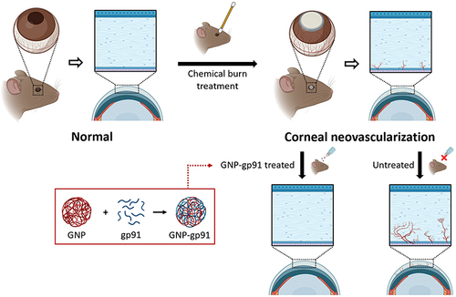
Evaluation of Corneal Neovascularization Treatment
Seventy-two mice were used in this study for three repeated tests, with six mice in each group (Control, PBS, gp91, and GNP-gp91). The corneal NV mice model was induced as previous study.Citation34 Briefly, the mice were anesthetized and immobilized before examination. Next, topical administration of Alcaine (Alcon, Puurs, Belgium) for local anesthesia, followed by pressing the tip of an applicator containing silver nitrate/potassium (25%/75%, Grafco, Atlanta, GA, USA) to the center of the cornea steadily for eight seconds. Excess nitrate was washed with PBS after cauterization. Each mouse was treated for only one eye. Eye drops containing gp91 or GNP-gp91 were diluted in PBS to a final gp91 peptide concentration of 100 μg/mL. Five microliters of eye drops were applied to the mouse ocular surface once every two days for 7 days. The burn stimulus response and severity of corneal neovascularization were observed using a handheld portable slit lamp (SL-17, Kowa Company Ltd., Torrance, CA, USA) and photographed. The corneal neovascularization area was quantified using ImageJ software and presented as the percentage of the vessel area to the non-vessel area.
Histological Examination
After 1 week of treatment, the mouse eyes were harvested and fixed in 10% formalin. The fixed eyes were individually embedded in paraffin and cut into 5 µm-thick sections. The sections were stained with hematoxylin and eosin and observed by a slide-based tissue cytometry (Axio Observer Z1, Tissue Gnostics, Vienna, Austria).
Angiogenic Factors Examination from Cornea Lysate
The mouse corneas were isolated and weighed. Corneal tissues (four corneas/group from two batches) were harvested and homogenized with protein extraction buffer (Thermo Fisher Scientific). The mixture from each sample was centrifuged at 10,000 g for 3 minutes at 4 °C and the supernatant was collected. Total protein was quantified using Bradford assay (p010, GeneCopoeia, Rockville, MD, USA). Mouse nicotinamide adenine dinucleotide phosphate oxidase 2 (Nox2) ELISA Kit (My BioSource, cat no. MBS269961) was used to quantify Nox2 content in cornea. The angiogenic cytokines (MMP2, MMP9, and VEGF) content in each group was measured using the Quantikine® ELISA kit and Mouse VEGF/total MMP9/MMP2 Immunoassay (R&D Systems, Minneapolis, MN, USA) in triplicate. The experiments were conducted according to the manufacturer’s protocol.
Statistical Analysis
All data are presented as the mean ± standard deviation (SD). Experiments were additionally repeated to confirm reproducibility. Statistical differences between groups were analyzed using one-way ANOVA, followed by Tukey’s post-hoc test using SPSS 17.0 (SPSS, Inc., Chicago, IL, USA). Statistical significance was set at p < 0.05.
Results
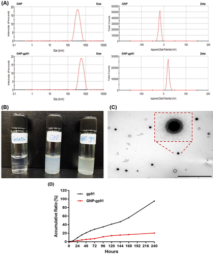
Characterization of GNP and GNP-gp91
and are the results of the characterization of GNP and GNP-gp91. As shown in , when using gelatin solutions with pH 8, the average size of GNP and GNP-gp91 were 349.6 ± 25.8 and 550.6 ± 61.2 nm and the average zeta (ζ) potential of GNP and GNP-gp91 were −18.9 ± 1.0 mV and 21.7 ± 1.8 mV. When using gelatin solutions with pH 9, the average size of GNP and GNP-gp91 were 393.0 ± 44.2 nm and 483.2 ± 41.9 nm and the average zeta potential of GNP and GNP-gp91 were −20.2 ± 0.8 mV and 21.7 ± 0.9 mV. The encapsulation efficiency (EE) of gp91 in the GNP-gp91 was higher at pH 8 (79.4% ± 8.0%) than at pH 9 (44.8% ± 39.5%). Therefore, gelatin solutions with pH 8 was chosen to prepare GNP-gp91 for the subsequent experiments. Both GNP and GNP-gp91 with low PDI values (0.08~0.15) had a narrow size distribution (). The Size and Zeta potential patterns of GNP and GNP-gp91 are shown in revealing single and narrow peak pattern in each group. The monodispersed colloidal solution of GNP and GNp-gp91 is shown in . From the TEM image, the synthesized GNP-gp91 showed no aggregation with spherical and smooth morphology, which also exhibited good dispersion (). In , the in vitro release profiles of gp91 and GNP-gp91 in PBS at pH 7 is revealed. The releases of gp91 and GNP-gp91 were 95.70% and 21.58% after 240 hours, respectively. This result demonstrated that gp91 was release faster than GNP-gp91. The gp91 loaded in GNP exhibited a slower peptide release behavior.
Table 1 Characterization of GNP and GNP-gp91 Prepared in Variant pH
Figure 2 Characterization of GNPs and GNP-gp91. (A) DLS results of size and zeta potential of GNP and GNP-gp91 at pH8. (B) Photograph of gelatin solution and GNP/GNP-gp91 colloidal solutions. (C) TEM image of GNP-gp91, scale bar: 10 µm. (D) The cumulative drug release profile of gp91 and GNP-gp91 in PBS at pH 7. Data are expressed as mean ± SD, n = 3.
GNP-gp91 Improves Uptake into Cells, Impairs Cell Viability, and Reduces ROS Production in the Cultured Endothelial Cells
To investigate the effect of gp91 in different formulations in vitro, the uptake rate of gp91 and GNP-gp91 was evaluated. FITC-labeled peptide (gp91FITC) and GNP-gp91FITC were tested in HUVECs and compared against the uptake of the peptide using flow cytometry (). In 0.5 and 2 hours, the fluorescence peak of GNP-gp91FITC was shifted to the right showing high fluorescent intensity (). The quantification result is revealed in . The mean fluorescence intensities (MFI) of HUVECs treated with GNP-gp91 for 0.5 and 2 hours were 76,945 and 138,545 MFI (), respectively. This is much higher in the GNP-gp91 treated cells compared in the gp91FITC- group (*p < 0.05).
Figure 3 Cell interaction with GNP-gp91. (A) Flow cytometric histograms of gp91FITC- and GNP-gp91FITC-treated cells, and (B) its quantification data of intracellular MFI (n = 3, *p < 0.05, compared with gp91FITC). (C) Cell viability of HUVECs incubated with variant formulation at gp91 concentration from 50 to 150 µg/mL on day 1 and day 3 (n = 5, *p < 0.05, **p < 0.01). (D) Representative images of Live/Dead staining of HUVECs cultured with variant peptide formula (100 μg/mL) acquired on day 3, scale bar: 250 µm. (E) ROS changes in LPS-induced inflamed cells. (n = 3).
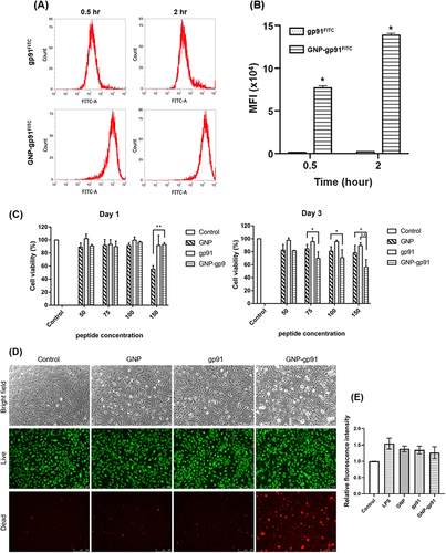
The angiogenic assays including cell viability, migration, and tube formation were performed using HUVECs. For cell viability test, the gp91 content was adjusted to the same concentration as in the gp91 and GNP-gp91 groups. The same gelatin concentration of GNP was used to elucidate the effects of the nanoparticles. At gp91 concentrations ranging from 50~150 μg/mL, cell viability of all groups was greater than 85% and no significant difference compared with the control group (100%) (, Day 1). According to the cytocompatibility standards of ISO 10993–5, it indicated that all formulations have good cytocompatibility (>70%) at gp91 peptide concentration of 100 μg/mL. Along with increased peptide concentration and extended culture period, the cell viability of GNP-gp91 treated group started to decrease significantly. After 3 days of treatment, the GNP-gp91 group at a peptide concentration of 150 μg/mL showed the highest reduction in cell viability at 56.6% ± 11.6%, which was significantly lower than the same particle concentration of GNP (79.6% ± 9.8%) and peptide concentration of gp91 (89.49 ± 4.56%) groups (, Day 3). At lower gp91 concentration (100 μg/mL), cell viability value (70.82 ± 12.49%) revealed that acceptable cyto-compatibility was observed in the GNP-gp91 group (, Day 3); therefore, this peptide concentration was selected for further experiments. Images of HUVECs labeled with the live/dead stain are shown in . Live cells exhibited green color and dead cells revealed in red fluorescence, respectively. A large number of live cells showed green fluorescence in all the tested groups at a peptide concentration of 100 µg/mL on day 3 including GNP-gp91. However, more red fluorescent spots were also observed in the GNP-gp91 treated one. These results indicated that GNP-gp91 (100 µg/mL) was a little bit toxic to influence HUVECs. This result is in line with the Cell viability result.
The ROS content in LPS-induced inflamed cells treated with the variant formula is shown in . The ROS level in the control cells (basal medium) was used as the standard (100%). After LPS stimulation, ROS levels increased (1.5 folds). The ROS content in the GNP-, gp91- and GNP-gp91-treated cells was reduced, and the ROS value of GNP-gp91 was the lowest one (1.2 folds). The GNP-gp91 possessed capacity to reduce ROS content in inflamed cells, although there is no obviously differences here.
GNP-gp91 Attenuates the Angiogenic Capacity in the Cultured Endothelial Cells
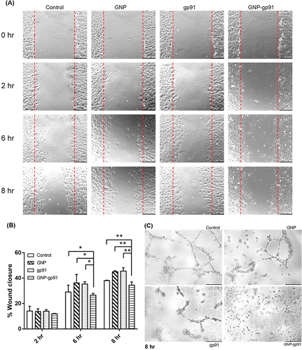
The scratch wound healing assay and MatrigelTM-based tube formation assay were used to evaluate the anti-angiogenic effects of the various formulations. As shown in , HUVECs migrated into the gap after 6–8 hours in the control group. The wound closure rate after 8 hours in the control, GNP, gp91, and GNP-gp91 groups were 43.9% ± 0.3%, 45.1% ± 0.8%, 45.5% ± 2.7%, and 34.4% ± 2.6%, respectively (). This result implied that GNP-gp91 significantly inhibited the migratory capability of HUVECs (*p < 0.001). When HUVECs were cultured on MatrigelTM for 8 hours, capillary-like tubular structures were observed in the control group and the GNP-treated group (). However, fewer capillary structures were observed in the gp91 treated cells. Almost no mesh-like structure was found in the GNP-gp91-treated cells (). Overall, these results demonstrate that GNP-gp91 can effectively inhibit angiogenic activity in vitro.
Figure 4 Effects of GNP-gp91 on cell migration and tube formation. (A) Representative photos from wound healing assay of HUVECs treated with GNP, gp91, and GNP-gp91. Scale bar: 250 µm. (B) Quantification of wound closure rate at the indicated times (n = 3), *p < 0.05, **p < 0.001 compared with GNP-gp91 group. (C) Representative images of tube formation after culturing for 8 h, scale bar: 250 µm.
The Retention and Location of GNP-gp91 on Mice Ocular Surface
To confirm whether nanoparticle-containing eye drops can prolong the drug retention on the ocular surface, TAMRA-SE, a red fluorescent dye, was conjugated with GNPs to explore the accumulation and distribution of nanoparticles on the ocular surface by tracking fluorescent signals. Representative photographs of eyes from mice treated with various eye drops with dye (TAMRA) examined by the IVIS imaging system are shown in and . In normal mice, the eye treated with GNP-gp91 contained eyedrops exhibited higher fluorescence intensity (55.2%) than those treated with GNP (30.8%) and free dye solution (22.5%) for 10 min (). Subsequently, the highest fluorescence intensity was still recorded in the GNP-gp91 group; even after 80 min of exposure, there is 36.4% retention on ocular surface (). However, the free dye (TAMRA) group was <10%. We performed the same test on the eyes of corneal NV mice on day 7 (). The relative fluorescent percentage examined from the corneal NV mice () is much higher than normal eyes. For example, the relative fluorescent intensity of GNP-gp91 is around 180% at 10 min, but the tendency of fluorescence intensity changes in these tested groups was similar to that in the normal and neovascularized cornea. The GNP-gp91 group showed the highest fluorescence intensity in the neovascularized cornea, whereas TAMRA (free dye) group was the lowest (). These data suggest that GNP-gp91 eyedrops can prolong the retention on the ocular surface.
Figure 5 Ocular retention and distribution of GNP-gp91. (A) Photo to show fluorescence accumulated on normal mice eye treated by GNPTAMRA, free dye (TAMRA), and GNP-gp91TAMRA after 10 minutes dosing, and (B) its intensity variation curve changed with time. (C) Photo of neovascularized mice eye treated by variant fluorescent formulation after 10 minutes dosing, and (D) its intensity variation curve changed with time. Data are presented as mean ± SD (n = 3). (E) Corneal cryosections revealing the distribution of free dye, GNPTAMRA, and GNP-gp91TAMRA in cornea (yellow arrow in TAMRA images indicated dye or NPs location). Scale bar: 250 µm. Corneal epithelium: Ep, Stroma: St, endothelium: Ed.
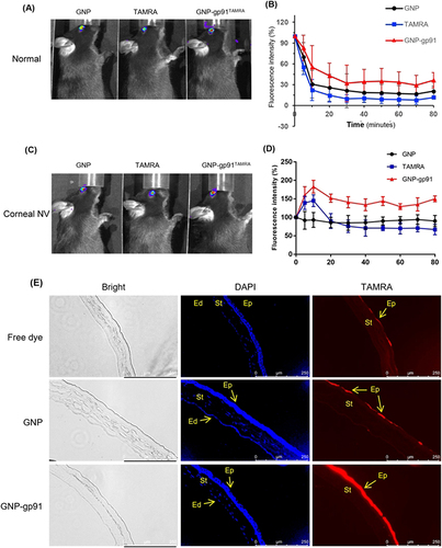
To confirm the location of NPs, corneal cryosections were examined using a fluorescence microscope. Strong blue belt staining shows the cell nuclei of the corneal epithelial layers (, DAPI). Intensive red fluorescence was observed in the corneal epithelium (Ep) of the GNP-gp91 group (, TAMRA), weak signals were observed in the GNP group, and no red fluorescence signal was observed in the free dye (TAMRA) group (). The results provided direct evidence of high accumulation and longer retention of GNP-gp91 on the ocular surface and proofed its localization in the corneal epithelium.
Therapeutic Efficacy of GNP-gp91 for Treating Corneal Neovascularization
Chemically burned corneal neovascularization models of mice were performed to evaluate the therapeutic effect of GNP-gp91 eye drops. Various eye drops were administered once every two days for seven days. The photographs captured under the slip lamp showed a smooth and transparent cornea in the normal eye (, control). Radial growth of blood vessels arising from the limbus towards the central burn area was observed in the PBS group. In contrast, fewer and thinner vessels were observed in both the gp91 and GNP-gp91 groups (), and the corneal area in the GNP-gp91 treated eye showed better transparency with almost no vessel formation (). The area of newly grown vessels was quantified and represented as the neovessel’s ratio compared to the entire eyeball area (). Treatment group treated by GNP-gp91 showed a significantly lower neovessel percentage (7.89 ± 0.76%) than PBS (33.99 ± 2.94%) and gp91 (19.67 ± 1.04%) groups (**p < 0.001). The GNP-gp91 can significantly inhibit vessel formation in chemical-burned cornea even in the dosing frequency of every two-day interval.
Figure 6 Eyedrops containing GNP-gp91 can inhibit vessel formation in chemical cauterization-induced corneal neovascularization. (A) Representative images of cornea from normal eye, and PBS, gp91, and GNP-gp91NP-treated corneal NV. (B) The area of blood vessels in the cornea was quantified using Image J software. Data are presented as mean ± SD (Normal: n = 3. PBS, gp91, and GNP-gp91: n = 6). **p < 0.001 compared with GNP-gp91 group. (C) Histological assessment of central corneal sections after 7 days treatment. Inflammatory cell infiltrating into the stroma (red arrows) and exhibiting defective corneal epithelium (black arrows) was observed in the PBS group compared with the control group. Epithelium: Ep, stroma: St, endothelium: Ed.
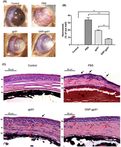
Histopathology of the corneal tissues was examined under a microscope by H&E staining. Representative images of the central cornea are shown in . The corneal surface of normal mice was smooth, and the epithelium (Ep) was integral. In the damaged cornea, the epithelial layer became thinner, the epithelium was exfoliated, and many inflammatory cells were found in the stroma (St) in the PBS- and gp91-treated groups after seven days. In the GNP-gp91-treated eye, cauterized corneas with relatively intact epithelium and fewer inflammatory cells in the stroma were observed (), which is similar to the normal cornea, were observed.
Reduction of Angiogenic Factors in Cornea
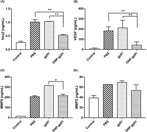
The gp91 peptide can mediate Nox2 and ROS, then influencing angiogenesis; therefore, the Nox2 content in cornea was examined. Corneal neovascularization is associated with several angiogenic factors such as VEGF and MMPs, and these factors were also quantified. The protein level of VEGF, MMP2, and MMP9 in control cornea were 9.20 ± 3.81 pg/mL, 38.99 ± 4.93 ng/mL, and 15.57 ± 0.27 ng/mL (), respectively. The PBS-treated cornea showed higher concentration of Nox2 (1.01 ± 0.01 ng/mL), VEGF (183.21 ± 3.47 pg/mL), MMP2 (65.21 ± 0.02 ng/mL), and MMP9 (207.23 ± 9.74 ng/mL), which were higher than the control group (normal cornea). The gp91 group showed similar results to those of the PBS group, and it had no effect on the reduction in MMP9 and MMP2 levels ( and Figure ). In the GNP-gp91-treated group, the Nox2 level (0.53 ± 0.03 ng/mL) and VEGF level (72.22 ± 0.04 pg/mL) were the lowest in all tested groups ( and ), and these'’re statistical difference with PBS- or gp91-treated groups (**p < 0.05). The MMP2 concentration in the GNP-gp91-treated one is the lowest (53.81 ± 10.86 ng/mL) in all tested groups ().
Figure 7 Quantification of angiogenic cytokines from cornea protein lysate. Mice received chemical cauterization and were treated with PBS, gp91, and GNP-gp91 eye drops. Quantification of (A) Nox2, (B) VEGF, (C) MMP9, and (D) MMP2 levels from the cornea lysates. Data are presented as mean ± SD (n = 3), *p < 0.05, **p < 0.001 compared with GNP-gp91 group.
Discussion
Corneal neovascularization is also a common cause of blindness worldwide.Citation3,Citation4,Citation35 The majority of corneal neovascularization cases involve inflammatory conditions at an early stage, and VEGF is the major factor involved in the development of neovascularization. Although steroids and anti-VEGF agents have been used to suppress corneal neovascularization in clinics, serious side effects such as hypertension, cataracts, or delayed wound healing have revealed that alternative therapeutic agents are needed. In this study, we demonstrated that the nanomedicine, gp91 tat peptide loaded in gelatin nanoparticles (GNP-gp91), can effectively inhibit the migration and tube formation capacity of HUVECs and can also be used as eye drops to reduce vessel formation in mice with corneal neovascularization induced by chemical injury.
Gelatin can be extracted using acidic/alkaline processes to prepare type A and type B gelatin from collagen. At pH 6∼7, type A gelatin has a positive net charge, while type B gelatin is negatively charged.Citation36 The gp91 tat peptide with high amount of arginine and lysine residues possesses positively charged;Citation37 therefore, using type B gelatin can have charge interaction to form strong attraction between gelatin and gp 91 peptides. For gelatin nanoparticle (GNP) preparation, pH value of the prepared gelatin solution is considered as one of the critical factors that affects the structure, conformation, and charge properties of gelatin,Citation38–40 further influencing the formation and stability of GNP.Citation39,Citation40 In addition, glutaraldehyde was used as a crosslinker in the study, and it is reported that a better cross-linking degree will be obtained under higher pH conditions (alkaline conditions: pH 8–10).Citation41,Citation42 Owing to the isoelectric point (PI) of gp91 is around 12.5–13.0 (calculated by the website of Expasy and Prot pi-Peptide Tool); therefore, to maintain the amino group on gp91 protonated lead to non-participating in the crosslinking reaction, we just investigated the synthesis under pH 8 and pH 9. As shown in , the GNP fabricated using gelatin solution at pH 8 had a smaller particle size than at pH 9 and a negative zeta potential at −18.9 mV. However, after loading the gp91 peptide into GNP, a substantial change in size was observed, accompanied by a positive zeta potential at 21.7 mV at pH 8. Of these 18 amino acids of the gp91ds-tat peptide, 11 were positively charged arginine and lysine residues,Citation37 resulting in this peptide becoming a positively charged peptide. Therefore, the zeta potential of GNP changed from negative (−19 mV) to positive after gp91 loading (GNP-gp91, approximately 22 mV). As shown in , the encapsulation efficiency (EE) under pH 8 and pH 9 is 79.4% and 44.8%, respectively. Owing to non-participating in the crosslinking reaction, the encapsulation efficiency of gp91 will depend on the compactness of GNP and ionic interaction. In theory, a better crosslinking efficiency will be obtained under pH 9. Therefore, the result here indicated that a higher encapsulation efficiency under pH 8 is because the charge of gp91 will be more positive resulting in the ionic interaction between gp91 and crosslinker gelatin being stronger. The release of peptide in solution (gp91) was faster, and slow release of peptide from GNP-gp91 was observed (). The drug release mechanism from GNP is reported including 3 steps: (1) simple diffusion of drug particles, (2) degradation release, and (3) cleavage of the gelatin matrix by proteolytic enzymes.Citation28 In this study, gp91 peptide was mixed with a gelatin solution before GNP preparation. Therefore, the gp91 peptide was homogeneously distributed in the nanoparticle matrix ( and , low PDI) and was not easily released by simple diffusion with no burst release () compared to the release profile of human serum albumin-loaded GNP.Citation43 When the diffusion of the drug is lower than matrix degradation, the mechanism of drug release mainly depends upon degradation.Citation28 Gelatin is enzymatically degraded into amino acids as a function of parameters such as pH, temperature, or concentration.Citation27 A controllable system with constant release of GNP-gp91via gelatin degradation in buffer for a longer time, as well as cleavage by lysozyme in cells can be predicted.
For cellular uptake, generally, gp91, a cell-penetrating peptide, could also exhibit a favorable cellular uptake efficiency. However, in and , a higher cellular uptake efficiency of GNP-gp91 was founded, and this is facilitated by gelatin which could further specifically bind to collagen receptors and enter into cells through clathrin-dependent endocytosis.Citation44 The gp91ds-tat has shown to suppress both oxidative stress and cytokine production, which may have significant neuroprotective effects.Citation12,Citation45 It also effectively attenuated ischemia/reperfusion-induced myocardial injury by inhibiting ROS release.Citation45,Citation46 Hachisuka et al reported that gp91ds-tat treatment reduced new blood vessel formation in a tissue-engineering chamber.Citation47 However, whether gp91ds-tat plays an important role in the treatment of corneal NV is unknown. We found that gp91ds-tat nanoparticles (GNP-gp91) at a peptide concentration <100 µg/mL had no toxic effects on HUVECs via cell viability assay on day 1 and day 3 (). But when gp91 peptide concentration reaching 150 μg/mL, the cell viability of GNP-gp91 on day 3 was down to around 50%. This may result from the endocytosis of GNP-gp91 getting high amount of drug intracellular ( and ) and slow-release of gp91 to cause long-term effect ( and -day 3). The Live/Dead staining results showed that after 3 days of treatment with GNP-gp91 (100μμg/mL), some dead cells were observed () due to the effect of gp91. Nox is an effector involved in VEGF signaling in pathological angiogenesis.Citation48,Citation49 Nox2 is known to be abundantly expressed in vascular and inflammatory cells and is important for angiogenesis through the production of ROS.Citation50,Citation51 Despite there is no significant difference between these formulations, the average ROS amount for the GNP-gp91 was the lowest () showing this gp91-nanoformulation has potential to reduce ROS content in cells.
To further confirm its ability to inhibit angiogenesis, GNP-gp91 was evaluated in in vitro via wound healing and tube formation assays. GNP-gp91 group remarkably suppressed cell recovering to the gap area ( and ) and tube formation () of HUVECs compared to GNP group and gp91 group. These results indicated that gp91 carried by GNP-gp91 remained higher peptide concentration and certain peptide activity for working on HUVEC cells and influencing its function.
Positively charged NPs can interact with the negatively charged mucin layer of the tear film, which extends the retention time and absorption on the ocular surface.Citation29,Citation36,Citation52–54 The IVIS results showed that the GNP-gp91 group had higher retention properties than the free dye (TAMRA) and GNP group in normal and corneal NV mice (). We also observed the distribution via fluorescence microscopy, and the GNP-gp91 group demonstrated obvious fluorescence in the corneal epithelium compared with the GNP group and free dye group (), which is in line with expectations.
Recent studies demonstrate that Nox1, Nox2, and Nox4 modulate retinal and choroidal neovascularization via VEGF signaling.Citation49,Citation55–57 Alkali-burned corneas were found to markedly upregulate the transcription and expression of NOX2 and NOX4 in human and mouse corneas.Citation58,Citation59 Chan et al found that cauterizing a mouse cornea with a nitrate stick to cause vascularization led to an increase in the mRNA expression of NOX2, which also causes an increase in VEGFA, VEFGR1, and VEGFR2.Citation59 A similar result was observed in the present study. Interestingly, we have observed a high level of Nox2 and VEGF protein () in the alkali-burned mice cornea treated with gp91. This may result from the lower amount of peptide accumulating on the ocular surface () and not easily get intracellular to effect cells ( and ), then no effective function for inhibiting tube formation () and vessel formation in cornea (, gp91 group). Similarly, topically administered bevacizumab shows poor penetration through the corneal epithelium owing to tight intracellular junctions.Citation60 Therefore, the topical administration of gp91ds-tat requires a more efficient drug delivery system such as GNP to improve therapeutic efficacy. For the ocular retention test, relative higher fluorescence intensity (especially GNP-gp91) accumulated on the damaged ocular surface (Corneal NV, and ) than normal cornea ( and ). This tendency is in accordance with the study of L.D, Huang et al revealed in 2021, that large amount of bromo-heptadecafluorooctane and tetrandrine which loaded in liposome can be remained on ocular surface and accumulated in the corneal epithelium of dry eye rabbit ; this apperance was caused by inflamed corneal cells uptaken more liposome nanoparticles into damage cornea.Citation61
In the present study, we further showed that block NOX2 signaling by GNP-gp91 substantially reduced the protein expression of Nox2, VEGF, MMP2, and MMP9 (). Activation of Nox2 requires a combination of p47phox, p67phox, p40phox, and Ras-related C3 botulinum toxin substrate 1 (Rac1).Citation62 gp91ds-tat prevents the interaction of p47phox with Nox2 and membrane subunits (gp91phox and p22phox) and inhibits O2− formation.Citation37,Citation62 High oxidative stress can impair endothelial cell function and angiogenesis as well as Nox2 and ROS production.Citation63 ROS production by Nox2 activation contributes to various pathologies, such as diabetes and hypertension.Citation64 gp91ds-tat has been reported to reverse insulin resistance-induced impairment of endothelial cells.Citation64 Zuo et al reported that targeting VEGF therapy in alkali burn-induced mouse corneal NV was beneficial in reducing the expression of vascular areas and the number of new vessels.Citation65 Kvanta et al revealed high expression of MMP2, MMP9, and VEGF in a rat model of inflammation-associated corneal neovascularization.Citation66 The effective inhibition of NV in the cornea of GNP-gp91 eye drops is attributed to 1) high retention and accumulation of cationic NPs in the cornea and uptake by cells () and 2) slow release to lasting peptide bioavailability in the cornea even at low dosing frequency (every two days) (). The last, released gp91 peptide affected Nox2 and ROS activity, thereby inducing a cascade inhibition of angiogenesis ().
From the presented study’s perspective, topical application of gp91 peptide in nanoparticle formulations could be a feasible way for the treatment of corneal neovascularization.
Conclusions
In summary, gp91ds-tat peptide-loaded GNP (GNP-gp91) was successfully synthesized at a size of 550 nm and zeta potential of approximately 22 mV under colloidal conditions (PDI < 0.15) prepared from gelatin solution at pH 8. It showed a slow-release profile of GNP-gp91. High cell viability and non-toxicity of HUVECs when gp91 concentration <100 μg/mL in variant peptide formulation was proofed even GNP-gp91. This nanomedicine, GNP-gp91, substantially inhibited HUVEC wound healing and tube formation capacity at a peptide concentration of 100 μg/mL after 8 h treatment. GNP-gp91 can be easily captured on the ocular surface and possesses a longer retention time to reduce the dosing frequency when delivered topically (eye drops) every two days. Topical application of GNP-gp91 as eye drops can suppress the corneal neovascularization in a mouse model of chemically burned eye, and result with less vessel area formation in cornea (8%) was obtained when compared with PBS group (34%) and gp91 group (20%). Overall, our data indicate that GNP-gp91 nanomedicine contained eye drops has the potential to effectively treat corneal neovascularization in a low dosing frequency, which is effective and friendly to patients for clinical us.
Disclosure
The authors declare no conflicts of interest.
Additional information
Funding
References
- Zhang C, Yin Y, Zhao J, et al. An update on novel ocular nanosystems with possible benefits in the treatment of corneal neovascularization. Int J Nanomedicine. 2022;17:4911–4931. doi:10.2147/IJN.S375570
- Britton Anna K, Crayford Basil B. Resolution of refractory corneal neovascularization with subconjunctival bevacizumab. Case Rep Ophthalmol. 2020;11:652–657. doi:10.1159/000510114
- Sharif Z, Sharif W. Corneal neovascularization: updates on pathophysiology, investigations & management. Romanian J Ophthalmol. 2019;63:15–22. doi:10.22336/rjo.2019.4
- Nicholas MP, Mysore N. Corneal neovascularization. Exp Eye Res. 2021;202:108363. doi:10.1016/j.exer.2020.108363
- Brunner M, Romano V, Steger B, et al. Imaging of corneal neovascularization: optical coherence tomography angiography and fluorescence angiography. Invest Ophthalmol Vis Sci. 2018;59:1263–1269. doi:10.1167/iovs.17-22035
- Hsu C-C, Chang H-M, Lin T-C, et al. Corneal neovascularization and contemporary antiangiogenic therapeutics. J Chin Med Assoc. 2015;78:323–330. doi:10.1016/j.jcma.2014.10.002
- Roshandel D, Eslani M, Baradaran-Rafii A, et al. Current and emerging therapies for corneal neovascularization. Ocul Surf. 2018;16(4):398–414. doi:10.1016/j.jtos.2018.06.004
- Al-Debasi T, Al-Bekairy A, Alkatheri A, Al Harbi S, Mansour M, Albekairy A. Topical versus subconjunctival anti-vascular endothelial growth factor therapy (Bevacizumab, Ranibizumab and Aflibercept) for treatment of corneal neovascularization. Saudi J Ophthalmol. 2017;31(2):99–105. doi:10.1016/j.sjopt.2017.02.008
- Barry Z, Park B, Corson TW. Pharmacological potential of small molecules for treating corneal neovascularization. Molecules. 2020;25(3468):1–40. doi:10.3390/molecules25153468
- Feizi S, Azari A, Safapour S. Therapeutic approaches for corneal neovascularization. Eye Vision. 2017;4(28):1–10. doi:10.1186/s40662-017-0094-6
- Rojas M, Lemtalsi T, Toque H, et al. NOX2-induced activation of arginase and diabetes-induced retinal endothelial cell senescence. Antioxidants. 2017;6(43):1–13. doi:10.3390/antiox6020043
- Khayrullina G, Bermudez S, Byrnes K. Inhibition of NOX2 reduces locomotor impairment, inflammation, and oxidative stress after spinal cord injury. J Neuroinflammation. 2015;12:172. doi:10.1186/s12974-015-0391-8
- Singh PK, Saadi A, Sheeni Y, Shekh-Ahmad T. Specific inhibition of NADPH oxidase 2 modifies chronic epilepsy. Redox Biol. 2022;58:102549. doi:10.1016/j.redox.2022.102549
- Hu CG, Wu ZF, Huang ZH, et al. Nox2 impairs VEGF-A-induced angiogenesis in placenta via mitochondrial ROS-STAT3 pathway. Redox Biol. 2021;45:102051. doi:10.1016/j.redox.2021.102051
- Ucgul RK, Celebi S, Yilmaz NS, Bukan N, Ucgul AY. Intrastromal versus subconjunctival anti-VEGF agents for treatment of corneal neovascularization: a rabbit study. Eye. 2021;35(11):3123–3130. doi:10.1038/s41433-020-01347-3
- Fraunfelder FT. Chapter 2 - Ocular drug delivery and toxicology. In: Fraunfelder FFT, Fraunfelder FRW, editors. Drug-Induced Ocular Side Effects. 8th ed. London: Elsevier; 2021:3–11.
- Loftsson T. Topical drug delivery to the retina: obstacles and routes to success. Expert Opin Drug Deliv. 2022;19(1):9–21. doi:10.1080/17425247.2022.2017878
- Tian B, Bilsbury E, Doherty S, et al. Ocular drug delivery: advancements and Innovations. Pharmaceutics. 2022;14(9):1931. doi:10.3390/pharmaceutics14091931
- Srividya G, Rapalli VK, Waghule T, et al. Nanocarriers for ocular drug delivery: current status and translational opportunity. RSC Adv. 2020;10:27835–27855. doi:10.1039/D0RA04971A
- Joseph R, Venkatraman S. Drug delivery to the eye: what benefits do nanocarriers offer? Nanomedicine. 2017;12:683–702. doi:10.2217/nnm-2016-0379
- Mun E, Morrison P, Williams A, Khutoryanskiy V. On the barrier properties of the cornea: a microscopy study of the penetration of fluorescently labeled nanoparticles, polymers, and sodium fluorescein. Mol Pharm. 2014;11:3556–3564. doi:10.1021/mp500332m
- Zimmer A, Kreuter J. Microspheres and nanoparticles used in ocular delivery systems. Adv Drug Deliv Rev. 1995;16:61–73. doi:10.1016/0169-409X(95)00017-2
- Yadav T, Jat RK. Microspheres as an ocular drug delivery system – a review. J Drug Deliv Therap. 2013;3:114–123. doi:10.22270/jddt.v3i1.343
- Nagarwal R, Kant S, Singh P, Maiti P, Pandit J. Polymeric nanoparticulate system: a potential approach for ocular drug delivery. J Control Release. 2009;136:2–13. doi:10.1016/j.jconrel.2008.12.018
- Zhu S, Gong L, Li Y, Xu H, Gu Z, Zhao Y. Safety assessment of nanomaterials to eyes: an important but neglected issue. Adv Sci. 2019;6(16):1802289. doi:10.1002/advs.201802289
- Mobaraki M, Soltani M, Zare Harofte S, et al. Biodegradable nanoparticle for cornea drug delivery: focus review. Pharmaceutics. 2020;12(12):1–26. doi:10.3390/pharmaceutics12121232
- Sahoo N, Sahoo R, Biswas N, Guha A, Kuotsu K. Recent advancement of gelatin nanoparticles in drug and vaccine delivery. Int J Biol Macromol. 2015;81:317–331. doi:10.1016/j.ijbiomac.2015.08.006
- Yasmin R, Shah M, Khan S, Ali R. Gelatin nanoparticles: a potential candidate for medical applications. Nanotechnol Rev. 2017;6:191–207. doi:10.1515/ntrev-2016-0009
- Huang H-Y, Wang M-C, Chen Z-Y, et al. Gelatin–epigallocatechin gallate nanoparticles with hyaluronic acid decoration as eye drops can treat rabbit dry-eye syndrome effectively via inflammatory relief. Int J Nanomedicine. 2018;13:7251–7273. doi:10.2147/IJN.S173198
- Chuang YL, Fang HW, Ajitsaria A, et al. Development of Kaempferol-loaded gelatin nanoparticles for the treatment of corneal neovascularization in mice. Pharmaceutics. 2019;11(635):1–16. doi:10.3390/pharmaceutics11120635
- Coester C, Langer K, Briesen H, Kreuter J. Gelatin nanoparticles by two step desolvation - A new preparation method, surface modifications and cell uptake. J Microencapsul. 1999;17:187–193.
- Ofokansi K, Winter G, Fricker G, Coester C. Matrix-loaded biodegradable gelatin nanoparticles as new approach to improve drug loading and delivery. Eur J Pharm Biopharm. 2010;76(1):1–9. doi:10.1016/j.ejpb.2010.04.008
- Kalyanaraman B, Darley-Usmar V, Davies K, et al. Measuring reactive oxygen and nitrogen species with fluorescent probes: challenges and limitations. Free Radic Biol Med. 2012;52:1–6. doi:10.1016/j.freeradbiomed.2011.09.030
- Wang J-H, Tseng C-L, Lin F-L, et al. Topical application of TAK1 inhibitor encapsulated by gelatin particle alleviates corneal neovascularization. Theranostics. 2022;12:657–674. doi:10.7150/thno.65098
- World Health Organization. Blindness and vision impairment; 2022. Available from: https://www.who.int/news-room/fact-sheets/detail/blindness-and-visual-impairment. Accessed March 10, 2023.
- Tseng C-L, Chen K-H, Su W-Y, Lee Y-H, Wu -C-C, Lin F-H. Cationic gelatin nanoparticles for drug delivery to the ocular surface: in vitro and in vivo evaluation. J Nanomater. 2013;2013:1–11. doi:10.1155/2013/238351
- Rey F, Cifuentes-Pagano E, Kiarash A, Quinn M, Pagano P. Novel competitive inhibitor of NAD(P)H oxidase assembly attenuates vascular O2− and systolic blood pressure in mice. Circ Res. 2001;89:408–414. doi:10.1161/hh1701.096037
- Li Z, Gu L. Effects of mass ratio, pH, temperature, and reaction time on fabrication of partially purified pomegranate ellagitannin-gelatin nanoparticles. J Agric Food Chem. 2011;59:4225–4231. doi:10.1021/jf200024d
- Ahsan S, Rao C. The role of surface charge in the desolvation process of gelatin: implications in nanoparticle synthesis and modulation of drug release. Int J Nanomedicine. 2017;12:795–808. doi:10.2147/IJN.S124938
- Vinjamuri BP, Papachrisanthou K, Haware R, Chougule M. Gelatin solution pH and incubation time influences the size of the nanoparticles engineered by desolvation. J Drug Deliv Sci Technol. 2021;63:102423. doi:10.1016/j.jddst.2021.102423
- Zhang T, Yu Z, Ma Y, Chiou B-S, Liu F, Zhong F. Modulating physicochemical properties of collagen films by cross-linking with glutaraldehyde at varied pH values. Food Hydrocoll. 2022;124:107270. doi:10.1016/j.foodhyd.2021.107270
- Slusarewicz P, Zhu K, Hedman T. Kinetic characterization and comparison of various protein crosslinking reagents for matrix modification. J Mater Sci Mater Med. 2010;21(4):1175–1181. doi:10.1007/s10856-010-3986-8
- Solanki P, Sajwan R. Gelatin nanoparticles as a delivery system for proteins. J Nanomed Res. 2015;2:18–20. doi:10.15406/jnmr.2015.02.00018
- Potapovitch A, Suhan T, Shutava T, Kostyuk V. Receptor-mediated endocytosis is an important way for gelatin nano-particles penetration into cells. J Belarusian State Univ Biol. 2020;3–10. doi:10.33581/2521-1722-2020-1-3-10
- Cifuentes-Pagano E, Meijles DN, Pagano PJ. The quest for selective nox inhibitors and therapeutics: challenges, triumphs and pitfalls. Antioxid Redox Signal. 2014;20(17):2741–2754. doi:10.1089/ars.2013.5620
- DelBaugh R, Chen Q, Devine I, et al. Nox2ds-Tat, A Peptide Inhibitor of NADPH Oxidase, exerts cardioprotective effects by attenuating reactive oxygen species during ischemia/reperfusion injury. Am J Biomed Sci. 2016;8:208–227.
- Hachisuka H, Dusting G, Abberton K, Morrison W, Jiang F. Role of NADPH oxidase in tissue growth in a tissue engineering chamber in rats. J Tissue Eng Regen Med. 2008;2:430–435. doi:10.1002/term.115
- Xia C, Meng Q, Liu L-Z, Rojanasakul Y, Wang X-R, Jiang B-H. Reactive oxygen species regulate angiogenesis and tumor growth through vascular endothelial growth factor. Cancer Res. 2007;67:10823–10830. doi:10.1158/0008-5472.CAN-07-0783
- Chan E, Wijngaarden P, Liu G-S, Jiang F, Peshavariya H, Dusting G. Involvement of Nox2 NADPH oxidase in retinal neovascularization. Invest Ophthalmol Vis Sci. 2013;54:7061–7067. doi:10.1167/iovs.13-12883
- Fukai M. Redox signaling in angiogenesis: role of NADPH oxidase. Cardiovasc Res. 2006;71:226–235. doi:10.1016/j.cardiores.2006.04.015
- Bedard K, Krause K-H. The NOX family of ROS-generating NADPH oxidases: physiology and pathophysiology. Physiol Rev. 2007;87:245–313. doi:10.1152/physrev.00044.2005
- Tsai C-H, Wang P-Y, Lin IC, Huang H, Liu G-S, Tseng C-L. Ocular drug delivery: role of degradable polymeric nanocarriers for ophthalmic application. Int J Mol Sci. 2018;19(2830):1–20. doi:10.3390/ijms19092830
- Castro B, Fulgêncio G, Domingos L, Cotta O, Silva-Cunha A, Fialho S. Positively charged polymeric nanoparticles improve ocular penetration of tacrolimus after topical administration. J Drug Deliv Sci Technol. 2020;60:101912. doi:10.1016/j.jddst.2020.101912
- Des Rieux A, Fievez V, Garinot M, Schneider Y-J, Preat V. Nanoparticles as potential oral delivery systems of proteins and vaccines: a mechanistic approach. J Control Release. 2006;116:1–27. doi:10.1016/j.jconrel.2006.08.013
- Li Q, Dinculescu A, Shan Z, et al. Downregulation of p22phox in retinal pigment epithelial cells inhibits choroidal neovascularization in mice. Mol Ther. 2008;16:1688–1694. doi:10.1038/mt.2008.164
- Vogel J, Kruse C, Zhang M, Schröder K. Nox4 supports proper capillary growth in exercise and retina neo-vascularization: nox4 in exercise. J Physiol. 2015;593:2145–2154. doi:10.1113/jphysiol.2014.284901
- Wilkinson-Berka J, Deliyanti D, Rana I, et al. NADPH oxidase, NOX1, mediates vascular injury in ischemic retinopathy. Antioxid Redox Signal. 2014;20:2726–2740. doi:10.1089/ars.2013.5357
- Gu X-J, Liu X, Zhao Y, et al. Involvement of NADPH oxidases in alkali burn-induced corneal injury. Int J Mol Med. 2016;38:75–82. doi:10.3892/ijmm.2016.2594
- Chan E, Wijngaarden P, Chan E, et al. NADPH oxidase 2 plays a role in experimental corneal neovascularisation. Clin Sci. 2016;130:683–696.
- Dastjerdi M, Sadrai Z, Saban D, Zhang Q, Dana R. Corneal penetration of topical and subconjunctival Bevacizumab. Invest Ophthalmol Vis Sci. 2011;52:8718–8723. doi:10.1167/iovs.11-7871
- Huang L, Gao H, Wang Z, Zhong Y, Hao L, Du Z. Combination nanotherapeutics for dry eye disease treatment in a rabbit model. Int J Nanomedicine. 2021;16:3613–3631. doi:10.2147/IJN.S301717
- Brandes R. A radical adventure: the quest for specific functions and inhibitors of vascular NAPDH oxidases. Circ Res. 2003;92:583–585. doi:10.1161/01.RES.0000066880.62205.B0
- Fukai M. VEGF signaling through NADPH oxidase-derived ROS. Antioxid Redox Signal. 2007;9:731–739. doi:10.1089/ars.2007.1556
- Fukai T, Fukai M. Cross-talk between NADPH oxidase and mitochondria: role in ROS signaling and angiogenesis. Cells. 2020;9:1849. doi:10.3390/cells9081849
- Zuo L, Fan Y, Wang F, Gu Q, Xu X. A SiRNA targeting vascular endothelial growth factor- A inhibiting experimental corneal neovascularization. Curr Eye Res. 2010;35:375–384. doi:10.3109/02713681003597230
- Kvanta A, Sarman S, Fagerholm P, Seregard S, Steen B. Expression of matrix metalloproteinase-2 (MMP-2) and vascular endothelial growth factor (VEGF) in inflammation-associated corneal neovascularization. Exp Eye Res. 2000;70:419–428. doi:10.1006/exer.1999.0790