
Abstract
Background
Silica (SiO2) nanoparticles (NPs) are widely used in diverse industrial and biomedical applications. Their applicability depends on surface modifications, which can limit potential health problems.
Objective
To assess the potential impact of SiO2 NP exposure and NPs chemical modifications in allergic airway inflammation.
Methods
Mice were sensitized by five repetitive intraperitoneal injections of ovalbumin/aluminum hydroxide (1 μg) over 42 days, then intratracheally instilled with plain or modified SiO2 NPs (50 μg/mouse), and subsequently aerosol challenged for 20 minutes with ovalbumin. One or 5 days later, allergic inflammation was evaluated by cell differentiation of bronchoalveolar lavage fluid, lung function and gene expression and histopathology, as well as electron and confocal microscopy of pulmonary tissue.
Results
Plain SiO2 NPs induced proinflammatory and immunomodulatory effects in vivo, highlighted by enhanced infiltration of inflammatory cells in the bronchoalveolar lavage fluid, induction of a pulmonary T helper type 2 (Th2) cytokine pattern, differentiation of type 2 macrophages, and by morphological changes in the lung of sensitized mice. These effects were dramatically attenuated using surface-functionalized NPs with amino and phosphate groups, but not with polyethylene glycol. The role of macrophages in taking up SiO2 NPs was confirmed by flow cytometry, confocal microscopy, and gene expression analysis.
Conclusion
Our data suggest that amino and phosphate surface modifications, but not polyethylene glycol (PEG), mitigate the proinflammatory and immunomodulatory effect of SiO2 NPs in allergic airway inflammation, paving the way for new strategies in the production of nanomaterials with lower health impact for humans.
Introduction
Engineered nanoparticles (NPs) are frequently used in commercial products due to their unique physical and chemical characteristics, which depend on their specific dimensions (100 nm or less in one or more dimension).Citation1 Silicon dioxide (silica [SiO2]) nanomaterials are of great importance in the fabrication of electric and thermal insulators, catalyst supports, and drug carriers, as well as in gene delivery, media for coating processes, and filler materials.Citation2 They are also produced on an industrial scale as additives for cosmetics, drugs, printer toners, varnishes, and food packing.Citation3 The same properties that mediate the advantages of nanotechnology also confer their potential toxic effects, resulting in a major health concern regarding the use of nanomaterials.Citation1,Citation4
NPs may enter the body via different routes, such as the gastrointestinal tract,Citation5 skin,Citation6,Citation7 and lung.Citation8,Citation9 Due to their small size, NPs can translocate from entry portals into the circulatory and lymphatic systems, causing long-lasting cell damage.Citation10,Citation11 Several studies show that SiO2 NPs induce dose-dependent cytotoxicity in in vitro systems, including human lung epithelial cellsCitation12 and bronchoalveolar carcinoma-derived cellsCitation13 and human alveolar barrier,Citation14 as well as murine macrophages.Citation15 Short-term instillations or inhalation exposure to amorphous SiO2 NPs showed that these particles possess the potential to cause toxic effects in the lung.Citation16–Citation18 Immunomodulatory effects by particle exposure during the sensitization phase have also been demonstrated.Citation19,Citation20 Recently, surface modifications of SiO2 NPs were synthe-sized to study their influence on the biological activity of SiO2 NPs. In vitro studies using various mammalian cells have demonstrated that unmodified SiO2 NPs exhibit higher cytotoxic and genotoxic properties compared to surface-modified NPs.Citation18,Citation21,Citation22
The aim of this study was to investigate the effects of amorphous SiO2 NPs and their surface modifications in the elicitation phase in a murine model of ovalbumin-specific allergic airway inflammation. SiO2 and polyethylene glycol (PEG) SiO2 (SiO2-PEG) exerted proinflammatory and immunomodulatory activity in allergic airway inflammation, while modifying SiO2 NPs with amino and phosphate groups markedly decreased these effects. Alternative activation of pulmonary macrophages that take up SiO2 NPs and distinct chemokine patterns in the lung were identified as underlying mechanisms.
Materials and methods
Animals
Female 6- to 10-week-old BALB/c mice were obtained from Charles River (Wilmington, MA, USA), housed under specific pathogen-free conditions in individually ventilated cages (VentiRack; BioZoneGlobal Ltd, Ramsgate, UK) and fed a standard diet and water ad libitum. The study was conducted under federal guidelines for the use and care of laboratory animals and was approved by the Government of the District of Upper Bavaria and the Animal Care and Use Committee of the Helmholtz Center Munich, Munich, Germany.
NP preparation
Plain SiO2 (SiO2), PEGylated SiO2 (SiO2-PEG), phosphate-coated SiO2 (SiO2-P), and amino-coated SiO2 (SiO2-NH2) NPs were obtained from BASF SE, Ludwigshafen, Germany, and fluorescein isothiocyanate (FITC)-conjugated SiO2 (SiO2-FITC) NPs from Bayer Technology Services GmbH, Leverkusen, Germany. All NPs were amorphous with a single particle/agglomerate size between 5 and 50 nm, with the exception of SiO2-FITC (23–30 nm). NPs were freshly diluted in water (aqua ad iniectabilia; B Braun Melsungen AG, Melsungen, Germany) and buffered with phosphate-buffered saline (PBS) to physiological pH, to the final concentration of 1 mg/mL, just before intratracheal instillation. Supernatant controls (SUP) were obtained by hard sedimentation (24,000 rpm, 15 hours). Additional NP characteristics are disclosed in the “Supplementary materials” section.
Study design
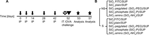
To evaluate the immunomodulatory effect of NPs, a protocol of mild allergic inflammation in the lung was used, as previously described ().Citation23 Briefly, mice were sensitized by repetitive intraperitoneal injections of 1 μg ovalbumin (OVA) (grade VI; Sigma-Aldrich, St Louis, MO, USA) in PBS adsorbed to 2.5 mg aluminum hydroxide (alum) (Thermo Fisher Scientific, Waltham, MA, USA) on days 0, 7, 14, 28, and 42. Blood samples were taken before and after sensitization. OVA/alum-sensitized mice (S mice), compared to non-sensitized mice (NS mice), were characterized by high titers of OVA-specific immuno-globulin E (8.05±1.64 versus 0.1±0.03 μg/mL). At day 52, mice were intratracheally instilled with 50 μg (1 mg/mL) of SiO2 NPs, or with their correspondent SUP (). For dose-response studies, S mice were instilled with 12.5, 25, or 50 μg of SiO2 and SiO2-PEG NPs. NP instillation was followed by OVA aerosol challenge for 20 minutes with 1% OVA in PBS delivered by a PARI BOY nebulizer (PARI GmbH, Starnberg, Germany). Lung function, flow cytometry, and confocal analysis were performed on day 53. Bronchoalveolar lavage (BAL), histology, scanning electron microscopy (SEM), and messenger (m)RNA expression analysis were performed 5 days after OVA challenge (day 57).
Figure 1 Experimental protocol.
Notes: (A) BALB/c mice were intraperitoneally sensitized (S) with OVA/aluminum hydroxide or non-sensitized (NS) with phosphate-buffered saline/aluminum hydroxide (black arrows). On day 52, S and NS mice were intratracheally instilled with SiO2 nanoparticles or with SUP (white arrow) and subsequently challenged with OVA aerosol (arrowhead). Sacrifice was on day 53 or 57, according to the analysis performed (stars). (B) Experimental groups.
Abbreviations: FITC, fluorescein isothiocyanate; IP, intraperitoneal; IT, intratracheal; OVA, ovalbumin; PEG, polyethylene glycol; SiO2, silicon dioxide; SiO2-NH2, amino-coated SiO2; SiO2-P, phosphate-coated SiO2; SiO2-PEG, PEGylated SiO2; SUP, supernatant controls.
Flow cytometry
On day 53, single cell suspensions were generated from lungs of NS and S mice exposed to SiO2-FITC or to corresponding SUP (n=5–7), dissociated with a lung dissociation kit (kit number 130-065-927; Miltenyi Biotec GmbH, Bergisch Gladbach, Germany), and analyzed by flow cytometry, monitoring the FITC conjugate. Untreated mice were used as negative controls. To investigate the interaction of SiO2-FITC with different lung cell populations, cells were stained and gated as described previously.Citation24 anti-mouse CD45 PE-Cyanine7, 7AAD, and anti-mouse F4/80 PE were obtained from eBioscience (San Diego, CA, USA), anti-mouse CD11b APC-Cy™7 from BD Biosciences (San Jose, CA, USA), anti-mouse CD11c APC from BioLegend (San Diego, CA, USA), and GR1 (LY6C/G) Pacific Orange™ from Invitrogen (Life Technologies, Carlsbad, CA, USA). Samples were measured in a BD FACSCanto II flow cytometer and data were analyzed using a FlowJo graphics application V9.7.2 (TreeStar Inc., Ashland, OR, USA).
Interstitial macrophages were defined as CD45+, Gr-1Low, F4/80+, CD11b+ but CD11c−; alveolar macrophages as CD45+, Gr-1Low, F4/80+, CD11c+ but CD11b−; neutrophils as CD45+, Gr-1High, and CD11b+; and lymphocytes as CD45+, but CD11b− and SSCLow.
Confocal microscopy
On day 53, lungs were embedded in, Tissue-Tek™ CRYO-OCT Compound (Thermo Fisher Scientific), removed, and snap frozen in liquid nitrogen (n=2/group). Pulmonary tissue sections (20 μm) were fixed in 4% paraformaldehyde (Merck KGaA, Darmstadt, Germany) and subsequently blocked for 2 hours with 3% bovine serum albumin/PBS. The primary antibody for detecting macrophages (anti-CD68 marker, 1:100; AbD Serotec, Kidlington, UK) was incubated overnight and the secondary antibody ([anti-rat immunoglobulin G] Alexa Fluor® 555, 1:7000; Cell Sig-naling Technology, Inc., Danvers, MA USA) for 1 hour. Antibodies were diluted in 1% bovine serum albumin/PBS. Nuclei were stained with 4′,6-diamidino-2-phenylindole (DAPI) (0.1 μg/mL in PBS; Sigma-Aldrich) for 5 minutes. Sections were embedded in Moviol 4-88 (10% polyvinyl alcohol, 20% glycerol in PBS; Merck). Images were acquired with an inverted confocal laser scanning microscope (LSM 510 Meta; Carl Zeiss Meditec AG, Jena, Germany) with 40× objective, Z-Stack size 6 μm, and collapsed two stacks. For detailed images, a 100× objective was used, with Z-Stack size 2 μm. Orthogonal sections of single cells were generated with the LSM Image Browser software (Carl Zeiss Meditec AG).
Histology and SEM
On day 57, after BAL, the lungs were excised and the left lobe fixed in 4% buffered formalin and embedded in paraffin (n=4–5/group). Sections of 3 μm thickness were stained with hematoxylin and eosin and periodic acid–Schiff. For electron microscopy, lungs were processed as previously described.Citation25
Lung function analysis
Lung function analysis was performed on day 53, 24 hours after allergen challenge in intubated, mechanically ventilated animals (n=5–8/group; Buxco® Research Systems, Wilmington, NC, USA). Anesthesia was induced by an intraperitoneal injection of ketamine (100 mg/kg) and xylazine (5 mg/kg) in PBS. After cannulation of the trachea, the animals were ventilated with room air at rates of 130 breaths/minute. A protocol for measuring airway hyperreactivity was initiated by challenging the mice with increasing methacholine concentrations by inhalation, using an in-line nebulizer (5 μL methacholine solution in PBS delivered for 30 seconds at the following concentrations: 0, 1.25, 2.5, 5, 10, 20, and 40 mg/mL). Data were recorded using FinePoint software version 1.0 (Buxco® Research Systems). The highest value of respiratory system resistance and the lowest value of dynamic compliance were recorded every 5 seconds during the data-recording interval, which was set at 3 minutes after each methacholine level. The heart rate of each animal was continuously monitored using an electrocardiography device connected with three subcutaneous electrodes throughout the entire experiment (Buxco® Research Systems).
Polymerase chain reaction arrays and real-time polymerase chain reaction
Total RNA was extracted from snap-frozen lung tissue as previously described.Citation25 For polymerase chain reaction (PCR) arrays (n=4/group of experiment) (SABiosciences, Hilden, Germany), 0.4 μg RNA was converted into complementary (c)DNA with the RT2 First Strand Kit (Qiagen) and real-time PCR was performed with RT2 SYBR Green ROX qPCR Mastermix (Qiagen) and ViiA™7 thermocycler (Applied Biosystems; Life Technologies). Relative expression levels were calculated using the 2ΔΔct method, normalized to the arithmetic mean of glyceraldehyde-3-phosphate dehydrogenase (GAPDH) and beta-actin (ACTB). For the dose-response experiment, 0.4 μg of RNA was converted to cDNA following the manufacturer’s instructions (RevertAid First Strand cDNA Synthesis Kit #K1622; Fermentas, Thermo Fisher Scientific). Real-time PCR was performed with SYBR Green and ViiA™7 thermocycler (n=3/group) (Applied Biosystems). Relative expression levels were calculated using the 2ΔΔct method, normalized to ACTB. Primer sequences are listed in . Data were considered significant with a P-value ≤0.05 with a confidence interval of 95%.
Data analysis
Results are shown as boxplots indicating minimum, 25th percentile, median, 75th percentile, and maximum, or as mean ± standard deviation. Statistical significance among groups was determined by the Mann–Whitney U-test for boxplots, two-way analysis of variance with post hoc least significant difference test for lung function studies, and one-way analysis of variance with Bonferroni post hoc test for PCR and flow cytometry (GraphPad Prism; GraphPad Software, Inc., La Jolla, CA, USA). Statistically significant values were categorized as follows: P≤0.05, P≤0.01, and P≤0.001.
Results
SiO2 NPs exert proinflammatory and Th2 immunomodulatory activity in vivo
In order to investigate possible proinflammatory and immunomodulatory effects of SiO2, increasing concentrations of SiO2 NPs (0, 12.5, 25, and 50 μg/mouse) were intratracheally instilled in the lungs of S mice prior to OVA challenge. Bronchoalveolar lavage fluid (BALF) was analyzed for differential cell count 5 days after OVA challenge. The lowest NP concentration used (12.5 μg) did not evoke a significant increase of inflammatory cells into the BALF compared to the respective SUP. Increasing NP dose to 25 μg led to a significant increase of macrophages (P<0.01) and neutrophils (P<0.01) (). The highest dose of 50 μg SiO2 NPs led to a significant increase of macrophages, lymphocytes, neutrophils, and eosinophils (P<0.001) in the BALF compared to sensitized SUP (). Similar effects were shown following SiO2-PEG instillation (). Intratracheal instillation of 50 μg/mouse SiO2 NPs in NS mice exerted a proinflammatory effect, leading to significantly increased neutrophils and lymphocytes (P<0.001) and to a slight increase of eosinophils (P<0.05) compared to non-sensitized SUP (). These results suggest that SiO2 NPs exert both a strong proinflammatory and immunomodulatory effect in allergic airway inflammation.
Figure 2 SiO2 NPs exert proinflammatory and immunomodulatory activity.
Notes: Inflammatory cell infiltration in bronchoalveolar lavage fluid of sensitized (A) and non-sensitized (B) mice 5 days after intratracheal instillation with increasing concentrations of SiO2 NPs (12.5–50 μg/mouse, (A) or 50 μg/mouse (B) SiO2 NPs [pattern and red filled bar] or with respective SUP [white bars]). Data are displayed in boxplots (five to ten mice per bar). *P≤0.05; **P≤0.01; ***P≤0.001 versus SUP (0 μg).
Abbreviations: NP, nanoparticle; ns, not significant; SiO2, silicon dioxide; SUP, supernatant controls.
![Figure 2 SiO2 NPs exert proinflammatory and immunomodulatory activity.Notes: Inflammatory cell infiltration in bronchoalveolar lavage fluid of sensitized (A) and non-sensitized (B) mice 5 days after intratracheal instillation with increasing concentrations of SiO2 NPs (12.5–50 μg/mouse, (A) or 50 μg/mouse (B) SiO2 NPs [pattern and red filled bar] or with respective SUP [white bars]). Data are displayed in boxplots (five to ten mice per bar). *P≤0.05; **P≤0.01; ***P≤0.001 versus SUP (0 μg).Abbreviations: NP, nanoparticle; ns, not significant; SiO2, silicon dioxide; SUP, supernatant controls.](/cms/asset/ee7b1f57-a2ea-4f9e-83ad-467b73326f15/dijn_a_57396_f0002_c.jpg)
Pulmonary macrophages take up SiO2 NPs
To determine whether OVA sensitization affects pulmonary retention of NPs, mice were instilled with SiO2-FITC or respective SUP prior to OVA challenge. Twenty-four hours later, a minor, but not significant, increase in the percentage of FITC+ cells was observed in the lung of S mice, compared to NS mice. FITC+ cells in the negative control and in S and NS mice instilled with SUP were not higher than background level ().
Figure 3 Macrophage populations take up nanoparticles.
Notes: (A) Percentage of FITC+ cells in lung (a) and within AMs (b) and IMs (c) of NS and S mice 24 hours after 50 μg SiO2-FITC and OVA challenge. Neg CTL: untreated mouse; SUP: instillation of SiO2-FITC-supernatant. Mean ± standard deviation (n=5–7/group). **P≤0.01 versus SUP. (B) NS (a) and S (b) lungs 24 hours after SiO2-FITC (green) and OVA challenge. Macrophages: red; nuclei: blue. Alexa 555: Alexa Fluor® 555 (1:7000; Signaling Technology, Inc., Danvers, MA USA). Left: 40× and right: 100× orthogonal sections of single cell (arrowed cells 1 and 2).
Abbreviations: AM, alveolar macrophage; DAPI, 4′,6-diamidino-2-phenylindole; FITC, fluorescein isothiocyanate; IM, interstitial macrophage; Neg CTL, negative controls; NS, non-sensitized; OVA, ovalbumin; S, sensitized; SiO2, silicon dioxide; SUP, supernatant controls.
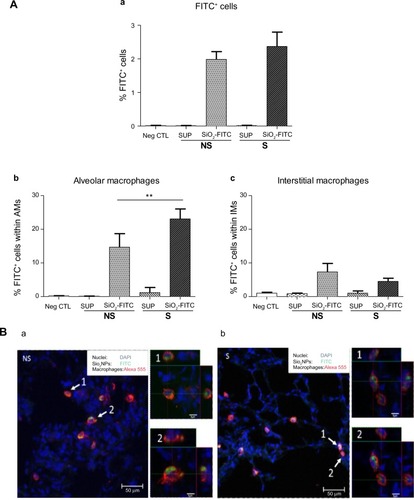
Flow cytometry of lung cell subtypes showed that macrophages displayed the highest percentage of FITC positivity. In detail, in the lungs of NS mice, 15.3% of alveolar macrophages and 8.2% of interstitial macrophages were positive for SiO2-FITC NPs. In the lungs of S mice, a significantly increased percentage of alveolar macrophages (23.2%, P<0.01 versus NS mice) and a reduced percentage of interstitial macrophages (4.3%) were positive for SiO2-FITC (). Pulmonary suspensions of negative controls displayed minimal FITC autofluorescence, comparable to the lungs instilled with FITC-SUP. A minimal percentage of T-cells and neutrophils were FITC positive (data not shown).
To evaluate whether SiO2 NPs interacted only with the surface of macrophages or were internalized by these cells, confocal analysis was performed 24 hours after OVA challenge. shows representative lung sections of NS and S mice stained with the macrophage marker CD68 (red), and with DAPI for the nuclei (blue), suggesting that pulmonary macrophages take up SiO2 NPs (green) in both NS and S mice. The 100× orthogonal sections of single cells show that NPs were present inside the macrophages.
Specific surface modifications of SiO2 NPs significantly reduce their immunomodulatory activity
In order to evaluate the possible reduction of immunomodulatory effects on the allergic response by the functionalization of SiO2 NPs, we evaluated the BAL cell infiltration in NS and S mice after intratracheal instillation of SiO2 NPs with PEG, amino, and phosphate surface modifications.
Similarly to SiO2 plain NPs, SiO2-PEG NPs exerted proinflammatory effects, with a significant increase of neutrophils and lymphocytes (P<0.001) and a minor increase of eosinophils (P<0.05), in NS mice. Moreover, functionalizing SiO2 plain NPs with PEG did not impair the immunomodulatory effect of SiO2 NPs in the BALF of S mice. Lymphocyte, neutrophil, and eosinophil total cell numbers were still significantly increased in the BALF of S mice (P<0.001) ().
Figure 4 Surface modifications of SiO2 nanoparticles influence their proinflammatory and immunomodulatory effects in vivo.
Notes: Bronchoalveolar lavage fluid inflammatory infiltrate of NS and S mice 5 days after intratracheal instillation of 50 μg SiO2-PEG (A), SiO2-P (B), or SiO2-NH2 (C), or respective SUP. Data are displayed as boxplots (n=6–10/bar). *P≤0.05; **P≤0.01; ***P≤0.001.
Abbreviations: ns, not significant; NS, non-sensitized; PEG, polyethylene glycol; S, sensitized; SiO2, silicon dioxide; SiO2-NH2, amino-coated SiO2; SiO2-P, phosphate-coated SiO2; SiO2-PEG, PEGylated SiO2; SUP, supernatant controls.
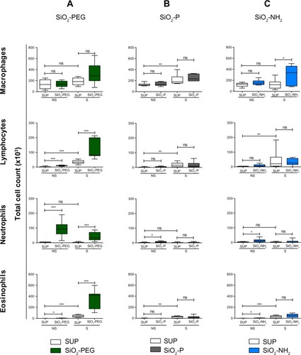
In contrast, mice treated with SiO2-P or SiO2-NH2 () showed minimal proinflammatory effects in NS mice, which reached significance only for neutrophil and eosinophil counts in SiO2-NH2-treated mice (P<0.05). In S mice, no significant effects on the BALF total cell count were shown compared to control, apart from a slight increase in the macrophage cell number after SiO2-NH2 instillation (P<0.05).
SiO2 and SiO2-PEG increase pulmonary inflammation and goblet cell necrosis
To further elucidate the effects of SiO2 NPs and their surface coatings, we evaluated pulmonary inflammation in the lung. Intratracheal instillation of the supernatants of all four NP preparations did not affect pulmonary histopathology (). In S mice, SiO2 and SiO2-PEG NPs increased the inflammatory lung infiltrate and, although in higher degree SiO2-PEG, mucus hypersecretion when compared to sensitized SUP mice (). In contrast, mice treated with SiO2-P and SiO2-NH2 NPs showed minimal pulmonary inflammation when compared with sensitized SUP mice; these observations were in accordance with the effects observed in the BALF differential cell count in S mice. Irrespective of functionalization, SiO2 NPs did not induce pulmonary pathological alterations in NS mice (data not shown).
Figure 5 SiO2 and SiO2-PEG NPs exacerbate goblet cell hyperplasia and induce necrosis.
Notes: Representative NS and S lungs 5 days after SUP (a and b, respectively) or 50 μg nanoparticles (c: S SiO2; d: S SiO2-PEG; e: S SiO2-P; f: S SiO2-NH2) and ovalbumin challenge. (A) Periodic acid–Schiff staining: mucus hypersecretion (arrowheads) and inflammatory infiltrate (arrows). Bar: 100 μm. (B) Scanning electron microscopy of bronchiolar epithelium. (a) Clara cells (arrowheads); (b) goblet cells (arrowheads); (c and d) necrosis of goblet cells (insets, arrows). Bar: 5 μm (insets: 3 μm).
Abbreviations: NS, non-sensitized; PEG, polyethylene glycol; S, sensitized; SiO2, silicon dioxide; SiO2-NH2, amino-coated SiO2; SiO2-P, phosphate-coated SiO2; SiO2-PEG, PEGylated SiO2; SUP, supernatant controls.
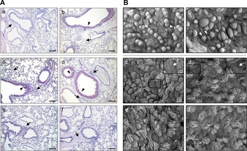
To evaluate structural changes of the airways following SiO2 NP instillation, SEM was performed. NS mice instilled with the four different particles and respective supernatants showed no significant ultrastructural modifications of the lung (data not shown), apart from sporadic hypotrophy of Clara cells present in all samples (). S mice instilled with the four different sensitized SUP developed the classical pulmonary alterations due to OVA sensitization and challenge, ie, metaplasia of Clara cells to mucus-secreting goblet cells (). Lungs of S mice instilled with SiO2 and SiO2-PEG NPs showed morphological alterations of goblet cells, including the perforation of cell membrane, indicating cell death (). In contrast, no additional effects due to particle exposure were observed in S mice instilled with SiO2-NH2 or SiO2-P ().
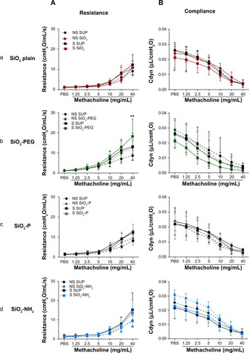
SiO2 and SiO2-PEG NPs, but not SiO2-P and SiO2-NH2 NPs, induce moderate alterations in lung function
Lung function tests performed 24 hours after intratracheal instillation of SiO2 NPs and OVA challenge in S mice showed different results for the four particles tested. SiO2 plain did not have an effect on respiratory system resistance when compared to SUP; however, dynamic compliance was significantly decreased in S mice instilled with SiO2 already, before methacholine challenge (P<0.05), maintaining the lower trend at 1.25 and 2.5 mg mL methacholine concentrations (P=0.05 and P=0.06, respectively) (). The instillation of SiO2-PEG in S mice led to an increase of resistance following the highest methacholine concentration, namely 40 mg/mL (P<0.01). Dynamic compliance was lower in SiO2-PEG with respect to sensitized SUP, but this difference was not statistically significant (). On the contrary, SiO2-P and SiO2-NH2 NPs had no effect on either lung resistance or compliance in S mice (). The effect of the instillation of NPs in NS mice was minimal for all NPs tested ().
Figure 6 SiO2 and SiO2-PEG nanoparticles induce moderate alterations in pulmonary function.
Notes: Lung resistance (A) and dynamic compliance (B) of NS and S mice exposed to increasing concentrations of methacholine (1.25–40 mg/mL) 24 hours after 50 μg SiO2 plain (a), SiO2-PEG (b), SiO2-P (c), or SiO2-NH2 (d) (solid lines) or respective SUP (dashed lines) and ovalbumin challenge. Mean ± standard deviation (n=6–8/group). *P≤0.05; **P≤0.01 versus S SUP.
Abbreviations: NS, non-sensitized; PBS, phosphate-buffered saline; PEG, polyethylene glycol; S, sensitized; SiO2, silicon dioxide; SiO2-NH2, amino-coated SiO2; SiO2-P, phosphate-coated SiO2; SiO2-PEG, PEGylated SiO2; SUP, supernatant controls.
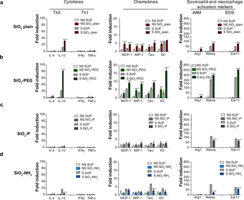
SiO2 and SiO2-PEG NPs, but not SiO2-P and SiO2-NH2 NPs, augment the Th2 proinflammatory milieu in the lungs of OVA-sensitized mice
To characterize the distinct effects of the different surface modification of SiO2 NPs on immune responses, we performed PCR arrays on total RNA from lung homogenates. A distinct increase of T helper type 2 (Th2) cytokines (interleukin [IL]-4/IL-13) was observed in S mice instilled with SiO2-PEG and, to a lesser extent, by SiO2, with no effect on Th1 (interferon-γ/tumor necrosis factor-α) cytokines (). Different Th2 chemokines (MCP-1/CCL2, MIP-1/CCL3, Tarc/CCL17, KC/CXCL1) involved in recruitment of macrophages, eosinophils, lymphocytes, and neutrophils were exclusively upregulated by SiO2-PEG and SiO2, and downregulated or unregulated by SiO2-NH2 and SiO2-P. Moreover, SiO2 and SiO2-PEG only increased markers of Th2 alternative activation of macrophage (Arg1 and Retnla) and eosinophil (Ear11) activation, and had no effect on the markers for classical activation (CXCL9, Nos2) ( and ). These effects of NPs were observed also for other chemotactic chemokines (MIP-3/CCL20, MDC/CCL22, MIP-2/CXCL2) as well as for asthma markers (Retnlg, Chi3l4). Muc5ac was only upregulated by SiO2-PEG in S mice (). The effects of SiO2 and SiO2-PEG NP instillation on selective Th2 gene expression levels were dose dependent, and significant upregulation of IL-13, MIP-1/CCL3, and Arg1 was detected at the highest concentration (50 μg/mouse) ().
Figure 7 SiO2 nanoparticles differentially modulate inflammatory genes.
Notes: Representative gene regulations in NS and S lungs 5 days after intratracheal instillation with 50 μg SiO2 plain (a), SiO2-PEG (b), SiO2-P (c), or SiO2-NH2 (d), or respective SUP and ovalbumin challenge. Mean ± standard deviation (n=4/group). *P≤0.05; **P≤0.01; ***P≤0.001 versus respective SUP.
Abbreviations: AAM, alternatively activated macrophages; EOS, eosinophils; IFN, interferon; IL, interleukin; NS, non-sensitized; PEG, polyethylene glycol; S, sensitized; SiO2, silicon dioxide; SiO2-NH2, amino-coated SiO2; SiO2-P, phosphate-coated SiO2; SiO2-PEG, PEGylated SiO2; SUP, supernatant controls; TNF, tumor necrosis factor; Th1, T helper type 1; Th2, T helper type 2.
Discussion
In this study, we used a well-characterized, murine model of mild allergic airway inflammationCitation23 to investigate SiO2 NP-mediated, surface-dependent proinflammatory and immunomodulatory effects in the lung. SiO2 plain and SiO2-PEG exerted potent Th2-immunomodulatory activities that involved alternatively activated alveolar macrophages that presumably phagocytized SiO2 NPs. Moreover, functionalization of SiO2 NPs with amino or phosphate groups conferred a higher NP tolerance, with a minimal inflammatory milieu in OVA-sensitized mice.
Only a few studies have investigated the modulation of allergic disease by NPs.Citation19,Citation20,Citation26 Our results showed significant immunomodulatory effects exerted by SiO2 NPs in OVA-sensitized mice, with a significant increase in BALF inflammatory infiltrate, mainly comprising macrophages and eosinophils. Our data are in line with a previous study testing titanium dioxide and gold NPs in the elicitation phase of the allergic response.Citation26 We focused our read-out to day 5 after challenge. The reason for this choice was that this time point is representative for the elicitation phase of the allergic response in our mouse model, since it is characterized by strong recruitment of inflammatory cells into the lung.Citation27 Since the Th2-adjuvant effects of SiO2 NPs applied during the sensitization phase have been previously demonstrated,Citation19,Citation20 we now can assume that SiO2 NPs also exacerbate the elicitation phase of allergic airway inflammation.
SiO2 NPs showed a strong proinflammatory effect in NS mice, leading to neutrophil and lymphocyte recruitment after intratracheal instillation, which is in agreement with a previous workCitation16 showing neutrophil recruitment in the early inflammation stages (up to 1 week after instillation) following intratracheal instillation of SiO2 NPs. We also demonstrated an increase of eosinophils in NS mice exposed to SiO2 NPs, a phenomenon also observed in rats after titanium NP exposure.Citation10
The observation that, 24 hours after intratracheal instillation, the percentage of FITC+ cells in S mice was higher compared to NS mice, is in agreement with our earlier study.Citation28 NP uptake occurred mainly by pulmonary macrophages, as has been observed in healthy animals.Citation29 Confocal analysis confirmed that NPs were located inside the phagocytes, and did not just interact with the cell surface. Moreover, we demonstrated that, in S mice, more alveolar macrophages phagocytize NPs compared to in NS mice and, consequently, particle translocation and retention in the interstitium by interstitial macrophages occurs to a higher extent in NS mice. Since these cells exert immunoregulatory properties, this could explain the short-term NP-induced inflammation in NS mice.Citation16 The higher percentage of phagocytizing alveolar macrophages in S mice relates to our observation of SiO2-dependent enhancement of expression of markers for alternatively activated macrophages, like arginase 1 and mannose receptors in NP-treated S mice. These cells show increased phagocytic activityCitation30 and aggravate allergic airway inflammation.Citation31 The SiO2-dependent increase in alternatively activated macrophages is of high clinical importance and could account, at least in part, for the differential localization of SiO2 NPs in S and NS mice.
A few groups have investigated the effects of surface modifications of SiO2 NPs in vitro and in vivo;Citation21,Citation22,Citation32,Citation33 however, to our knowledge, this is the first study that explores the effect of SiO2 NP functionalization on their proinflammatory and immunomodulatory effects in allergic airway inflammation in vivo. Our data showed that coating NPs with short-chain PEG was not a successful strategy for modifying biological properties of SiO2 NPs: in fact, both NS and S mice treated with SiO2-PEG responded similarly in terms of inflammation to those treated with SiO2 plain. Moreover, both SiO2 plain and SiO2-PEG enhanced mucus production and exerted cytotoxic effects. The ultrastructural damages are very likely mediated by SiO2 NP-induced oxidative stress and apoptosis.Citation34 Possibly, the 500 Da PEG chain used to coat the NPs was not long enough to mask the surface of the NPs. Lipka et al suggested that the PEG chain length is essential for determining the biodistribution of NPs.Citation11 Moreover, both molecular weight and chain density of PEG influence mesoporous SiO2 NP binding to serum proteins and phagocytosis from THP-1 macrophages, proposing 10 kDa as the minimal molecular weight to inhibit phagocytosis.Citation35 In light of these studies, our PEG chain might have been too small to mask the effect of SiO2 NPs. In contrast, our results showed that coating with amino or phosphate groups successfully mitigated the toxic and immunomodulatory effects of SiO2 NPs. These effects were observed at histopathological and functional levels, with less BALF cell infiltrate, absence of goblet cell necrosis, and normal lung function in both NS and S mice instilled with SiO2-P and SiO2-NH2 versus SiO2 plain or SiO2-PEG. It has been demonstrated that coating of NPs can affect their interaction with biological fluids.Citation36 Proteins bind to the surface of NPs and form a coating known as the “protein corona,” which can critically affect the interaction of the NPs with living systems. Speculation can be made as to whether our surface modifications of SiO2 NPs affected the thermodynamic characteristics of lipid monolayers, including lung surfactant or cell membrane, as previously observed in 1,2-dipalmitoyl-sn-glycerol-3-phosphocholine–cholesterol Langmuir monolayers.Citation37,Citation38
Different cellular uptake mechanisms may be involved when NPs are functionalized; in fact, it has been previously shown that SiO2 NPs coated with amine or carboxyl groups display a different intracellular distribution compared to those that are uncoated.Citation39 In our model, both amino and phosphate functionalization failed to regulate markers of macrophage activation, suggesting a different compartmentalization of SiO2-P and SiO2-NH2 within macrophages. Further studies should investigate these aspects.
The successful mitigation of effects of SiO2 NPs coated with amino groups is in agreement with the results of Yamashita et al,Citation32 which showed less fetotoxicity of amino-coated NPs when compared to plain NPs. To the best of our knowledge, this is the first study showing mitigation of side effects of SiO2 NPs after phosphate coating.
Our findings on the expression of key genes of inflammation support the hypothesis that functionalization of NPs modify their biological activity in vivo. The immunomodulatory NPs (SiO2 plain and SiO2-PEG), but not the counterpart NPs (SiO2-P and SiO2-NH2), increased the inflammatory milieu in the pulmonary tissue, with upregulation of Th2 cytokines, as well as sensitization-induced chemokines, the latter responsible for chemoattraction of granulocytes (MIP-1/CCL3 and MIP-2/CXCL2, KC/CXCL1), lymphocytes (Tarc/CCL17, MIP-3/CCL20, and MDC/CCL22), and macrophages (MCP-1/CCL2).Citation40 These data support the concept of recruitment of inflammatory cells observed in the BALF of OVA-sensitized mice by immunomodulatory NPs only. Furthermore, our gene expression analysis showed that only SiO2 plain and SiO2-PEG NPs could lead to increased marker of eosinophils activation (Ear11)Citation41 and alternative activation of macrophages, with no modulation of markers of classical activation of macrophages (Arg1/Retnla versus Nos2/CXCL9).Citation31 The asthmatic profile present in the lungs of S mice was further confirmed by the upregulation of genes involved in allergic airway inflammation: Chitinase 3-like 4 (Chi3l4)Citation42 was modulated in our experiments by both SiO2 and SiO2-PEG NPs in S mice. Among the resistin family, FIZZ1 (Retnla) and FIZZ2 (Retnlb), but not FIZZ3 (Retnlg), mRNAs were found to be upregulated in the lungs of S mice 6 hours after the OVA challenge:Citation43 in this study we still found modulation of FIZZ1 and FIZZ3 in S mice 5 days after OVA challenge, which was significantly enhanced by both SiO2 and SiO2-PEG NPs and downregulated by SiO2-NH2. The prolongation of FIZZ1 and FIZZ3 expression after challenge may have been due to our relatively long sensitization protocol.
In most of the genes evaluated in our array, SiO2-PEG had a greater effect than SiO2. A clear example regards the regulation of the mucin Muc5ac gene, responsible for mucus production,Citation44 which was selectively upregulated by SiO2-PEG NPs but not by SiO2 NPs. Since IL-13 is a known modulator of Muc5ac,Citation45 the lower expression of IL-13 mRNA upon SiO2 compared to SiO2-PEG could explain this finding.
Conclusion
Our observations indicate the need for selecting surface modifications of engineered NPs for commercial purposes, in order to improve their biological activity and reduce or abolish their side effects. Moreover, our results can inform and help in the choice of drug delivery strategy, as the selection of the surface modification of NPs is crucial for targeted application.
Supplementary materials
Table S1 Murine primers for real-time polymerase chain reaction
Figure S1 Dose-dependent effects of SiO2 and SiO2-PEG nanoparticles.
Notes: (A) Bronchoalveolar lavage fluid cellular infiltration 5 days after intratracheal instillation with increasing concentrations of SiO2-PEG and ovalbumin challenge. Data are displayed as boxplots (per bar =5 mice/group). (B) Pulmonary messenger RNA expression of sensitized lungs 5 days after intratracheal instillation with the same concentrations of SiO2 plain (red) or SiO2-PEG (green). Data are expressed as mean ± standard deviation (n=3/group). *P≤0.05; **P≤0.01; ***P≤0.001 versus SUP (0 μg).
Abbreviations: IL, interleukin; ns, not significant; PEG, polyethylene glycol; SiO2, silicon dioxide; SiO2-NH2, amino-coated SiO2; SiO2-P, phosphate-coated SiO2; SiO2-PEG, PEGylated SiO2; SUP, supernatant controls.
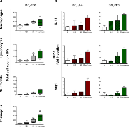
Figure S2 SiO2 nanoparticles differentially modulate inflammatory genes.
Notes: Representative gene regulations in NS and S lungs 5 days after intratracheal instillation with 50 μg SiO2, SiO2-PEG, SiO2-P or SiO2-NH2, or respective SUP (white) and ovalbumin challenge. Mean ± standard deviation (n=4/group). *P≤0.05; **P≤0.01; ***P≤0.001 versus respective SUP.
Abbreviations: NS, non-sensitized; PEG, polyethylene glycol; S, sensitized; SiO2, silicon dioxide; SiO2-NH2, amino-coated SiO2; SiO2-P, phosphate-coated SiO2; SiO2-PEG, PEGylated SiO2; SUP, supernatant controls.
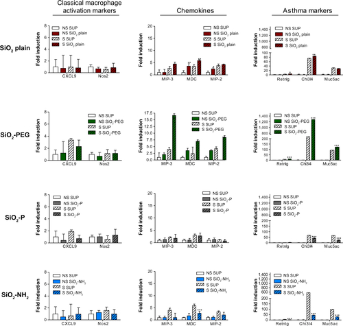
Figure S3 Physical-chemical characterization.
Notes: (A) Size distributions by analytical ultracentrifugation (AUC), plotted as differential distribution of concentration dc per diameter interval dD. The area under the curve is proportional to the mass within this diameter range; (B) Surface charge by electrophoretic mobility with pH titration; (C) Surface chemistry by SIMS (SiO2-P), showing in the top 1nm layer of the particles both the added PO3 functionality and the intrinsic SiO2 fragments of the intrinsic surface chemistry.
Abbreviations: SiO2, silicon dioxide; SiO2-NH2, amino-coated SiO2; SiO2-P, phosphate-coated SiO2; SiO2-PEG, PEGylated SiO2; PEG, polyethylene glycol; dC/dD, distribution of concentration per diameter interval.
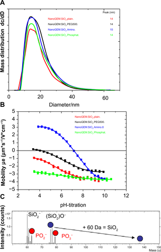
Figure S4 Size and agglomeration characterization of SiO2 NPs by transmission electron microscopy.
Note: (A) Silicon dioxide (plain) and (B) silicon dioxide–fluorescein isothiocyanate nanoparticles.
Abbreviations: SiO2, silicon dioxide; NPs, nanoparticles.
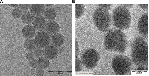
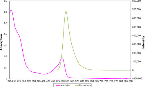
Figure S5 Absorption and fluorescence spectra of silica nanoparticles.
Nanoparticle characteristics
Covalent functionalization of nanoparticles (NPs) was obtained with three different low-molar-mass silanes:
PEGylated silicon dioxide (silica [SiO2]) NPs: polyethylene glycol of chain length molecular weight =500 g/mol.
Amino-coated SiO2 NPs: carry a positively charged amino end group on a flexible, but short C3 linker.
Phosphate-coated SiO2 NPs: carries a negatively charged PO3 end group on the same C3 linker.
Successful functionalization was confirmed by transmission electron microscopy, which showed that NPs maintained a spherical structure with no aggregates, agglomerates, or secondary nucleation. Size distribution in water as measured by analytical ultracentrifugationCitation1 was not significantly changed by the surface modification, with a peak diameter at 14 nm as shown in . Surface charge was measured by electrophoretic mobility with pH titration (), showing that the non-functionalized SiO2 NPs (plain) retained their negative charge across the physiological pH range; phosphate-coated SiO2 NPs strongly negatively charged; and the charge was reduced in PEGylated SiO2 and inverted to positive values in amino-coated SiO2.
Moreover, surface chemistry was evaluated by secondary ion mass spectrometry measurements, showing the presence of free SiOx groups and free functionalized groups, underlining a partial coverage of the NPs ( shows an example of one of the NPs used in this study, phosphate-coated SiO2 NPs). All NPs used in this study were tested for lipopolysaccharide concentration. This was <0.25 EU/mL NPs suspension, which resulted in <0.000055 EU/intratracheal instillation for the highest NP concentrations as determined by limulus amebocyte lysate assay (PyroGene rFC Endotoxin Detection system, product number 50-658U; Lonza Group Ltd, Basel, Switzerland).
Supernatants for each NP were obtained by hard sedimentation (24,000 rpm, 15 hours), with successful removal of 95%–98% of NPs in solution. summarizes the characteristics of the NPs.
SiO2-FITC NPs were prepared as described previously.Citation2 The FITC was covalently bound to the SiO2 core and an SiO2 shell was then produced with the addition of tetraethoxysilane. This shell prevented the influence of the FITC dye on the charge of the NP cores. Successful synthesis of the fluorescent SiO2 NPs (SiO2-FITC) was confirmed by transmission electron microscopy, which showed spherical NPs with no aggregates, agglomerates, or secondary nucleation. The size of the particles was determined by transmission electron microscopy and dynamic light scattering (: plain SiO2; B: SiO2-FITC). The surface charge was measured by electrophoretic mobility with pH titration. The results showed no significant difference between the plain SiO2 and the fluorescent SiO2 NPs. The fluorescent properties were confirmed by fluorescence spectroscopy (). shows the absorption and the emission spectra of the SiO2 particles excited at 460 nm.
References
- WohllebenWValidity range of centrifuges for the regulation of nanomaterials: from classification to as-tested coronasJ Nanopart Res201214130023239934
- HerzEOwHBonnerDBurnsAWiesnerUDye structure–optical property correlations in near-infrared fluorescent core-shell silica nanoparticlesJ Mater Chem20091963416347
Acknowledgments
We thank Johanna Grosch, Benjamin Schnautz, Christine Weil, Katja Haslauer, and Alexandra Seisenberger for excellent technical assistance. We thank Claudia Traidl-Hoffmann, Tanja Seher, Antje Vennemann, and Sebastian Öder for fruitful discussions. We thank Thomas Kuhlbusch for the excellent coordination of the NanoGEM Project. This study was supported by the German Federal Ministry of Education BMBF, NanoGEM Project, FKZ 03X0105, and CK Care, Christine Kühne Center for Allergy Research and Education.
Disclosure
The authors report no conflicts of interest in this work.
References
- AroraSRajwadeJMPaknikarKMNanotoxicology and in vitro studies: the need of the hourToxicol Appl Pharmacol201225815116522178382
- NapierskaDThomassenLCLisonDMartensJAHoetPHThe nanosilica hazard: another variable entityPart Fibre Toxicol201073921126379
- HansenSFMichelsonESKamperABorlingPStuer-LauridsenFBaunACategorization framework to aid exposure assessment of nanomaterials in consumer productsEcotoxicology20081743844718454314
- NelAXiaTMädlerLLiNToxic potential of materials at the nanolevelScience200631162262716456071
- FröhlichERobleggEModels for oral uptake of nanoparticles in consumer productsToxicology2012291101722120540
- MortensenLJOberdörsterGPentlandAPDelouiseLAIn vivo skin penetration of quantum dot nanoparticles in the murine model: the effect of UVRNano Lett200882779278718687009
- YuKOGrabinskiCMSchrandAMToxicity of amorphous silica nanoparticles in mouse keratinocytesJ Nanopart Res2009111524
- KreylingWGSemmler-BehnkeMSeitzJSize dependence of the translocation of inhaled iridium and carbon nanoparticle aggregates from the lung of rats to the blood and secondary target organsInhal Toxicol200921Suppl 1556019558234
- HoMWuKYCheinHMChenLCChengTJPulmonary toxicity of inhaled nanoscale and fine zinc oxide particles: mass and surface area as an exposure metricInhal Toxicol20112394795622122307
- GustafssonÅLindstedtEElfsmarkLSBuchtALung exposure of titanium dioxide nanoparticles induces innate immune activation and long-lasting lymphocyte response in the Dark Agouti ratJ Immunotoxicol2011811112121309687
- LipkaJSemmler-BehnkeMSperlingRABiodistribution of PEG-modified gold nanoparticles following intratracheal instillation and intravenous injectionBiomaterials2010316574658120542560
- BergJMRomoserAAFigueroaDESpencer WestCSayesCMComparative cytological responses of lung epithelial and pleural mesothelial cells following in vitro exposure to nanoscale SiO2Toxicol In Vitro201327243322985735
- LinWHuangYWZhouXDMaYIn vitro toxicity of silica nanoparticles in human lung cancer cellsToxicol Appl Pharmacol200621725225917112558
- FarcalLRUboldiCMehnDMechanisms of toxicity induced by SiO2 nanoparticles of in vitro human alveolar barrier: effects on cytokine production, oxidative stress induction, surfactant proteins A mRNA expression and nanoparticles uptakeNanotoxicology2013761095111022769972
- ParkMDZLynchIRamírez-GarcíaSIn vitro evaluation of cytotoxic and inflammatory properties of silica nanoparticles of different sizes in murine RAW 264.7 macrophagesJ Nanopart Res20111367756787
- ChoWSChoiMHanBSInflammatory mediators induced by intratracheal instillation of ultrafine amorphous silica particlesToxicol Lett2007175243317981407
- KleinCLWienchKWiemannMMa-HockLvan RavenzwaayBLandsiedelRHazard identification of inhaled nanomaterials: making use of short-term inhalation studiesArch Toxicol2012861137115122532024
- LandsiedelRMa-HockLKrollATesting metal-oxide nanomaterials for human safetyAdv Mater2010222601262720512811
- HanBGuoJAbrahaleyTAdverse effect of nano-silicon dioxide on lung function of rats with or without ovalbumin immunizationPLoS One20116e1723621359146
- BrandenbergerCRowleyNLJackson-HumblesDNEngineered silica nanoparticles act as adjuvants to enhance allergic airway disease in micePart Fibre Toxicol2013102623815813
- LankoffAArabskiMWegierek-CiukAEffect of surface modification of silica nanoparticles on toxicity and cellular uptake by human peripheral blood lymphocytes in vitroNanotoxicology20137323525022264124
- MorishigeTYoshiokaYInakuraHThe effect of surface modification of amorphous silica particles on NLRP3 inflammasome mediated IL-1beta production, ROS production and endosomal ruptureBiomaterials2010316833684220561679
- AlessandriniFSchulzHTakenakaSEffects of ultrafine carbon particle inhalation on allergic inflammation of the lungJ Allergy Clin Immunol200611782483016630940
- BedoretDWallemacqHMarichalTLung interstitial macrophages alter dendritic cell functions to prevent airway allergy in miceJ Clin Invest20091193723373819907079
- AlessandriniFWeichenmeierIvan MiertEEffects of ultrafine particles-induced oxidative stress on Clara cells in allergic lung inflammationPart Fibre Toxicol201071120420656
- HussainSVanoirbeekJALuytsKLung exposure to nanoparticles modulates an asthmatic response in a mouse modelEur Respir J20113729930920530043
- AlessandriniFBeck-SpeierIKrappmannDRole of oxidative stress in ultrafine particle-induced exacerbation of allergic lung inflammationAm J Respir Crit Care Med200917998499119264975
- AlessandriniFSemmler-BehnkeMJakobTSchulzHBehrendtHKreylingWTotal and regional deposition of ultrafine particles in a mouse model of allergic inflammation of the lungInhal Toxicol20082058559318444011
- MorfeldPTreumannSMa-HockLBruchJLandsiedelRDeposition behavior of inhaled nanostructured TiO2 in rats: fractions of particle diameter below 100 nm (nanoscale) and the slicing bias of transmission electron microscopyInhal Toxicol20122493995123216155
- AlexisNESoukupJNierkensSBeckerSAssociation between airway hyperreactivity and bronchial macrophage dysfunction in individuals with mild asthmaAm J Physiol Lung Cell Mol Physiol2001280L369L37511159017
- MoreiraAPHogaboamCMMacrophages in allergic asthma: fine-tuning their pro- and anti-inflammatory actions for disease resolutionJ Interferon Cytokine Res20113148549121631355
- YamashitaKYoshiokaYHigashisakaKSilica and titanium dioxide nanoparticles cause pregnancy complications in miceNat Nanotechnol2011632132821460826
- YoshidaTYoshiokaYMatsuyamaKSurface modification of amorphous nanosilica particles suppresses nanosilica-induced cytotoxicity, ROS generation, and DNA damage in various mammalian cellsBiochem Biophys Res Commun201242774875223044420
- AhamedMSilica nanoparticles-induced cytotoxicity, oxidative stress and apoptosis in cultured A431 and A549 cellsHum Exp Toxicol20133218619523315277
- HeQZhangJShiJThe effect of PEGylation of mesoporous silica nanoparticles on nonspecific binding of serum proteins and cellular responsesBiomaterials2010311085109219880176
- MonopoliMPAbergCSalvatiADawsonKABiomolecular coronas provide the biological identity of nanosized materialsNat Nanotechnol201271277978623212421
- GuzmánEFerrariMSantiniELiggieriLRaveraFInfluence of silica nanoparticles on phase behavior and structural properties of DPPC – Palmitic acid Langmuir monolayersColloids Surf A Physicochem Eng Asp2012413280287
- GuzmánELiggieriLSantiniEFerrariMRaveraFMixed DPPC-cholesterol Langmuir monolayers in presence of hydrophilic silica nanoparticlesColloids Surf B Biointerfaces201310528429323384691
- NabeshiHYoshikawaTArimoriAEffect of surface properties of silica nanoparticles on their cytotoxicity and cellular distribution in murine macrophagesNanoscale Res Lett201169321711578
- PalmqvistCWardlawAJBraddingPChemokines and their receptors as potential targets for the treatment of asthmaBr J Pharmacol200715172573617471178
- CormierSAYuanSCrosbyJRT(H)2-mediated pulmonary inflammation leads to the differential expression of ribonuclease genes by alveolar macrophagesAm J Respir Cell Mol Biol20022767868712444027
- ZhangQHitchinsVMSchrandAMHussainSMGoeringPLUptake of gold nanoparticles in murine macrophage cells without cytotoxicity or production of pro-inflammatory mediatorsNanotoxicology2011528429520849214
- StützAMPickartLATrifilieffABaumrukerTPrieschl-StrassmayrEWoisetschlägerMThe Th2 cell cytokines IL-4 and IL-13 regulate found in inflammatory zone 1/resistin-like molecule alpha gene expression by a STAT6 and CCAAT/enhancer-binding protein-dependent mechanismJ Immunol20031701789179612574343
- RoseMCVoynowJARespiratory tract mucin genes and mucin glycoproteins in health and diseasePhysiol Rev20068624527816371599
- KanohSTanabeTRubinBKIL-13-induced MUC5AC production and goblet cell differentiation is steroid resistant in human airway cellsClin Exp Allergy2011411747175622092504