
 ?Mathematical formulae have been encoded as MathML and are displayed in this HTML version using MathJax in order to improve their display. Uncheck the box to turn MathJax off. This feature requires Javascript. Click on a formula to zoom.
Abstract
A rapid synthesis of silver nanoparticles (AgNPs) using Agrimoniae herba extract as reducing agent and stabilizer (A. herba-conjugated AgNPs [AH-AgNPs]) were designed, characterized, and evaluated for antitumor therapy feasibility. In this study, critical factors in the preparation of silver nanoparticles, including extraction time, reaction temperature, the concentration of AgNO3, and A. herba extract amount, were investigated using ultraviolet-visible spectroscopy. AH-AgNPs with well-defined spherical shape, homogeneous distributional small size (30.34 nm), narrow polydispersity index (0.142), and high negative zeta potential (−36.8 mV) were observed by transmission electron microscopy and dynamic light scattering. Furthermore, the results of X-ray diffraction and Fourier-transform infrared spectroscopy further indicated successful preparation of AH-AgNPs. Acceptable long-term storage stability of AH-AgNPs was also confirmed. More importantly, AH-AgNPs displayed significantly higher antiproliferative effect against a human lung carcinoma cell line (A549 cells) compared with A. herba extract and bare AgNPs prepared by sodium citrate. The half-maximal inhibitory concentrations of AH-AgNPs, bare AgNPs, and A. herba extract were 38.13 μg · mL−1, 184.87 μg · mL−1, and 1.147 × 10⁴ μg · mL−1, respectively. It is suggested that AH-AgNPs exhibit a strong antineoplastic effect on A549 cells, pointing to feasibility of antitumor treatment in the future.
Introduction
As a consequence of their unique physical and surface properties, silver nanoparticles (AgNPs) have been shown to have a wide range of applications in the fields of catalysis, photonics, biology, pharmaceutics, and drug-delivery systems in recent years.Citation1–Citation4 In the clinic, AgNPs offer an alternative strategy to reverse the increasing spread of multidrug resistance resulting from the abuse of antibiotics.Citation5–Citation9 Notably, AgNPs have also received great attention because of their antimicrobial and anticancer activities.Citation10–Citation12 Owing to these distinct functions, a myriad of studies have emerged that focus on the rapid and efficient synthesis of AgNPs for incorporation into medical dressings and devices in the past few decades.Citation13–Citation16 Compared with some chemical and physical methods, natural systems capable of reducing silver ions and fabricating NPs at moderate conditions exhibit the promise of generating large amounts of AgNPs with limited energy input and impact on the environment.Citation17 To date, bacteria, fungi, and plant extracts have been applied to establish silver nanosized systems, and such synthetic AgNP-preparation techniques involving the reduction of silver ions in the presence of a protective agent can prevent the AgNPs from aggregation and readily allow isolation of the NPs.Citation18,Citation19 From an economic perspective, plant extracts with various reductive groups can act as reducing and capping agents for the synthesis of NPs, which could realize more advantages over microbial synthesis in industrial production by way of cost reduction.Citation14,Citation15 Furthermore, natural component-mediated AgNPs utilizing reductive and pharmacological active plant extracts can integrate the advantages of bioactive components and AgNPs,Citation20 which also displayed less toxicity against mammalian health cells.Citation21
A number of plants are suitable for the preparation of AgNPs. For example, some reductive plants, including Aloe vera,Citation22 Manilkara zapota,Citation23 and Alternanthera sessilis,Citation24 have been used for the synthesis of AgNPs with stable structure and uniform distribution. Theoretically, Agrimoniae herba contains various reductive groups, including flavonoids, phenols, and tannin, which are capable of reducing Ag+ to AgNPs through specific technology. As a famous traditional herbal medicine, A. herba containing various active components is widely used in anticancer,Citation25 antibacterial,Citation26 and antiinflammatoryCitation27 treatment. Herein, the traditional herbal medicine A. herba extract was utilized to develop a rapid synthesis of AgNPs for in vitro antineoplastic evaluation. In this study, we investigated the influence of various reaction conditions, such as extraction time, temperature, AgNO3 concentration and A. herba extract amount, on the yield of A. herba-conjugated AgNPs (AH-AgNPs) by monitoring the conversion of Ag+ using ultraviolet (UV)-visible spectroscopy. Furthermore, we characterized the structure and morphology of AgNPs by means of transmission electron microscopy (TEM), dynamic light scattering (DLS), X-ray diffraction (XRD), and Fourier-transform infrared spectroscopy (FTIR). More importantly, the advantage of AH-AgNPs over A. herba extract and bare AgNPs in cytotoxicity against a human lung cancer cell line (A549 cells) was also evaluated using the 3-(4,5- dimethylthiazol-2-yl)-2,5-diphenyltetra-zoliumbromide (MTT) assay.
Materials and methods
Materials
A. herba was purchased from Bozhou Huqiao Pharmaceutical (Bozhou, People’s Republic of China), and was authenticated by Shihui Qian, a professor at the Jiangsu Provincial Academy of Chinese Medicine. Aluminum chloride, anhydrous sodium carbonate, silver nitrate, and phosphoric acid were provided by Nanjing Chemical Reagent (purity >99.8%, analytical reagent; Nanjing, People’s Republic of China). Folin-Ciocaileu reagent was obtained from Nanjing Duly Biotech (Nanjing, People’s Republic of China). Rutin and gallic acid reference substances (purity >99.5%) were purchased from the National Institutes for Food and Drug Control (Beijing, People’s Republic of China). Trisodium citrate was provided by Kunshan Jincheng Reagent (Kunshan, People’s Republic of China). MTT, dimethyl sulfoxide (DMSO), Roswell Park Memorial Institute (RPMI) 1640 medium, trypsin, and phosphate-buffered saline were provided by Nanjing KeyGen Biotech (Nanjing, People’s Republic of China). Fetal bovine serum (FBS) was bought from Zhejiang Tianhang Biological Technology (Hangzhou, People’s Republic of China). Water in this study was provided by the Milli-Q water-purification system (EMD Millipore, Billerica, MA, USA). All other chemicals and solvents were of analytical or reagent grade.
Methods
Preparation of A. herba extract
Fresh A. herba (30 g) was culled and boiled with 300 mL of deionized water for 15 minutes in a 500 mL Erlenmeyer flask, followed by filtration in a reduced-pressure condition. At the end of this time, the volume of extract was adjusted to 300 mL with deionized water and stored at 4°C until further use.
Content determination
Total flavonoid content
In this experiment, the concentration of total flavonoid was detected at 408 nm by UV-visible spectroscopy using rutin as the reference substance.Citation28 Briefly, 1 mL of A. herba extract was mixed with 2 mL of 1% AlCl3 in methanol and incubated at room temperature. After coincubation for 10 minutes, UV-visible absorbance was recorded at 408 nm, and the total flavonoid content was calculated according to a calibration curve of rutin.
Total phenol content
The total phenol content was measured at 765 nm by UV-visible spectroscopy using gallic acid as the reference substance.Citation29 Briefly, 1 mL of Folin-Ciocaileu reagent was added dropwise into the 5 mL of fivefold-diluted A. herba extract. Subsequently, the mixture was incubated for 5 minutes at room temperature, followed by the addition of 10 mL of 10% Na2CO3 solution. UV-visible absorbance was recorded at 765 nm. Total phenol content was estimated by a calibration curve of gallic acid.
Synthesis of AH-AgNPs
A. herba extract (10 mL) was added to 100 mL of an aqueous solution of AgNO3 (1 mM) with vigorous stirring at room temperature until crimson was observed. Afterward, the reaction mixture was subjected to centrifugation at 13,000 rpm for 15 minutes, followed by the removal of excessive silver ions through rinsing the precipitate of AH-AgNPs thrice. Finally, a crimson AH-AgNP solution was obtained when nanoparticles were redispersed in deionized water.
Synthesis of bare AgNPs
Due to the reductive ability, sodium citrate has been widely used for the preparation of AgNP systems in previous papers.Citation30,Citation31 Briefly, 10 mL of 1% sodium citrate solution was added dropwise to 300 μg · mL−1 of AgNO3 aqueous solution at 100°C. After vigorous stirring for 10 minutes, the bare AgNPs were obtained under a pH environment of 4.0–6.0 using phosphoric acid as a regulator.
Optimization of AH-AgNP studies
Based on the AH-AgNP preparation, the extraction time of A. herba was predetermined at 2, 5, 15, 30, and 60 minutes to evaluate the influence on the yield of AH-AgNPs. Likewise, the reaction temperature was preset at 4°C, 25°C, 40°C, 50°C, and 60°C to investigate the time-dependent influence on the synthesis of AH-AgNPs. Furthermore, in order to observe the concentration-related influences on the synthesis of AH-AgNPs, various concentration gradients of AgNO3 – set at 0.1 mM, 0.5 mM, 1 mM, and 2 mM – were also employed as the critical parameters. Finally, 2.5, 5, 10, 25, 50, and 75 mL of A. herba extract (equal to 0.25, 0.5, 1, 2.5, 5, and 7.5 g crude drug, respectively) were added to 100 mL of 1 mM aqueous AgNO3 solution, namely the volume ratio of A. herba extract to AgNO3 solution (1 mM) varied in the range of 1:40–3:4.
The UV-visible absorption spectrum was recorded to evaluate the optimal characterization and analyze the synthesis rate of AH-AgNPs obtained under different reaction conditions at a wavelength of 300–800 nm using a UV-1800 PC UV-visible spectrophotometer (AoXi Technology Instrument, Shanghai, People’s Republic of China).
Characteristics of AH-AgNPs
TEM and DLS measurements
The morphology of the optimal AH-AgNP solution was observed by TEM (Tecnai 12; Philips, Amsterdam, the Netherlands). Briefly, 15 μL of the AH-AgNP solution was dropped on a carbon-coated copper grid and dried prior to measure. Ten fields of vision (including 400 NPs in all) were selected randomly from TEM images of AH-AgNPs, and the average particle size was obtained after statistical analysis. In addition, DLS (Nano ZS; Malvern Instruments, Malvern, UK) was also used to measure particle size and zeta potential of AH-AgNPs. Accordingly, morphologic characterization of bare AgNPs was performed as per the method described earlier.
X-ray diffraction measurement
Crystallization of the AH-AgNP dry powder obtained from freeze-drying the solution of AH-AgNPs was characterized by XRD (D8 Advance; Bruker Optik, Ettlingen, Germany) with CuKα radiation (λ=1.5406 Å) and working at 40 kV/40 mA in the range of 10°–80° with a 2°-per-minute scanning rate.
Fourier-transform infrared spectroscopy
The characteristic groups of A. herba extract conjugated with the surface of AgNPs were characterized by FTIR (Nicolet IS10; Thermo Fisher Scientific, Waltham, MA, USA) spectroscopy in the range of 4,000–500 cm−1 at 2° · cm−1 resolution. Each sample was recorded with KBr pellets on an FTIR spectroscope at room temperature.
A549 cell line culture
A human lung cancer cell line (A549) was obtained from KeyGen Biotech. The cells were cultured in RPMI 1640 medium supplemented with 10% FBS, 1% nonessential amino acids, 100 U · mL−1 streptomycin, and 100 μg · mL−1 penicillin at 37°C under an atmosphere of 5% CO2 and 90% relative humidity. The cells were subcultivated every 3–4 days (at 70% confluence) using trypsin at a split ratio of 1:4.
In vitro cytotoxicity studies
A549 cells were used to investigate the cytotoxicity of AH-AgNPs in vitro by MTT assay. The A549 cells were seeded into 96-well culture plates at a density of 5 × 10³ cells per well. After 80% confluence, the culture medium was removed, and 200 μL of each solution (100 μL of FBS-free culture medium with 100 μL of AH-AgNPs, bare AgNPs, and A. herba solution at various concentrations) and a negative control (100 μL of FBS-free culture medium with 100 μL of culture medium) were added to the wells. After further cell culture for 24 hours, 20 μL of sterile MTT solution (5 mg · mL−1) was added to each well, and then the cells were cultured for a further 4 hours at 37°C. Afterward, the culture medium was removed, and 100 μL of DMSO was added to dissolve the produced formazan crystals. The optical density (OD) at 490 nm was measured by using a Multiskan MK3 microplate reader (Thermo Fisher Scientific). Cell viability (%) was calculated according to the following formula: cell viability (%) = (OD of test group/OD of control group) × 100%. The drug concentration causing 50% cell-growth inhibition (IC50) was calculated using SPSS 17.0 (SPSS, Inc., Chicago, IL, USA) software.
Statistical analysis
All experiments were conducted in triplicate unless otherwise specified. Statistical analysis was performed with SPSS 17.0 software. All the results were expressed as means plus or minus standard deviation. Student’s t-test was used to evaluate the differences between groups. A value of **P<0.01 was considered extremely significant.
Results
Content determination
A. herba is known to be rich in diverse bioactive components (such as flavonoids and phenols) that might play a significant role in the bioreduction of AgNPs. Phytochemical analysis of A. herba extract (0.1 g crude drug · mL−1) revealed a high level of total flavonoid content up to 4.0 mg · mL−1. Likewise, A. herba extract (0.1 g crude drug · mL−1) was found to be rich in total phenols (2.6 mg · mL−1). Accordingly, the high level of total flavonoids and phenols showed the potential capability of reducing Ag+ to AgNPs.
Effect of extraction time of A. herba on the synthesis of AH-AgNPs
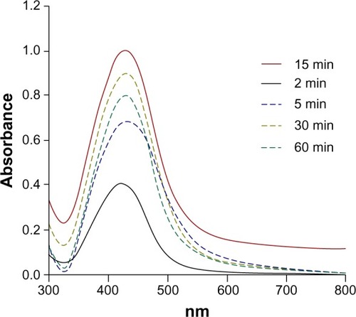
The absorption spectrum of AH-AgNPs was recorded at different extracted times of A. herba (). The maximum amount of A. herba conjugated with AgNPs was found after extraction for 15 minutes. Interestingly, a longer extraction time had a comparatively lower yield. Consequently, the extraction time played a key role in the synthesis of AH-AgNPs, the main reason might be connected with different extraction time due to various active components.
Figure 1 Ultraviolet-visible spectra of AH-AgNPs obtained via Agrimoniae herba extract and 1 mM AgNO3 for various extraction times.
Notes: The full line represents the optimum reaction time, and the dashed lines represent assayed reaction conditions. The ordinate represents the maximal ultraviolet absorption within a range of 300–800 nm.
Abbreviation: AH-AgNPs, Agrimoniae herba-conjugated Ag nanoparticles.
Effect of reaction temperature and time on the synthesis of AH-AgNPs
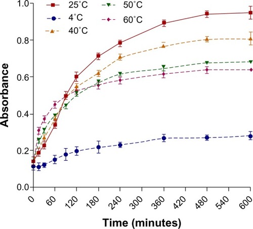
We further prearranged various temperatures from 4°C to 60°C and different reaction times to observe the peak wavelength of AH-AgNPs and investigate the influence of temperature and reaction time on the yield of AH-AgNPs. As shown in , an expansion of reaction time was accompanied with an obvious improvement in the yield of AH-AgNPs before 8 hours, but no significant enhancement was found after 8 hours. Also, temperatures below or above 25°C were unable obtain the maximal synthesis rate of AH-AgNPs.
Figure 2 Time course of AH-AgNPs prepared via Agrimoniae herba extract and 1 mM AgNO3 under different reaction temperatures.
Notes: The full line represents the optimum reaction temperature, and the dashed lines represent assayed reaction temperatures. Data represent means ± standard deviation. The ordinate represents the maximal ultraviolet absorption within a range of 300–800 nm.
Abbreviation: AH-AgNPs, Agrimoniae herba-conjugated Ag nanoparticles.
Effect of Ag ion concentration on the synthesis of AgNPs
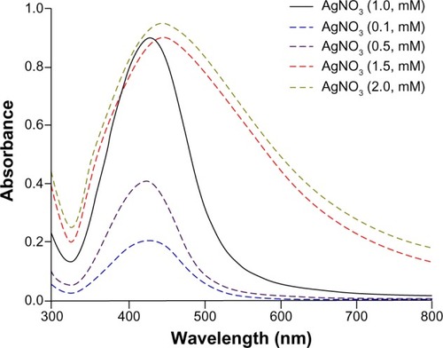
Ag+ bioreduction mediated by A. herba extract was also monitored by recording absorption spectra at different AgNO3 concentrations. As shown in , the absorbance at 447 nm increased obviously as the concentration increased from 0.1 to 1.0 mM. However, the absorption wavelength was slightly shifted when AgNO3 concentrations were higher than 1.0 mM, at which the comparative agglomeration was also speculated to be due to the broadening of peak width at half height.Citation32
Figure 3 Ultraviolet-visible spectra of AH-AgNPs obtained via Agrimoniae herba extract at various concentrations of AgNO3.
Notes: The full line represents the optimum concentration of AgNO3, and the dashed lines represent assayed concentrations of AgNO3. The ordinate represents the maximal ultraviolet absorption within a range of 300–800 nm.
Abbreviation: AH-AgNPs, Agrimoniae herba-conjugated Ag nanoparticles.
Effect of A. herba extract amount on the synthesis of AH-AgNPs
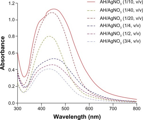
Optimization of the amount of A. herba extract was carried out by recording UV-visible absorption of AH-AgNPs. In this experiment, the reaction was monitored by varying ratios of extract solution to silver nitrate (1:40, 1:20, 1:10, 1:4, 1:2, and 3:4, v/v). As shown in , the maximal absorbance (represented yield) was displayed when the ratio of extract to silver nitrate was predetermined at 1:10 (v/v). According to the results of visual observation, with the amount of A. herba extract increased, consequent changes in the color of AH-AgNPs were observed from yellow to reddish-yellow, and further crimson.
Figure 4 Ultraviolet-visible spectra of AH-AgNPs obtained at various volume ratios of Agrimoniae herba extract to AgNO3.
Notes: The full line represents the optimum amount of A. herba extract, and the dashed lines represent the assayed amount of A. herba extract. The ordinate represents the maximal ultraviolet absorption within a range of 300–800 nm.
Abbreviation: AH-AgNPs, Agrimoniae herba-conjugated Ag nanoparticles.
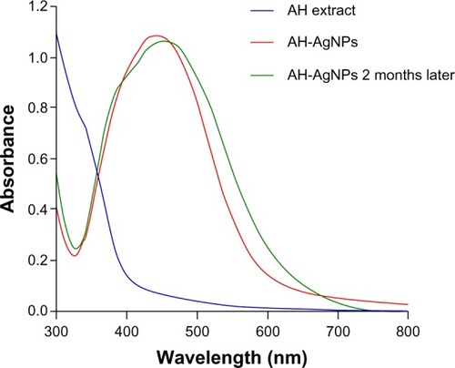
Stability of AH-AgNPs
In order to evaluate the stability of the AH-AgNPs in an aqueous system, the absorption spectrum of each optimized sample was detected after storage at room temperature for 2 months. As shown in , the absorption peak of AH-AgNPs shifted slightly from 445 to 450 nm, but no significant changes in peak intensity were observed. This suggested that AgNPs mediated by A. herba extract prepared under optimum condition might have acceptable stability even after 60 days.
Figure 5 Evaluation of stability of Agrimoniae herba extract, AH-AgNPs, and AH-AgNPs 2 months later by using an ultraviolet-visible instrument.
Abbreviation: AH-AgNPs, Agrimoniae herba-conjugated Ag nanoparticles.
TEM and DLS analysis of AH-AgNPs
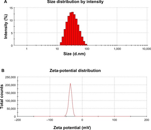
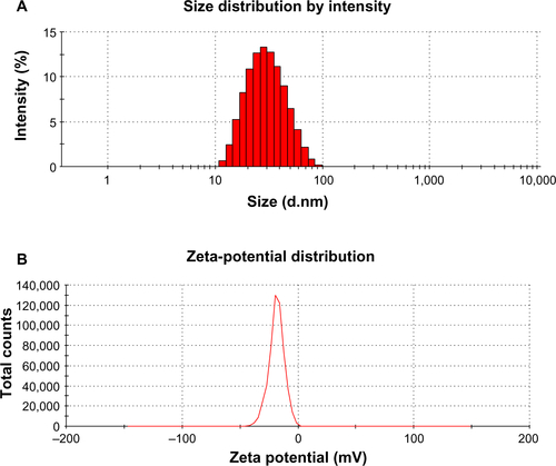
In order to investigate the microstructure of AH-AgNPs and evaluate the relevant preparation technology, AH-AgNP morphology was assessed by TEM. As shown in , well-defined spherical particles with homogeneous distributional size were observed clearly, which confirmed that the AH-AgNPs were successfully prepared. Meanwhile, the average particle size of ~11 nm was calculated after statistical analysis (see ). On the other hand, small size (30.34±5.9 nm), narrow polydispersity index (0.142), and highly negative zeta potential (−36.8±2.5 mV) were also measured by DLS (see ). This suggested a superior and highly efficient preparation technology of AH-AgNPs and a stable system contributed to by highly negative potential. In addition, the morphology of bare AgNPs also showed similar results for size (26.42±3.2 nm) and zeta potential (−28.3±4.7 mV, and ).
Figure 6 (A and B) Morphologic characteristics. (A) TEM image and (B) particle-size distribution of AH-AgNPs synthesized via Agrimoniae herba extract and AgNO3.
Abbreviations: TEM, transmission electron microscopy; AH-AgNPs, Agrimoniae herba-conjugated Ag nanoparticles.
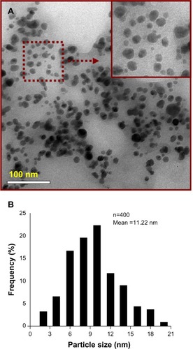
Figure 7 (A and B) Structure characteristics by DLS patterns. (A) Size distribution of AH-AgNPs; (B) zeta-potential analysis of AH-AgNPs.
Abbreviations: DLS, dynamic light scattering; AH-AgNPs, Agrimoniae herba-conjugated Ag nanoparticles.
Powder X-ray diffraction analysis
The crystalline nature of AgNPs was confirmed by XRD analysis (). XRD patterns showed four distinct diffraction peaks at 38.52°, 44.30°, 64.23°, and 77.56°, which can be attributed to the (111), (200), (220), and (311) crystallographic planes of the face-centered cubic silver crystals.Citation33,Citation34 The broadening of Bragg’s peaks indicated the formation of AgNPs, which conformed to the database of the Joint Committee on Powder Diffraction Standards file 04-0783. Average size was calculated using the Debye–Scherrer equation by determining the width of the Bragg’s reflection:
Figure 8 X-ray diffraction patterns of AH-AgNPs.
Abbreviation: AH-AgNPs, Agrimoniae herba-conjugated Ag nanoparticles.
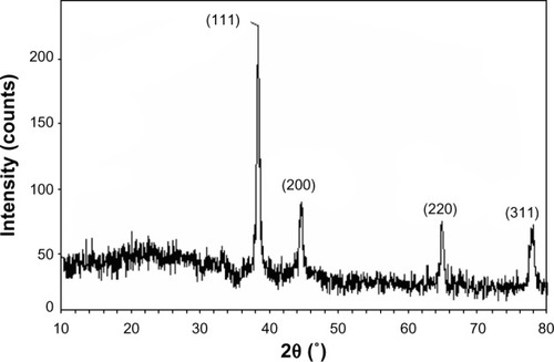
where D represents the mean crystal size of NPs, K represents the Scherer coefficient (0.89), λ is the wavelength of the X-ray radiation source (1.5406 Å), β represents the width of the XRD peak at half height, and θ represents the Bragg angle. According to the Debye–Scherrer equation, the average crystallite size of AH-AgNPs was calculated to be about 11.53 nm (), which was in line with the result offered by TEM.
Table 1 X-ray diffraction measurement of AH-AgNPs for evaluating the average crystallite size
FTIR spectroscopy analysis
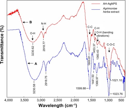
With the purpose of identifying the characteristic groups of A. herba extract and AH-AgNP freeze-dried powder, the FTIR absorption spectrum of each sample is displayed in . Compared with A. herba extract, obvious weakness in characteristic peaks at 1,360 cm−1 and 3,230 cm−1, which were assigned to the stretching and bending vibrations of hydroxyl groups, was observed from the FTIR spectra of AH-AgNPs, suggesting that the consumption of hydroxyl groups was responsible for the reduction of Ag+.35,36 The vibrational bands corresponding to the bonds were derived from water-soluble extracts, such as flavonoids and polyphenols present in A. herba.Citation21,Citation37 In addition, the peaks at 2,918.77 cm−1, 1,600.81 cm−1, and 1,021.10 cm−1 can be attributed to the stretching vibration of the –N–H–, –C=C–, and –C–O–C groups, respectively.Citation1,Citation38–Citation40 On the basis of these results, it is suggested that the AgNPs were coated with reductive components mentioned earlier, and hence could feasibly inhibit further aggregation in an aqueous environment.
Figure 9 Fourier-transform infrared absorption spectra of Agrimoniae herba extract (A), and (B) AH-AgNP freeze-dried power.
Abbreviation: AH-AgNPs, Agrimoniae herba-conjugated Ag nanoparticles.
Cytotoxicity of AH-AgNPs against A549 cells
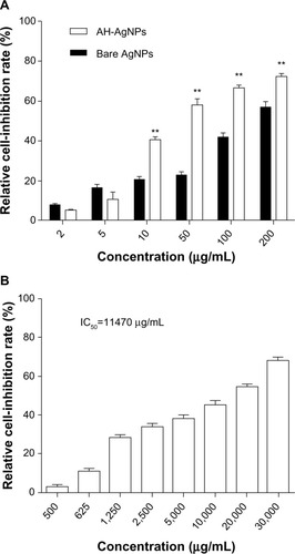
In order to confirm the advantage of AH-AgNPs over A. herba extract in antitumor effects in vitro, A549 cell-viability assays were performed by conventional MTT. As shown in , AH-AgNPs had a predominant cytotoxic effect against A549 cells, suggesting a significant enhancement on in vitro antitumor efficacy compared with A. herba extract. Also, bare AgNP cytotoxicity evaluation allowed us to further define the feasibility of AH-AgNPs in antitumor therapy. The IC50 values of AH-AgNPs, bare AgNPs, and A. herba extract after coincubation with A549 cells for 24 hours were 38.13, 184.87, and 1.147 × 10⁴ μg · mL−1, respectively.
Figure 10 Cytotoxicity of (A) AH-AgNPs and bare-AgNPs, and (B) Agrimoniae Herba extract against A549 cells.
Notes: **P<0.01 versus bare AgNPs. Data represent means ± standard deviation.
Abbreviations: AH-AgNPs, Agrimoniae herba-conjugated Ag nanoparticles; IC50, half-maximal inhibitory concentration.
Discussion
Malignant tumors are fatal diseases that threaten human health. Therefore, tremendous efforts have been put into exploring effective therapeutic modalities for antitumor treatment. As two approved effective anticancer media, A. herba and AgNPs have been applied in the clinical setting recently.Citation41 In this study, AH-AgNPs were rapidly synthesized for the first time, and their more potent antineoplastic effect in vitro was confirmed compared with that of A. herba extract and bare AgNPs.
A. herba extract containing rich flavonoids, phenols and tannin played a key role in the process for bioreduction of Ag+ to AgNPs.Citation42 Preparation-optimization studies showed that the yield of AH-AgNPs decreased when the extraction time was longer than 15 minutes, probably because the reductive compositions were destroyed in A. herba extract after enduring boiling for a long time. Likewise, the synthesis rate of AH-AgNPs decreased when the temperature was higher than 25°C in the process of preparation. Without good dispersion or lack of A. herba extract as a stabilizing agent, AgNPs were easily agglomerated. To some extent, A. herba extract also played a role in the stabilization of AH-AgNPs. On the other hand, particle size, polydispersity index, and zeta potential were important characteristic parameters for testing the long-term storage stability and structure characterization of a drug-delivery system. In DLS measurements, AH-AgNPs were dissolved in deionized water and water molecules encapsulated in silver nanoparticles surface, leading to the different sizes between DLS and TEM measurements. According to a previous paper, the development of crimson color in AH-AgNP solution might be attributed to the occurrence of surface-plasmon resonance, further identifying the successful preparation of AH-AgNPs.Citation35 According to the results of FTIR and DLS, the advantages of optimum AH-AgNPs, such as accepted stability and uniform dispersion, were attributed to A. herba extract conjugation with Ag+. With regard to synthetic rate, A. herba-mediated reduction of Ag+ was completed within 8 hours, which was about threefold improved in comparison with some other plants, such as Curcuma longa,Citation38 which needed 24 hours for complete synthesis of AgNPs. The obtained results indicated a rapid, economical, and effective preparation.
In this study, the strengths of AH-AgNPs over A. herba extract and bare AgNPs in cytotoxicity was evaluated by MTT assay. On the basis of IC50 of AH-AgNPs and bare AgNPs, AgNPs with or without A. herba extract exhibited evident cytotoxicity against A549 cells, suggesting that AgNPs, as a drug carrier, also can act as an effective anticancer agent. Furthermore, it was reasonable that the cell-growth inhibition of AH-AgNPs was about fivefold higher than that of bare AgNPs, which demonstrated that the antitumor potential of AH-AgNPs was more promising than that of bare AgNPs. The noteworthy cytotoxicity of AH-AgNPs further indicated a clear conjugation between A. herba extract and the surface of AgNPs. The mechanism of cell death by AH-AgNPs in A549 cells will need to be researched in the future.
Conclusion
In summary, A. herba-conjugated AgNPs were synthesized using a green and highly effective preparation technology. This synthesis approach has potential in the industrial process, and offers an alternative to traditional physical and chemical methods. More importantly, the cytotoxicity of AH-AgNPs against A549 cells had an advantage over that of bare AgNPs and A. herba extract. In conclusion, this study suggests that AH-AgNPs exhibit the potential to be a promising drug-delivery system in antitumor therapy.
Acknowledgments
This work was financially supported by the Jiangsu Provincial Chinese Medicine Leading Talent Project (LJ200913).
Supplementary materials
Figure S1 TEM image of bare Ag nanoparticles synthesized by a chemical method.
Abbreviation: TEM, transmission electron microscopy.
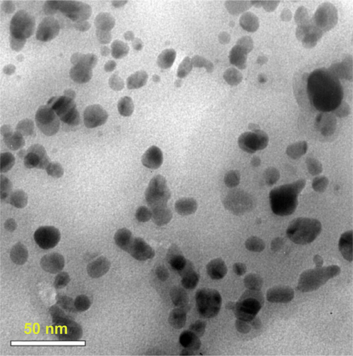
Figure S2 (A and B) DLS patterns. (A) Size distribution of bare AgNPs; (B) zeta-potential analysis of bare Ag nanoparticles.
Abbreviation: DLS, dynamic light scattering.
Disclosure
The authors report no conflicts of interest in this work.
References
- OberdörsterGMaynardADonaldsonKPrinciples for characterizing the potential human health effects from exposure to nanomaterials: elements of a screening strategyPart Fibre Toxicol20052816209704
- GittinsDIBethellDSchiffrinDJNicholsRJA nanometre-scale electronic switch consisting of a metal cluster and redox-addressable groupsNature20004086808676911081506
- DarroudiMAhmadMBAbdullahAHIbrahimNAGreen synthesis and characterization of gelatin-based and sugar-reduced silver nanoparticlesInt J Nanomedicine2011656957421674013
- XuJXiaoXRenFEnhanced photocatalysis by coupling of anatase TiO2 film to triangular Ag nanoparticle islandNanoscale Res Lett20127123922548875
- NeuHCThe crisis in antibiotic resistanceScience19922575073106410731509257
- PitoutJDSandersCCSandersWEJrAntimicrobial resistance with focus on beta-lactam resistance in Gram-negative bacilliAm J Med1997103151599236486
- CraigWAAntimicrobial resistance issues of the futureDiagn Microbiol Infect Dis19962542132178937847
- ShakibaieMRKapadnisBPDhakephalkerPChopadeBARemoval of silver from photographic wastewater effluent using Acinetobacter baumannii BL54Can J Microbiol19994512995100010696478
- PatwardhanRBDhakephalkarPKNiphadkarKBChopadeBAIncidence and prevalence of nosocomial pathogens in ICU with special reference to multiresistant Acinetobacter baumannii harboring multiple plasmidsIndian J Med Res2008128817818719001682
- ChenXSchluesenerHJNanosilver: a nanoproduct in medical applicationToxicol Lett20081411218022772
- HeQGLiuJHuangCWWuWSynthesis and characterization of a silver incorporated magnetic nanocomposite with enhanced antibacterial activitySci Adv Mater201461366376
- HeQHuangCLiuJPreparation, characterization and antibacterial activity of magnetic greigite and Fe3S4/Ag nanoparticlesNanosci Nanotechnol Lett2014611017
- LiYLeungPYaoLSongQWNewtonEAntimicrobial effect of surgical masks coated with nanoparticlesJ Hosp Infect2006621586316099072
- StrohalRSchellingMTakacsMJureckaWGruberUOffnerFNanocrystalline silver dressings as an efficient anti-MRSA barrier: a new solution to an increasing problemJ Hosp Infect200560322623015896880
- ZhangWZQiaoXLChenJGSynthesis of silver nanoparticles – effects of concerned parameters in water/oil microemulsionMater Sci Eng B20071421115
- DaiZGXiaoXHZhangYPIn situ Raman scattering study on a controllable plasmon-driven surface catalysis reaction on Ag nanoparticle arraysNanotechnology2012233333570122842646
- MandalDBolanderMEMukhopadhyayDSarkarGMukherjeePThe use of microorganisms for the formation of metal nanoparticles and their applicationAppl Microbiol Biotechnol200669548549216317546
- QuirosIYamadaMKuboKMizutaniJKuriharaMNishiharaHPreparation of alkanethiolate-protected palladium nanoparticles and their size dependence on synthetic conditionsLangmuir200218414131418
- ChenSHuangKStearnsJAAlkanethiolate-protected palladium nanoparticlesChem Mater200012540541
- GenganRMAnandKPhulukdareeAChuturgoonAA549 lung cell line activity of biosynthesized silver nanoparticles using Albizia adianthifolia leafColloids Surf B Biointerfaces2013105879123352951
- LansdownABSilver in health care: antimicrobial effects and safety in useCurr Probl Dermatol200633173416766878
- ChandranSPChaudharyMPasrichaRAhmadASastryMSynthesis of gold nanoparticles and silver nanoparticles using Aloe vera plant extractBiotechnol Prog200622257758316599579
- KamarajCRajakumarGRahumanAAFeeding deterrent activity of synthesized silver nanoparticles using Manilkara zapota leaf extract against the house fly, Musca domestica (Diptera: Muscidae)Parasitol Res201211162439244822033735
- NiraimathiKLSudhaVLavanyaRBrindhaPBiosynthesis of silver nanoparticles using Alternanthera sessilis (Linn.) extract and their antimicrobial, antioxidant activitiesColloids Surf B Biointerfaces201310228829123006568
- LiWYChanSWGuoDJYuHFCorrelation between antioxidative power and anticancer activity in herbs from traditional Chinese medicine formulae with anticancer therapeutic effectPharm Biol2007457541546
- ParkNYParkKNLeeSHAntimicrobial activities and food preservative effects of Agrimoniae herbaJ Korean Soc Food Sci Nutr2004332244248
- GongCGZhangGQWangXYLeiYHExperimental studies of anti-inflammatory and analgesic effects of extract from Herba agrimoniae [sic]J Pharm Pract200620066339341356
- JiangPBurczynskiFCCampbellCPierceGAustriaJABriggsCJRutin and flavonoid contents in three buckwheat species Fagopyrum esculentum, F. tataricum, and F. homotropicum and their protective effects against lipid peroxidationFood Res Int2007403356364
- AinsworthEAGillespieKMEstimation of total phenolic content and other oxidation substrates in plant tissues using Folin-Ciocalteu reagentNat Protoc20072487587717446889
- LinkSWangZLEl-SayedMAAlloy formation of gold-silver nanoparticles and the dependence of the plasmon absorption on their compositionJ Phys Chem B19991031835293533
- WeiYJinRCChadAMirkin DNA-modified core-shell Ag/Au nanoparticlesJ Am Chem Soc2001123327961796211493092
- JhaAKPrasadKKumarVPrasadKBiosynthesis of silver nanoparticles using Eclipta leafBiotechnol Prog20092551476147919725113
- AhmadMBShameliKDarroudiMYunusWMIbrahimNASynthesis and characterization of silver/clay nanocomposites by chemical reduction methodAm J Appl Sci200961119091914
- BorchertHShevchenkoEVRobertAKornowskiAHaaseMWellerHDetermination of nanocrystal sizes: a comparison of TEM, SAXS, and XRD studies of highly monodisperse CoPt3 particlesLangmuir20052151931193615723491
- ElavazhaganTArunachalamKDMemecylon edule leaf extract mediated green synthesis of silver and gold nanoparticlesInt J Nanomedicine201161265127821753878
- GeethalakshmiRSaradaDVLGold and silver nanoparticles from Trianthema decandra: synthesis, characterization, and antimicrobial propertiesInt J Nanomedicine201275375538423091381
- PhilipDUnniCExtracellular biosynthesis of gold and silver nanoparticles using Krishna tulsi (Ocimum sanctum) leafPhysica E Low Dimens Syst Nanostruct201143713181322
- ShameliKAhmadMBZamanianAGreen biosynthesis of silver nanoparticles using Curcuma longa tuber powderInt J Nanomedicine201275603561023341739
- ShaligramNSBuleMBhambureRBiosynthesis of silver nanoparticles using aqueous extract from the compactin producing fungal strainProcess Biochem2009448939943
- ValliJSVaseeharanBBiosynthesis of silver nanoparticles by Cissusquadrangularis extractsMater Lett201282171173
- KimSHLeeTHYangDCShiquandabutangjiaweibang inhibits tumor metastasis and angiogenesis via regulation of topoisomerase-1J Ethnopharmacol2005981–215716215763377
- EgorovaEMRevinaAASynthesis of metallic nanoparticles in reverse micelles in the presence of quercetinColloids Surf A Physicochem Eng Asp200016818796