
Abstract
Magnetic iron oxide nanoparticles (Fe3O4 MNPs) are among the most useful metal nanoparticles for multiple applications across a broad spectrum in the biomedical field, including the diagnosis and treatment of cancer. In previous work, we synthesized and characterized Fe3O4 MNPs using a simple, rapid, safe, efficient, one-step green method involving reduction of ferric chloride solution using brown seaweed (Sargassum muticum) aqueous extract containing hydroxyl, carboxyl, and amino functional groups mainly relevant to polysaccharides, which acts as a potential stabilizer and metal reductant agent. The aim of this study was to evaluate the in vitro cytotoxic activity and cellular effects of these Fe3O4 MNPs. Their in vitro anticancer activity was demonstrated in human cell lines for leukemia (Jurkat cells), breast cancer (MCF-7 cells), cervical cancer (HeLa cells), and liver cancer (HepG2 cells). The cancer cells were treated with different concentrations of Fe3O4 MNPs, and an MTT (3-[4,5-dimethylthiazol-2-yl]-2,5 diphenyl tetrazolium bromide) assay was used to test for cytotoxicity, resulting in an inhibitory concentration 50 (IC50) value of 23.83±1.1 μg/mL (HepG2), 18.75±2.1 μg/mL (MCF-7), 12.5±1.7 μg/mL (HeLa), and 6.4±2.3 μg/mL (Jurkat) 72 hours after treatment. Therefore, Jurkat cells were selected for further investigation. The representative dot plots from flow cytometric analysis of apoptosis showed that the percentages of cells in early apoptosis and late apoptosis were increased. Cell cycle analysis showed a significant increase in accumulation of Fe3O4 MNP-treated cells at sub-G1 phase, confirming induction of apoptosis by Fe3O4 MNPs. The Fe3O4 MNPs also activated caspase-3 and caspase-9 in a time-response fashion. The nature of the biosynthesis and therapeutic potential of Fe3O4 MNPs could pave the way for further research on the green synthesis of therapeutic agents, particularly in nanomedicine, to assist in the treatment of cancer.
Introduction
Nanoscience and nanotechnology have elegant potential across a broad spectrum of cancer research, including diagnostic, monitoring, and therapeutic strategies, and provide novel approaches in these areas.Citation1 Some nanocarriers, like liposomes, dendrimers, micelles, carbon nanotubes, and nanoparticles, have been used to help in the diagnosis and theranostics of certain types of cancers.Citation2,Citation3 The green approach to synthesis of nanoparticles using plant materials, such as reducing and capping agents, could be considered attractive in nanobiotechnology. When compared with mechanical strategies, this technology is safe, simple, nontoxic, efficient, and environmentally friendly, and also provides efficacious single-pot reactions without the need for additional surfactants or capping agents.Citation4
Due to their superparamagnetic behavior and surface modification properties, iron oxides are considered to be capable candidates in cancer therapy.Citation5 A novel approach to biosynthesis of iron oxide magnetic nanoparticles (Fe3O4 MNPs) is to make nanoparticles using natural products, such as plant extracts, to reduce metal ions, which are readily scalable and nontoxic compared with physical and chemical methods.Citation6
Synthesis of iron oxide in the presence of an oxidant creates several forms of iron oxides including magnetite (Fe3O4) and hematite (a-Fe2O3). Application of magnetite type iron oxide nanoparticles have been studied extensively in recent years for their potential beneficial effects.Citation7 The unique magnetic properties of iron oxide nanoparticles mean decreased drug expenditure and drug administration, and improved efficacy of diagnosis, targeting, and treatment of tumors.Citation8
The biocompatibility of Fe3O4 MNPs makes them suitable for biomedical application, such as in cellular therapy, tissue repair, drug delivery, and magnetofection.Citation9 Recent investigations have focused on developing methods that synthesize magnetic nanoparticles with vast potential including size, charge, stability, shape, and morphology.Citation10 In general, due to aggregation behavior, colloidal stability, and cytotoxicity, surface coatings of the nanoparticles are considered critical in nano research.Citation11 The biodistribution of these nanoparticles in vivo is greatly affected by their surface coating properties and the influence of an external magnetic field. In addition, a number of biocompatibility assays have been performed for polysaccharide-coated magnetic nanoparticles, due to concerns regarding the risks and benefits of magnetic nanoparticles and related technologies to human health and the environment.
Using Fe3O4 MNPs in the treatment of cancer, Ling et al reported on superparamagnetic iron oxide nanocrystals concurrently loaded with docetaxel, a chemotherapeutic anticancer drug, and assessed their efficacy as an anticancer agent. The targeted nanoparticles were seen to have an antiproliferative effect in PC3 prostate cancer cells on cytotoxicity assay. The IC50 value for iron nanoparticles loaded with docetaxel was 1.46-fold and 1.57-fold lower than that of docetaxel after 48 hours and 72 hours of treatment. Furthermore, the nanoparticles alone did not have any significant cytotoxic effect on PC3 cells, suggesting that a drug encapsulated in nanoparticles develops increased cytotoxicity in a time-dependent and dose-dependent manner.Citation12
Khan et al evaluated the cytotoxic effects of Fe3O4 MNPs in A549 human lung epithelial cancer cells and normal IMR-90 lung fibroblasts. In their study, cancerous and normal cells were exposed to various concentrations of Fe3O4 MNPs (1–100 μg/mL) for 24 hours and 48 hours. Their findings showed that Fe3O4 MNPs had significant cytotoxic effects on A549 human lung cancer cells, but not on normal IMR-90 human lung fibroblasts.Citation13 Recent studies have also confirmed the fact that bioactive compounds obtained from macroalgae can increases the chances of discovery of novel and versatile pharmaceutical agents with promise in cancer research, diagnostics, and treatment.Citation14
As mentioned in previous reports,Citation15 seaweed is a subgroup of macroalgae and an available food source in many countries, traditionally those in south-east Asia.Citation16 Seaweed contains a number of potentially biologically active ingredients, including polysaccharides, proteins, lipids, vitamins, soluble fiber, and minerals, with multiple medical applications in cancer,Citation17 inflammation,Citation18 allergy,Citation19 diabetes,Citation20 thrombosis,Citation21 and obesity (by bringing down the caloric value of the diet),Citation22 and may be useful in the reduction of lipid absorption and risk of cardiovascular disease,Citation23 hypertension,Citation24 and other degenerative diseases.Citation25 These biomedical applications of seaweed are mainly due to its functional groups, which act as capping agents in a green single-step process. Polysaccharides are the main constituent of biopolymers in seaweed water extract, and have been found to be strong stabilizers enabling increased biocompatibility. Further, they confer chemical functionality to nanostructures such as Fe3O4 MNPs.Citation26
In a previous study,Citation27 we synthesized and characterized Fe3O4 MNPs using a brown seaweed (Sargassum muticum) extract via the green method. The aim of this study was to investigate the cytotoxic effects of Fe3O4 MNPs prepared by green biosynthesis on various human cancer cell lines using a number of experimental methods.
Materials and methods
Materials
Specimens of the brown seaweed, Sargassum muticum, were obtained from coastal waters in the Persian Gulf. All aqueous solutions were made using distilled deionized water.
Cell cultures
All cancer cell lines were purchased from the American Type Culture Collection (ATCC, Rockville, MD, USA), maintained in Roswell Park Memorial Institute 1,640 medium (ATCC) supplemented with L-glutamine 2 mM and 10% fetal bovine serum (ATCC), with 100 U/mL penicillin and 100 μg/mL streptomycin (Sigma-Aldrich, St Louis, MO, USA) according to the ATCC protocol, and cultured and grown in 75 cm2 culture flasks (TPP Techno Plastic Products, Trasadingen, Switzerland) at 37°C in an incubator with a humidified atmosphere of 95% air and 5% CO2 (Binder GmbH, Tuttlingen, Germany). The cultures were frequently examined under an inverted microscope (MICROS, Sankt Veit an der Glan, Austria) for confluency and viability.
Methods
Preparation of S. muticum extract
As mentioned in our previous study,Citation27 the seaweed specimens were washed and stored at −20°C. To make the extract, seaweed samples (about 1 g) were ground, freeze-dried, and boiled with distilled deionized water (100 mL) in an Erlenmeyer flask, with constant stirring for 15 minutes. The extract was then cooled to room temperature, filtered, and stored at −20°C before use.
Preparation and characterization of Fe3O4 MNPs
Fe3O4 MNPs () have been prepared and well characterized previously by our group.Citation27
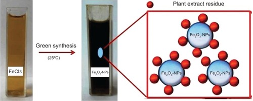
Figure 1 Magnetic iron oxide nanoparticles synthesized via brown seaweed extract.
Abbreviation: NPs; nanoparticles.
Cytotoxicity assay
The antiproliferative effect of Fe3O4 MNPs on various cancer cell lines and a normal Chang liver cell line was quantified using a 3-(4,5-dimethylthiazol-2-yl)-2,5 diphenyl tetrazolium bromide (MTT) kit (Sigma-Aldrich) according to the standard method.Citation28 Briefly, cells were allowed to grow in a 75 cm2 cell culture flask (TPP Techno Plastic Products, Trasadingen, Switzerland) until 95% confluent. The cells were then seeded into each well of a 96-well microculture plate (TPP Techno Plastic Products) at a concentration of 1×10⁵ cells/mL and treated with Fe3O4 MNPs at various concentrations. After incubation for 72 hours at 37°C in a 5% CO2 incubator (Binder GmbH), 25 μL of a 5.5 mg/mL MTT solution was added to each well, covered with aluminum foil, and incubated for a further 3 hours in the dark. Immediately afterwards, the medium was aspirated and the remaining purple formazan was lysed with MTT solution. The assay was performed in triplicate. The optical density was then measured at 570 nm using an enzyme-linked immunosorbent assay universal microplate reader (Bio Tek Instruments, Inc., VT, USA). The inhibitory concentration 50 (IC50) value was determined from the absorbance versus concentration curve. Dimethyl sulfoxide 0.1% was used as the negative control.
Acridine orange/propidium iodide assay
Fe3O4 MNP-induced death of Jurkat cells was investigated using the acridine orange/propidium iodide double-staining method according to the standard procedureCitation29 followed by examination under a fluorescence microscope (Leica, Tokyo, Japan) with Leica Q Fluoro software (Leica) installed. The Jurkat cells were plated at a concentration of 1×10⁶ cells/mL in a 25 cm2 culture flask (TPP Techno Plastic Products), exposed to Fe3O4 MNPs at the IC50, and incubated at 37°C in a 5% CO2 incubator for 24, 48, and 72 hours. Next, the cells were centrifuged at 200× g (Universal 320R Centrifuge, Andreas Hettich GmbH & Co.KG, Tuttlingen, Germany) for 10 minutes and the supernatant was discarded. The cells were then washed twice with phosphate-buffered saline after centrifuging at 200× g for 10 minutes to remove the remaining medium. About 10 μL of the cell pellets were stained with a 10 μL fluorescent dye mixture containing equal volumes (100 μg/mL) of acridine orange and propidium iodide for 2 minutes. Approximately 10 μL of freshly stained cell suspension was placed onto a glass slide, covered with a cover slip, and examined under a fluorescent microscope.
Annexin V-fluorescein isothiocyanate assay
Jurkat cells (1×10⁶ cells/mL) were exposed to the nanoparticles for 12, 24, and 48 hours, with untreated cells used as controls. The cells were then collected and centrifuged at 200× g (Universal 320R Centrifuge), (Andreas Hettich GmbH & Co.KG) for 10 minutes to remove the medium. The cell pellets were washed twice with 1 mL of ice-cold phosphate-buffered saline, recentrifuged, and resuspended in ice-cold 1× binding buffer. Exactly 500 μL of cell suspension was transferred to a 5 mL culture tube (TPP Techno Plastic Products), to which 5 μL of Annexin V-fluorescein isothiocyanate (FITC) conjugate and 10 μL of propidium iodide were added. The cells were incubated for 15 minutes at room temperature in the dark. Finally, flow cytometry was done under laser emitting excitation light at 488 nm using a BD FACSCalibur™ flow cytometer equipped with an argon laser (BD Biosciences, Franklin Lakes, NJ, USA). The data were analyzed using BD CellQuest™ Pro software (BD Biosciences).
Cell cycle assay
Flow cytometry was used to investigate the cytotoxicity of Fe3O4 MNPs in Jurkat cells. Briefly, Jurkat cells (2.0×10⁶ cells/mL) were cultured with the IC50 of Fe3O4 MNPs and incubated for 24, 48, and 72 hours. The cells were harvested by centrifugation at 1,500 rpm for 5 minutes and washed with phosphate-buffered saline. Next, 600 μL of 70% ice-cold ethanol were added to the cell pellets, which were then kept at −20°C overnight. A 1 mL volume of phosphate-buffered saline was added and spun down at 1,500 rpm for 5 minutes to remove the ethanol. The cell pellets were then washed twice with phosphate-buffered saline, stained with staining buffer containing 0.1% Triton X-100 (Sigma-Aldrich), 10 mM ethylenediaminetetraacetic acid (Sigma-Aldrich), 50 μg/mL Ribonuclease A (Sigma-Aldrich), and 3 μg/mL propidium iodide (Sigma-Aldrich), and incubated in the dark on ice for 30 minutes. Flow cytometry was performed with laser emitting excitation light at 488 nm using a FACSCalibur™ flow cytometer equipped with an argon laser (BD Biosciences). The data analysis was performed using CellQuest™ Pro software.
Caspase-3 and caspase-9 assays
The activity of caspase-3 and caspase-9 in Jurkat cells treated with Fe3O4 MNPs was determined after 24, 48, and 72 hours using a colorimetric assay kit (BD Biosciences) according to the manufacturer’s instructions without modification.
Statistical analysis
The experiments were done in triplicate, and the results are expressed as the mean ± standard deviation. The statistical analysis was done using IBM® SPSS® Statistics for Windows version 20.0 software (IBM Corp., Armonk, NY, USA). P-values <0.05 were considered to be statistically significant.
Results and discussion
Fe3O4 MNPs inhibit proliferation of human cancer cells
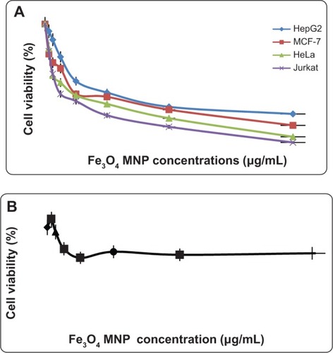
Observation of morphological changes in cells indicated that Fe3O4 MNPs inhibited proliferation of the various cancer cell lines in a dose-dependent and time-dependent manner (). No toxicity was seen in the normal Chang liver cell line (). The IC50 values calculated for Fe3O4 MNPs in the various cancer cell lines were 23.83±1.1 μg/mL (HepG2), 18.75±2.1 μg/mL (MCF-7), 12.5±1.7 μg/mL (HeLa), and 6.4±2.3 μg/mL (Jurkat) after treatment for 72 hours. Therefore, Jurkat cells were selected for further investigation.
Figure 2 (A) The cytotoxic effect of Fe3O4 MNPs on various cancer cell lines after 72 hours of treatment evaluated by mitochondrial activity using the MTT assay. Each point represents the mean value of three replicates. (B) The cytotoxic effect of Fe3O4 MNPs on a normal Chang human liver cell line after 72 hours of treatment evaluated with MTT assay. Each point represents the mean value of three replicates.
Abbreviations: Fe3O4 MNPs, magnetic iron oxide nanoparticles; MTT, 3-(4,5-dimethylthiazol-2-yl)-2,5 diphenyl tetrazolium bromide.
It is relevant to the evaluation toxicity of metal nanoparticles against cancer cells as many studies have already published. Coradeghini et al reported that BALB/C-3T3 mouse fibroblasts at higher doses than 50 μM for 72 hours of exposure to gold nanoparticles with a thickness of 5 nm, as compared with 15 nm, induced dose-dependent cell cytotoxicity. They reported that the size of gold nanoparticles is an important indicator of potential application in the biomedical field.Citation30 Another recent study reported that different types of iron oxide nanoparticles demonstrate diverse biological activity as a result of versatile surface coating. Novotna et al examined the effect of superparamagnetic iron oxide nanoparticles on human bone marrow mesenchymal stromal cells from two donors. Their results showed only hBMSCs-2 were sensitive to these nanoparticles; however, increased oxidative injury to lipids, proteins, and DNA was detected in cells from both donors.Citation31
Schweigera et al reported on Fe3O4 MNPs stabilized with poly(ethyleneimine)-g-poly(ethylene glycol) and branched poly(ethyleneimine). They also evaluated the cytotoxic effects of polymer-coated iron oxide nanoparticles in A549 epithelial adenocarcinoma cells. Their findings for cell viability showed that poly(ethyleneimine) layered onto iron oxide nanoparticles produced significant cytoxicity compared to free poly(ethyleneimine) polymer.Citation11
Quantification of apoptosis using acridine orange/propidium iodide double-staining
Induction of apoptosis is a useful strategy in the treatment of cancer. Several cellular and molecularbiological features can be demonstrated in apoptotic cells, including cell shrinkage, DNA fragmentation, and activation of the caspase cascade. The cytotoxicity of Fe3O4 MNPs was evaluated by inhibition of cell growth. When the growth-inhibited cells were stained with acridine orange/propidium iodide, apoptotic cell death was observed in a time-dependent and dose-dependent manner in all cultures ().
Figure 3 Fluorescent micrographs of acridine orange/propidium iodide double-stained Jurkat cells treated with Fe3O4 MNPs. (A) Untreated cells showing normal cell structure, (B) early apoptotic cells after 24 hours of treatment showing membrane blebbing and chromatin condensation, (C) blebbing and nuclear margination after 48 hours of treatment, and (D) DNA fragmentation and apoptotic body formation after 72 hours of treatment.
Abbreviations: VC, viable cells; EA, early apoptotic cells; CC, chromatin condensation; BL, membrane blebbing; MN, marginated nucleus; FN, fragmented nucleus; LA, late apoptotic cells; AB, apoptotic body; Fe3O4 MNPs, magnetic iron oxide nanoparticles; DNA, deoxyribonucleic acid.
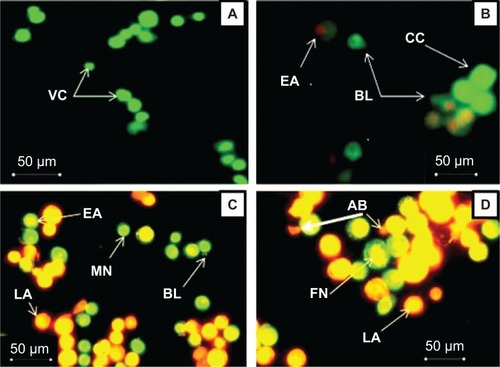
Intact membranes exclude propidium iodide, but allow uptake of acridine orange, which binds to double-stranded DNA and fluoresces green under 488 nm excitation. Untreated cells show diffuse green fluorescence, while apoptotic cells (containing condensed chromatin material) show clumps of intense green fluorescent spots within the cell. Characteristic condensation patterns were observed such as crescent shapes found at the nuclear periphery and an increase in the number of round clumps.
Annexin V-FITC assay
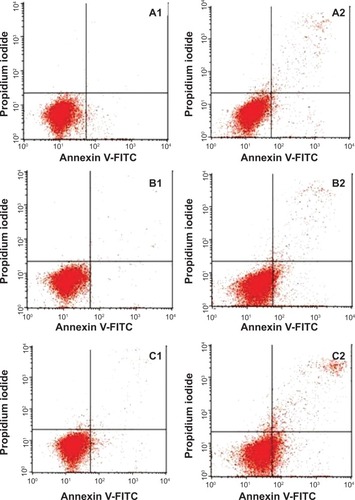
The apoptotic effect induced by the Fe3O4 MNPs was investigated further by determining the percentage of apoptotic cells using flow cytometric analysis with Annexin V/propidium iodide double-staining. Annexin V-positive/propidium iodide-negative staining indicated cells in early apoptosis due to the strong affinity of Annexin V-FITC for phosphatidylserine, which transports Annexin V protein from the inner leaflet of the plasma membrane to the outer surface of the membrane during early apoptosis. On the other hand, Annexin V-negative/propidium iodide-positive staining indicates necrotic cells, since propidium iodide, which cannot pass through an intact cell membrane, penetrates the compromised membranes of cells that are dead or in late apoptosis, and binds to nucleic acids. Meanwhile, Annexin V-negative/propidium iodide-negative staining indicates viable cells and Annexin V-positive/propidium iodide-positive staining indicates cells in late apoptosis. The representative dot plots for the flow cytometric analysis comparing untreated cells and treated cells (at 12, 24, and 48 hours) showed an increase in the percentages of treated cells in early apoptosis and late apoptosis ( and ). In addition, exposure to Fe3O4 MNPs resulted in a slight decrease in the amount of viable cells at 12, 24, and 48 hours. These results suggest that the antiproliferative effect of Fe3O4 MNPs on Jurkat cells results from induction of cell apoptosis.
Table 1 Flow cytometric analysis of Jurkat cells treated with Fe3O4 MNPs
Figure 4 Flow cytometric analysis of induction of apoptosis in Jurkat cells by Fe3O4 MNPs after staining with FITC-conjugated Annexin V and propidium iodide. (A1–C1) Untreated (control) Jurkat cells after 12, 24, and 48 hours of incubation, respectively. (A2–C2) Effects of 12, 24, and 48 hours of treatment with Fe3O4 MNPs, respectively.
Abbreviations: Fe3O4 MNPs, magnetic iron oxide nanoparticles; Annexin V-FITC, annexin V-conjugated fluorescein isothiocyanate.
Cell cycle assay
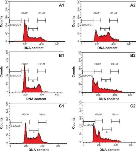
In cells treated with agents that induce apoptosis, a sub-population of cells appears before the G1 peak and is referred to as the sub-G1 peak. This is believed to be the result of endonuclease activation and subsequent leakage of DNA from the cells. Necrotic cells do not show an immediate reduction in DNA content, so a clear distinction could be made between necrotic and apoptotic cells. As shown in and , the sub-G1 population, indicating apoptotic cells, increased in a time-dependent manner from 10.10±0.28 24 hours after exposure to Fe3O4 MNPs to 20.0±0.20 after 48 hours and 23.85±0.56 after 72 hours. Regulation of the cancer cell cycle is a potential strategy in the development of anticancer drugs. Xia et al showed a shift of cell distribution into the G2/M phase, which was enhanced by Fe3O4 MNPsCitation32. Our data indicate that the sub-G1 population, ie, apoptotic cells, increased in a time-dependent manner, whereas other researchers have reported cells arrested in G1/S and in both G1/S and G2/M.Citation32 These conflicting results suggest that there may be fundamental differences between cell types.
Table 2 Flow cytometric analysis of Jurkat cells treated with Fe3O4 MNPs
Figure 5 Cell cycle analysis of Jurkat cells treated with Fe3O4 MNPs after staining with propidium iodide. (A1–C1) Untreated Jurkat cells treated for 24, 48, and 72 hours, respectively. (A2–C2) Effects of 24, 48, and 72 hours of exposure of Jurkat cells to Fe3O4 MNPs, respectively. G0/G1, G2/M, and S indicate the cell phase, and sub-G0/G1 refers to the proportion of apoptotic cells.
Abbreviations: Fe3O4 MNPs, magnetic iron oxide nanoparticles; DNA, deoxyribonucleic acid.
Caspase assay
The apoptotic effect of Fe3O4 MNPs was also examined by determining caspase-3 and caspase-9 activity relative to varying concentrations of protein content for cells treated with the IC50 of Fe3O4 MNPs. Our results show that enzyme activity increased in a time-dependent manner, as shown in and .
Table 3 Spectrophotometric analysis of caspases in Jurkat cells treated with Fe3O4 MNPs for 24, 48, and 72 hours
Figure 6 Effect of treatment with Fe3O4 MNPs on caspase-3 and caspase-9 activity in Jurkat cells. The values represent the mean ± standard deviation of three independent experiments. Significant differences (P<0.05) in caspase-3 and caspase-9 activity were found between the treated and control groups.
Abbreviation: Fe3O4 MNPs, magnetic iron oxide nanoparticles.
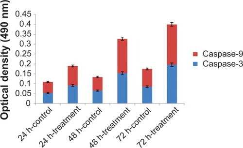
Apoptosis is orchestrated by a family of cysteine proteases known as caspases. The main effectors of apoptosis encompass proteases from the caspase family, which reside as latent precursors in most nucleated animal cells. Fourteen mammalian caspases have been identified, three of which (caspase-3, caspase-6, and caspase-7) are thought to coordinate the execution phase of apoptosis by cleaving multiple structural and repair proteins. Pathways to caspase-3 activation have been identified, which are either dependent on or independent of release of mitochondrial cytochrome c and caspase-9 function. Caspase-3 activation is the hallmark of apoptosis, and is necessary for apoptotic chromatin condensation and DNA fragmentation in all cell types examined. Thus, caspase-3 is essential for certain processes associated with dismantling of the cell and the formation of apoptotic bodies. However, it may also function before or at the stage when commitment to loss of cell viability is made. As shown in , Fe3O4 MNPs increased the activity of caspase-9 and caspase-3 in a time-dependent manner. According to studies of caspase activity, several distinct pathways exist, resulting in induction of apoptosis by Fe3O4 MNPs. In our study, we noted that Fe3O4 MNPs activated caspase-3 and caspase-9 in a time-response manner.
Conclusion
Fe3O4 MNPs with different sizes and characteristics have been developed and widely investigated in a number of biomedical applications. Stabilizing and preventing formation of aggregation are critical for controlling chemical reactions, which provide dependable, well-dispersed size and consistency. In this study, we investigated the toxicity of Fe3O4 MNPs in human leukemia (Jurkat) cells. Our results show that exposure of Jurkat cells to Fe3O4 MNPs results in significant cytotoxicity, with an apoptotic response, but not in a normal Chang liver cell line, providing new opportunities for safe delivery of Fe3O4 MNPs and application in anticancer therapy.
Acknowledgments
The authors are grateful to the staff of Universiti Putra Malaysia for their help with this research, and to the Department of Chemistry, Faculty of Science, Universiti Putra Malaysia, for allowing us to use its laboratory facilities and providing technical assistance.
Disclosure
The authors report no conflicts of interest in this work.
References
- MaengJHLeeDHJungKHMultifunctional doxorubicin loaded superparamagnetic iron oxide nanoparticles for chemotherapy and magnetic resonance imaging in liver cancerBiomaterials2010314995500620347138
- KimPSDjazayeriSZeineldinRNovel nanotechnology approaches to diagnosis and therapy of ovarian cancerGynecol Oncol201112039340321168905
- WangLSChuangMCHoJANanotheranostics – a review of recent publicationsInt J Nanomedicine201274679469522956869
- SalamHARajivPKamarajMJagadeeswaranPGunalanSSivarajRPlants: green route for nanoparticle synthesisInt Res J Biol Sci201218590
- SanthoshPBUlrihNPMultifunctional superparamagnetic iron oxide nanoparticles: promising tools in cancer theranosticsCancer Lett201333681723664890
- JayapaulJHodeniusMArnsSFMN-coated fluorescent iron oxide nanoparticles for RCP-mediated targeting and labeling of metabolically active cancer and endothelial cellsBiomaterials2011325863587121605902
- HyukWSuslickKSStuckyGDSuhYNanotechnology, nanotoxicology, and neuroscienceProg Neurobiol20098713317018926873
- IndhiraTKLakshmiPKMagnetic nanoparticles – a reviewInt J Pharmacol Sci Nanotechnol2010210351042
- GuoaJWangRTjiuWPanJLiuTSynthesis of Fe nanoparticles@graphene composites for environmental applicationsJ Hazard Mater20122252266373
- WangXWeiFLiuACancer stem cell labeling using poly (L-lysine)-modified iron oxide nanoparticlesBiomaterials2012333719373222342710
- SchweigerCPietzonkaCHeverhagenJKisselTNovel magnetic iron oxide nanoparticles coated with poly(ethyleneimine)-synthesis, stability, cytotoxicity and MR imagingInt J Pharm201140813013721315813
- LingYWeiKLuoYGaoXZhongSDual docetaxel/superparamagnetic iron oxide loaded nanoparticles for both targeting magnetic resonance imaging and cancer therapyBiomaterials2011327139715021726899
- KhanMIMohammadAPatilGNaqviSAChauhanLKAhmadIInduction of ROS, mitochondrial damage and autophagy in lung epithelial cancer cells by iron oxide nanoparticlesBiomaterials2012331477148822098780
- RajapakseNKimSKNutritional and Digestive Health Benefits of Seaweed1st edPhiladelphia, PA, USAElsevier Inc2011
- NamvarFBahararaJMahdiAAntioxidant and anticancer activities of selected Persian Gulf algaeInd J Clin Biochem2014291320
- NamvarFParidahMTMohamadRBiomedical properties of edible seaweed in cancer therapy and chemoprevention trials: a reviewNat Prod Commun201381811182024555303
- NamvarFSuhailaMGasemi FardSBehravanJPolyphenol-rich seaweed (Eucheuma cottonii) extract suppresses breast tumour via hormone modulation and apoptosis inductionFood Chem2012130376382
- KhanMChoiJLeeMKimENamTAnti-inflammatory activities of methanol extracts from various seaweed speciesEnviron Biol200829465469
- ZuercherAWFritschéRCorthésyBMercenierAFood products and allergy development, prevention and treatmentCurr Opin Biotechnol20061719820316481157
- PerezGRZavalaSMPerezGSPerezGAntidiabetic effect of compounds isolated from plantsPhytomedicine19985557523195700
- NishinoTFukudaANagumoTFujiharaMKajiEInhibition of the generation of thrombin and factor Xa by a fucoidan from the brown seaweed Ecklonia kuromeThromb Res199996374910554083
- MiyashitaKThe carotenoid fucoxanthin from brown seaweed affects obesityLipid Technol200921186190
- MohamedSHashimSNRahmanHSeaweeds: a sustainable functional food for complementary and alternative therapyTrends Food Sci Technol2012238396
- WadaKNakamuraKTamaiYSeaweed intake and blood pressure levels in healthy pre-school Japanese childrenNutr J201110838821827710
- NamvarFMohamadRBahararaJZafar-BalanejadSFargahiFRahmanHSAntioxidant, antiproliferative, and antiangiogenesis effects of polyphenol-rich seaweed (Sargassum muticum)Biomed Res Int2013201319
- DiasAMHussainAMarcosASRoqueAAA biotechnological perspective on the application of iron oxide magnetic colloids modified with polysaccharidesBiotechnol Adv20112914215520959138
- MahdaviMNamvarFAhmadMBMohamadRGreen biosynthesis and characterization of magnetic iron oxide (Fe3O4) nanoparticles using seaweed (Sargassum muticum) aqueous extractMolecules2013185954596423698048
- Van de LoosdrechtABeelenROssenkoppeleGJA tetrazolium-based colorimetric MTT assay to quantitate human monocyte mediated cytotoxicity against leukemic cells from cell lines and patients with acute myeloid leukemiaJ Immunol Methods19941743113208083535
- FanCWangWZhaoBZhangSMiaoJChloroquine inhibits cell growth and induces cell death in A549 lung cancer cellsBioorg Med Chem2006143218322216413786
- CoradeghiniRGioriaSGarcíaCPSize-dependent toxicity and cell interaction mechanisms of gold nanoparticles on mouse fibroblastsToxicol Lett201321720521623246733
- NovotnaBJendelovaPKapcalovaMOxidative damage to biological macromolecules in human bone marrow mesenchymal stromal cells labeled with various types of iron oxide nanoparticlesToxicol Lett2012210536322269213
- XiaGChenBDingJEffect of magnetic Fe3O4 nanoparticles with 2-methoxyestradiol on the cell-cycle progression and apoptosis of myelodysplastic syndrome cellsInt J Nanomedicine201161921192721931487