
Huang D, Huang C, Wang H, et al. Onco Targets Ther. 2021;14:1367–1376.
The authors have advised due to an error at the time of figure assembly, on page 1373 is incorrect. The correct is shown below.
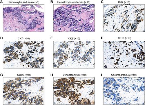
Figure 4 Histological analysis of the first external auditory canal biopsy. (A, B) Hematoxylin and eosin-stained slides showing some glandular structures in poorly differentiated tumor cells. (C–I) Immunohistochemical staining showing elevated proliferation rates (> 90% hotspot Ki-67) (C), and simultaneous expression of CK7 (D), CK8 (E), CK18 (F), CD56 (G), synaptophysin (H), and chromogranin A (I) in > 50% of the tumor cells. Magnifications: a, ×5; b–i, ×10.
The authors apologize for this error and advise it does not affect the results of the paper.