Abstract
Aim:
The aim of the study was to validate the expression of protein tyrosine kinase 6 (PTK6) in nonsmall cell lung cancer (NSCLC), and to evaluate its clinicopathological and prognostic significance.
Methods:
We first conducted a meta-analysis on the mRNA profiling data sets of NSCLC in the Oncomine database. Then, one of the most significantly upregulated tyrosine kinase targets, PTK6, was further validated by immunohistochemistry in 104 primary NSCLC tumors. Furthermore the association between PTK6 expression, the clinical parameters, and overall survival was further analyzed.
Results:
Using the Oncomine database, we identified a list of tyrosine kinase genes related to NSCLC, among which PTK6 was the second most overexpressed gene (median rank = 915, P = 2.9 × 10⁻⁵). We further confirmed that NSCLC tumors had a higher expression level of PTK6 than normal pulmonary tissues. Moreover, high PTK6 expression correlated positively with shorter overall survival time, but not with other clinicopathological characteristics. In the multivariate Cox regression model, high PTK6 expression was demonstrated to be an independent prognostic factor for NSCLC patients.
Conclusion:
Our results validated that PTK6 was found to be overexpressed in a proportion of NSCLC samples, and was associated with a poor prognosis, suggesting that this subgroup of NSCLC patients might benefit from PTK6 inhibitors in the future.
Introduction
Lung cancer ranks among the leading causes of cancer death globally. Mortality related to this malignant disease has risen by 465% during the last 30 years in People’s Republic of China.Citation1 Nonsmall cell lung carcinoma (NSCLC) accounts for approximately 85% of all lung cancer cases. Even among those who have received standard treatments including surgical resection, traditional chemotherapy, radiation therapy, and molecular targeted therapy, a significant proportion of NSCLC patients eventually develop resistance to chemotherapy and recurrence. Therefore, there is an urgent need to discover novel cancer-specific molecular targets and signaling pathways to develop new therapeutic strategies against this devastating malignancy.
Tyrosine kinases, being capable of phosphorylating specific target protein substrates, are involved in the modulation of multiple growth factor signaling during cancer progression.Citation2 In recent years, a list of tyrosine kinase inhibitors has been approved, while others are in clinical trials for NSCLC patients.Citation3,Citation4 Therefore, identifying new tyrosine kinases related to NSCLC would be of important significance in developing novel treatment strategies for this cancer.
In this study, we first performed a meta-analysis on the gene expression profiling data sets of NSCLC specimens from the Oncomine database (Compendia Bioscience, Inc, Ann Arbor, MI, USA), and identified a tyrosine kinase, protein tyrosine kinase 6 (PTK6), was specifically upregulated in NSCLC when compared to normal lung tissues, which might be a novel biomarker and potential therapeutic target for NSCLC. We further validated its expression in NSCLC samples and evaluated its clinicopathological and prognostic significance.
Materials and methods
Data mining
The Oncomine cancer microarray database was used to analyze the mRNA expression profiles of NSCLC tissues relative to their normal controls. Eleven publicly available datasets of gene expression profiles of were chosen in this study. The data mining strategy is based on a published methodology established by Oncomine.Citation5 First, significantly upregulated genes in cancer samples compared with the normal tissue counterpart (greater than twofold, P < 0.05) were selected. Then, concept filters in the Oncomine database were used to identify known tyrosine kinases differentially expressed in NSCLC.
Samples
Archived formalin-fixed paraffin-embedded NSCLC samples with corresponding adjacent nontumor tissues undergoing surgical resection were obtained from the 89th Hospital, Changhai Hospital, and from the Changzheng Hospital of People’s Republic of China between January 2006 and December 2008. General schemes for treatment choice in the sampled NSCLC patients were based on the National Comprehensive Cancer Network NSCLC Clinical Practice Guidelines (Chinese version). Overall survival was calculated from the date of surgery until the date of death or last follow-up. None of the patients had received neoadjuvant chemotherapy, radiation therapy, or immunotherapy. Ethics approval to start this study was obtained from the local Human Research Ethics Committees.
Immunohistochemistry
The immunostaining was performed according to standard SP methods according to the manufacturer’s protocol (Zymed®, Life Technologies, Carlsbad, CA, USA). Antigen retrieval was done by a combination of heat and pressure in sodium citrate buffer. Immunostaining was detected with a rabbit polyclonal antibody against PTK6 (Santa Cruz Biotechnology, Inc, Dallas, TX, USA). Detection was performed using diaminobenzidine as the chromogen. Nonspecific rabbit immunoglobulin G was used as negative control.
The immunostaining levels of PTK6 protein expression were assessed using a semiquantitative staining index method, as described previously with minor modifications.Citation6 The staining intensity results were graded to a four-scale (0 = no staining; 1 = staining obvious only at × 400; 2 = staining obvious at × 100 but not × 40; and 3 = staining obvious at × 40), while the percentages of positive cells were scored into four scales (0 = 0% positive cells; 1 = 1%–33% positive cells; 2 = 34%–66% positive cells; and 3 = 67%–100% positive cells). A staining index ranging from 0 to 9 was generated by multiplying staining intensity and the percentage of positive cells for each sample. The median staining index value was set as a cut-point to delineate PTK6-low and PTK6-high subgroups.
Statistics
The difference in clinicopathological characteristics between PTK6-low and PTK6-high subgroups was compared using the Chi-square test. Overall survival curves were estimated using the Kaplan–Meier method and evaluated using the Log-Rank test. Multivariate analysis on clinicopathological variables and PTK6 expression levels was performed using the Cox proportional hazard regression model. Statistical analyses were performed using the Statistical Package for the Social Sciences 16.0 (SPSS; IBM Corporation, Armonk, NY, USA), and a P-value less than 0.05 was considered statistically significant.
Results
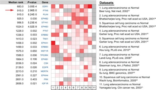
We first queried the Oncomine database to systematically compare the relative gene expression levels between NSCLC and normal pulmonary samples, then upregulating genes belonging to tyrosine kinase targets were further filtered out using a “tyrosine protein kinase, active site – InterPro Protein Domains and Families” concept. The top 20 upregulated tyrosine kinase genes in NSCLC are listed in . In the meta-analysis, discoidin domain receptor 1 (DDR1) was the highest ranked gene (median rank = 683) among all of the tyrosine kinase genes with a P-value of 3.05 × 10⁻⁴. Next on the list, with a median rank of 915 and a P-value of 2.96 × 10⁻⁵, was PTK6. Our bioinformatics analysis strongly suggested that PTK6 genes were closely associated with NSCLC, and might be a potential tyrosine kinase target for NSCLC.
Figure 1 Oncomine heat map of upregulated genes in clinical NSCLC samples compared with the normal pulmonary tissues filtered by the “tyrosine protein kinase, active site – InterPro Protein Domains and Families” concept.
Abbreviations: NSCLC, nonsmall cell lung cancer; PTK6, protein tyrosine kinase 6; EGFR, epidermal growth factor receptor.
We further applied a validation study of PTK6 expression in a panel of clinical samples. As for the staining pattern, the positive staining reaction for PTK6 in normal and cancerous lung tissue cells was observed in the cytoplasm, and in a combination of the nucleus and cytoplasm. Compared with normal lung tissues, NSCLC tissues had a significantly higher staining index of PTK6. For NSCLC tissues, the median staining index of 6 was set as the cut-point to delineate PTK6-low and PTK6-high subgroups. Representative immunostaining images were seen in . The associations between PTK6 expression and clinicopathological features of NSCLC are shown in . No significant association was observed between PTK6 expression with the clinicopathological traits observed in this study. Furthermore, we also found no significant correlation between PTK6 expression with another tyrosine kinase target epidermal growth factor receptor (EGFR) mutation status.
Figure 2 Representative images of PTK6 immunostaining in this study. (A) High level of PTK6 in NSCLC tumors; (B) low level of PTK6 expression in NSCLC tumors; (C) PTK6 expression in adjacent normal lung tissues; (D) negative control for immunostaining (IgG).
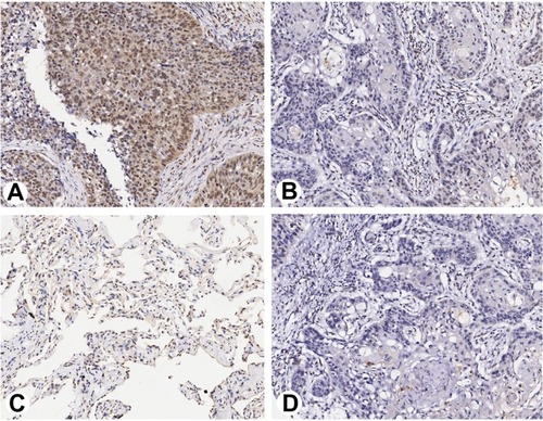
Table 1 Association between PTK6 expression and clinicopathological characteristics of the patients with NSCLC
Survival curves were plotted using the Kaplan–Meier method as seen in . The results indicated that the patients with a high PTK6 expression had a significantly shorter survival time, compared with those with low expression. High expression of PTK6 protein showed a statistically significant association with survival in univariate analysis (hazard ratio = 2.031, 95% confidence interval = 1.099–3.755, P = 0.024 by Log-Rank test). All the NSCLC clinical and pathological factors together with PTK6 expression status were included in a multivariate Cox regression model (). Our data demonstrated that high PTK6 expression retained a significant and independent prognostic factor for NSCLC patients (hazard ratio = 2.219, 95% confidence interval = 1.156–4.263, P = 0.017 by Log-Rank test).
Figure 3 Kaplan–Meier survival curves based on expression of PTK6.
Abbreviations: PTK6, protein tyrosine kinase 6; Cum, cumulative; OS, overall survival.
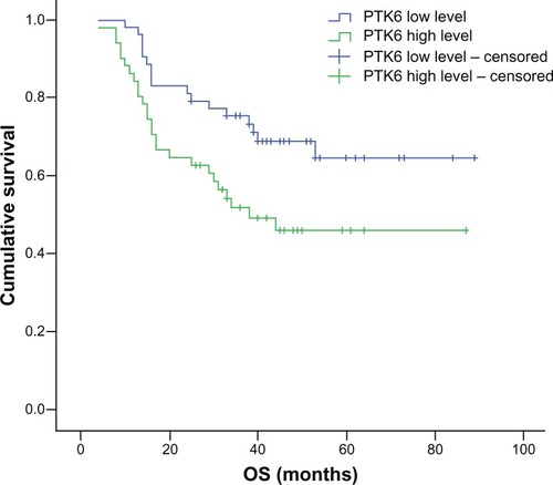
Table 2 Univariate and multivariate analysis for prognosis
Discussion
By meta-analysis on the Oncomine database, we screened and identified a list of upregulated tyrosine kinase genes in NSCLC, including some already confirmed therapeutic targets such as EGFR. The first ranking tyrosine kinase gene DDR1 has been demonstrated to contribute to the progression and poor prognosis of NSCLC in previous studies, and could serve as a potential therapeutic target.Citation7,Citation8 We further selected one of the top genes, PTK6, for further verification. This tyrosine kinase was chosen for the following reasons: firstly, in the gene expression microarray meta-analysis, PTK6 was the second ranked gene among all of the tyrosine kinase genes, and was shown to be significantly elevated in NSCLC tumors; secondly, the expression of PTK6 in NSCLC has not been widely investigated, and specifically its prognostic value has not been reported; thirdly, commercial antibodies for PTK6 immunohistochemistry are available.
We further evaluated the clinicopathological and prognostic significance of PTK6 expression in NSCLC tumors. Our immunostaining results validated that high expression of PTK6 was detectable in NSCLC tumor samples and was significantly correlated with decreased overall survival. Through multivariate analysis, high expression of PTK6 was shown to be an independent prognostic biomarker for poor overall survival. As far as we know, this is the first report of the prognostic significance of PTK6 expression in clinical NSCLC tissue samples. However, different from the previous study,Citation9 no significant association with tumor size, lymph node metastasis status, and TNM stage were found in this study. We think the difference between data from the current study and previous report might be a result of the different evaluation criteria of the immunohistochemistry staining adopted. Our findings suggest that high-PTK6 expressed NSCLC tumors could represent a distinct subgroup with more aggressive phenotypes and a poor prognosis, which might benefit from more intensive and targeted treatments including PTK-specific inhibitors.
PTK6 (also known as breast tumor kinase) is a nonreceptor tyrosine kinase, which has been originally cloned from human melanocytes and a metastatic breast tumor sample.Citation10,Citation11 High expression of PTK has been found in several cancer types, including breast cancer, melanoma, and colon cancer.Citation12,Citation13 Accumulating evidence indicated that PTK6 played an important regulatory role as a proto-oncogene in malignant settings, and was involved directly in the processes of proliferation, migration, and invasion in cancer cells.Citation14 Harvey et alCitation15 found that the proliferation of breast carcinoma cells was efficiently suppressed by specifically downregulated PTK6 protein levels using RNA interference. PTK6 has been demonstrated to interact with the ErbB family members EGFR, ErbR2, ErbR3, ErbR4, and enhanced EGF-induced proliferation and ERK1/2 activation.Citation16–Citation18 It has also been reported that PTK6 could also activate the PI3k/Akt and STAT pathway.Citation17 Moreover, potential PTK substrates also include a variety of several signaling molecules such as p190RhoGAP, paxillin, Akt, and KAP3A in most human cancer cells.Citation19–Citation21 Therefore, considering the overexpression of PTK6 in NSCLC, and its positive association with a poor prognosis, we proposed that specific inhibition of PTK6 kinase activity may provide a potentially novel approach to treat patients with NSCLC in the future. In particular, recently, Zeng et alCitation22 discovered imidazo [1,2-a] pyrazin-8-amines as potent PTK6 kinase inhibitors. The utility of these small molecular PTK6 inhibitors in NSCLC treatment deserves further investigation.
In conclusion, in this study, by meta-analysis on the gene microarray data of NSCLC, we screened and validated a novel tyrosine kinase, PTK6, overexpressed in NSCLC tumors, which was correlated positively to shorter survival. Our findings are, however, preliminary and require other validations on a larger and independent group of NSCLC patients. We also suggest that the exact mechanisms of PTK6 during the malignant progression of NSCLC and its relationship with other confirmed NSCLC tyrosine kinase targets such as ALK deserves further investigation in the future.
Acknowledgements
This research was supported by grants from the National Science Foundation of People’s Republic of China (81273282, 30972730), 973 Foundation (2013CB531606), Shanghai Municipal Commission for Science and Technology (11 JC1410902), and Changhai Hospital (CH125530300).
Disclosure
The authors report no conflicts of interest in this work.
References
- WenCDehnelTPeople’s Republic of China wrestles with lung cancerLancet Oncol20111211521249722
- AroraAScholarEMRole of tyrosine kinase inhibitors in cancer therapyJ Pharmacol Exp Ther2005315397197916002463
- PaoWMillerVAEpidermal growth factor receptor mutations, small-molecule kinase inhibitors, and non-small-cell lung cancer: current knowledge and future directionsJ Clin Oncol200523112556256815767641
- ThomasSMGrandisJRPharmacokinetic and pharmacodynamic properties of EGFR inhibitors under clinical investigationCancer Treat Rev200430325526815059649
- RhodesDRKalyana-SundaramSMahavisnoVOncomine 3.0: genes, pathways, and networks in a collection of 18,000 cancer gene expression profilesNeoplasia20079216618017356713
- TuxhornJAAyalaGESmithMJSmithVCDangTDRowleyDRReactive stroma in human prostate cancer: induction of myofibroblast phenotype and extracellular matrix remodelingClin Cancer Res2002892912292312231536
- FordCELauSKZhuCQAnderssonTTsaoMSVogelWFExpression and mutation analysis of the discoidin domain receptors 1 and 2 in non-small cell lung carcinomaBr J Cancer200796580881417299390
- YangSHBaekHALeeHJDiscoidin domain receptor 1 is associated with poor prognosis of non-small cell lung carcinomasOncol Rep201024231131920596615
- FanCZhaoYLiuDZhangXWangEDetection of Brk expression in non-small cell lung cancer: clinicopathological relevanceTumour Biol201132587388021603980
- MitchellPJBarkerKTMartindaleJECloning and characterisation of cDNAs encoding a novel non-receptor tyrosine kinase, brk, expressed in human breast tumoursOncogene199498238323908036022
- LeeSTStrunkKMSpritzRAA survey of protein tyrosine kinase mRNAs expressed in normal human melanocytesOncogene1993812340334108247543
- BarkerKTJacksonLECromptonMRBRK tyrosine kinase expression in a high proportion of human breast carcinomasOncogene19971577998059266966
- LlorXSerfasMSBieWBRK/Sik expression in the gastrointestinal tract and in colon tumorsClin Cancer Res1999571767177710430081
- HarveyAJCromptonMRThe Brk protein tyrosine kinase as a therapeutic target in cancer: opportunities and challengesAnticancer Drugs200415210711115075665
- HarveyAJCromptonMRUse of RNA interference to validate Brk as a novel therapeutic target in breast cancer: Brk promotes breast carcinoma cell proliferationOncogene200322325006501012902983
- XiangBChattiKQiuHBrk is coamplified with ErbB2 to promote proliferation in breast cancerProc Natl Acad Sci U S A200810534124631246818719096
- KamalatiTJolinHEFryMJCromptonMRExpression of the BRK tyrosine kinase in mammary epithelial cells enhances the coupling of EGF signalling to PI 3-kinase and Akt, via erbB3 phosphorylationOncogene200019485471547611114724
- AubeleMWalchAKLudygaNPrognostic value of protein tyrosine kinase 6 (PTK6) for long-term survival of breast cancer patientsBr J Cancer20089971089109518781181
- ChenHYShenCHTsaiYTLinFCHuangYPChenRHBrk activates rac1 and promotes cell migration and invasion by phosphorylating paxillinMol Cell Biol20042424105581057215572663
- IkedaOMiyasakaYSekineYSTAP-2 is phosphorylated at tyrosine-250 by Brk and modulates Brk-mediated STAT3 activationBiochem Biophys Res Commun20093841717519393627
- QiuHZappacostaFSuWAnnanRSMillerWTInteraction between Brk kinase and insulin receptor substrate-4Oncogene200524365656566415870689
- ZengHBelangerDBCurranPJDiscovery of novel imidazo[1,2-a]pyrazin-8-amines as Brk/PTK6 inhibitorsBioorg Med Chem Lett201121195870587521855335
- BeerDGKardiaSLHuangCCGene-expression profiles predict survival of patients with lung adenocarcinomaNat Med20028881682412118244
- PedersenNMortensenSSørensenSBTranscriptional gene expression profiling of small cell lung cancer cellsCancer Res20036381943195312702587
- GarberMETroyanskayaOGSchluensKDiversity of gene expression in adenocarcinoma of the lungProc Natl Acad Sci U S A20019824137841378911707590
- HouJAertsJden HamerBGene expression-based classification of non-small cell lung carcinomas and survival predictionPLoS One201054e1031220421987
- LandiMTDrachevaTRotunnoMGene expression signature of cigarette smoking and its role in lung adenocarcinoma development and survivalPLoS One200832e165118297132
- StearmanRSDwyer-NieldLZerbeLAnalysis of orthologous gene expression between human pulmonary adenocarcinoma and a carcinogen-induced murine modelAm J Pathol200516761763177516314486
- SuLJChangCWWuYCSelection of DDX5 as a novel internal control for Q-RT-PCR from microarray data using a block bootstrap re-sampling schemeBMC Genomics2007814017540040
- WachiSYonedaKWuRInteractome-transcriptome analysis reveals the high centrality of genes differentially expressed in lung cancer tissuesBioinformatics200521234205420816188928
- YamagataNShyrYYanagisawaKA training-testing approach to the molecular classification of resected non-small cell lung cancerClin Cancer Res20039134695470414581339