 ?Mathematical formulae have been encoded as MathML and are displayed in this HTML version using MathJax in order to improve their display. Uncheck the box to turn MathJax off. This feature requires Javascript. Click on a formula to zoom.
Abstract
Background
The upregulation of matrix metalloproteinase-1 (MMP-1) has been demonstrated to be correlated with lymph node metastasis of nasopharyngeal carcinoma (NPC), while the activation of protease-activated receptor-1 (PAR-1) mediates proliferation and invasion of NPC cells. The present study investigated the clinical significance of the coexpression of MMP-1 and PAR-1 in NPC patients in determining the prognosis.
Methods
Immunohistochemistry was performed to detect the expression of MMP-1 and PAR-1 in tumor tissue samples from 266 NPC patients.
Results
Overexpression of MMP-1 and PAR-1 proteins were, respectively, detected in 190 (71.43%) and 182 (68.42%) of the 266 NPC patients. In addition, the combined MMP-1 and PAR-1 expression was significantly associated with advanced T-stage (P = 0.01), advanced clinical stage (P = 0.002), positive recurrence (P = 0.01), and metastatic status (P = 0.01) of NPC. Moreover, the overall survival in NPC patients with MMP-1 and PAR-1 dual overexpression was significantly shorter than in those with dual low expression (P < 0.001). Furthermore, the multivariate analyses indicated that the combined MMP-1 and PAR-1 overexpression was an independent prognostic factor for overall survival (P = 0.001) in NPC patients, but the upregulation of MMP-1 and PAR-1 alone was, in each case, not an independent prognostic factor for this disease.
Conclusion
Our data provide convincing evidence, for the first time, that the activation of the MMP-1 and PAR-1 axis may be involved in the tumorigenesis and progression of NPC. The upregulation of MMP-1 in combination with PAR-1 overexpression is an unfavorable prognostic marker for NPC and might offer the possibility of future therapeutic targets.
Introduction
Nasopharyngeal carcinoma (NPC) is one of the most common malignant diseases, with a high prevalence in Southern China, Southeast Asia, and North Africa.Citation1 Worldwide, NPC accounts for 80,000 new cases and 50,000 deaths annually, predominantly in men.Citation2 NPC is an Epstein-Barr virus (EBV)-related cancer and differs from other head and neck cancers in its etiology, epidemiology, and potential therapeutic options.Citation3 Generally, enlarged lymph nodes in the neck region are the first sign of NPC; however, neck biopsy or neck dissection is not recommended, as this may reduce cure probability. The majority (75%–90%) of newly diagnosed NPC patients have locoregionally advanced disease, commonly with cervical nodal metastases.Citation4 Currently, the standard of care for NPC patients consists of concurrent chemoradiotherapy with cisplatin-based regimens, generally followed by adjuvant chemotherapy. This treatment approach leads to cure for the vast majority of patients, with 3-year disease-free and overall survival rates of approximately 70% and 80%, respectively.Citation5 Despite cure for the majority of the patients, it is still a challenge to prevent the disease relapse, to treat the patients with refractory or metastatic NPC, and to manage the long-term toxicities. Thus, there is an urgent need to identify the valuable factors for early diagnosis, prognosis, and novel therapeutic strategies.
Matrix metalloproteinases (MMPs) comprise a closely related family of 28 zinc-dependent endopeptidases that cleave extracellular matrix (ECM) proteins during tissue remodeling processes, such as wound healing, angiogenesis, and tumor invasion.Citation6 Among MMPs, MMP-1, together with MMP-8 and MMP-13, is known as the interstitial collagenase and is capable of initiating the degradation of fibrillar-type collagens by cleaving at the N-terminus. MMP-1 presents specific substrates for collagenases I, II, III, VII, VIII, and X, and for proteoglycans.Citation7,Citation8 It has been identified as one of the most highly upregulated proteins in various cancers. In particular, tumor expression of MMP-1 is associated with poor prognosis in malignant melanoma,Citation9 breast cancer,Citation10 ovarian cancer,Citation11 colorectal carcinoma,Citation12 pancreatic cancer,Citation13 and gastric cancer.Citation14 Recent studies have demonstrated that the collagenase activity of MMP-1 may be associated with tumor cell invasion and increased angiogenesis in xenograft models of malignant melanoma,Citation15 breast cancer,Citation16 and prostate cancer.Citation17 In NPC, Lu et alCitation18 showed clearly that the amounts of transcripts, proteins, and enzyme activities of MMP-1 were increased in cells expressing EBV proteins, suggesting that these viral proteins may be capable of regulating MMP-1 and may provide clues for the role of EBV in NPC progression; Ben Nasr et alCitation19 indicated that the expression of MMP-1 may be correlated with lymph node metastasis in NPC patients. Although the collagenolytic activity of MMP-1 may contribute to the tumor progression of NPC, its prognostic value in this carcinoma remains largely undefined.
Recently, a novel MMP-1 signaling axis (through activation of the protease-activated receptor [PAR]-1 to promote tumorigenesis and tumor invasion) has been identified in various cancers, such as breast cancer,Citation20 epithelial ovarian cancer,Citation21 esophageal squamous cell carcinoma,Citation22 glioma,Citation23 and hepatocellular carcinoma.Citation24 The PARs are known as the cellular seven transmembrane G protein-coupled receptors (GPCRs) for thrombin.Citation25 Four different PARs have been identified: PAR-1, PAR-2, PAR-3, and PAR-4. PARs are widely expressed in vascular and extravascular tissues and are involved in responses to vascular injury and in the regulation of inflammation.Citation26 PAR-1 and PAR-3 are activated by thrombin, PAR-2 is activated by tryptase or trypsin, and PAR-4 is activated by both thrombin and tryptase or trypsin. Among the PARs, the abnormal expression of PAR-1 has been demonstrated to be associated with the metastatic potentials of various malignancies, including breast cancer, prostate cancer,Citation27 and melanoma.Citation28 In particular, Zhu et alCitation29 indicated that the activation of PAR-1 may mediate the proliferation and invasion of NPC cells. Since the MMP-1/PAR-1 signal transduction axis facilitates tumor invasion, angiogenesis, and metastasis by inducing the expression of the genes associated with cell adhesion, invasion, and survival, it is of great significance to explore the expression and prognostic value of MMP-1 and PAR-1 in NPC. However, to date, there has been no investigation of the clinicopathologic relevance of combined MMP-1 and PAR-1 expression in NPC tissues. Therefore, the aim of the present study was to evaluate the potential association of the coexpression of MMP-1 and PAR-1 in NPC tissues with clinicopathologic findings and with postresectional survival, by an immunohistochemical analysis.
Materials and methods
Patients and tissue samples
The study was approved by the Research Ethics Committee of the Ministry of Public Health of the People’s Republic of China. Informed consent was obtained from all of the patients.
A total of 266 archival formalin-fixed, paraffin-embedded NPC specimens and 100 noncancerous nasopharyngeal specimens were obtained from Huai’an First People’s Hospital, Nanjing Medical University, People’s Republic of China during 1996–2006. In the 266 NPC group of patients, there were 166 males and 100 females, with age ranging from 16 to 80 years (median, 48 years). None of the patients recruited in this study had chemotherapy or radiotherapy before the surgery. Clinical information was obtained by reviewing the medical record of radiographic images, by telephone or written correspondence, and by review of death certificates. All specimens had a confirmed pathological diagnosis and were staged according to the 1992 NPC staging system of the People’s Republic of China.Citation31 The clinical characteristics of these patients are summarized in .
Table 1 Association of MMP-1 and/or PAR-1 expression patterns with clinicopathologic parameters of nasopharyngeal carcinoma patients
For the analysis of survival and follow up, the date of surgery was used to represent the beginning of the follow-up period. All the patients who died from diseases other than NPC or from unexpected events were excluded from the case collection. The study follow-up period terminated at March 8, 2012. The median follow-up period was 46 months (range, 3–126 months). Treatment modalities after relapse were given according to a uniform guideline, as described.Citation31
Immunohistochemistry analysis
The expression patterns of the MMP-1 and PAR-1 proteins in the 266 NPC specimens and 100 noncancerous nasopharyngeal specimens were detected by immunohistochemical staining. In brief, all tissue specimens were retrieved and cut into 4 μm sections and then, mounted on precoated slides. After deparaffinizing in xylene (Chemical Industry Co., Ltd., Shenzhen, People’s Republic of China) and washing in a graded series of ethanol (Chemical Industry Co., Ltd.), the sections were submerged into ethylenediaminetetraacetic acid (EDTA) antigenic retrieval buffer (Chemical Industry Co., Ltd.) and microwaved for antigenic retrieval. The slides were incubated with primary antibodies raised against MMP-1 (1:1000 dilution) (MMP-1 [3B6] Antibody; #sc-21731, Santa Cruz Biotechnology, Inc, Dallas, TX, USA) and against PAR-1 (1:1000 dilution) (Antibody; #sc-33732, Santa Cruz Biotechnology, Inc). All incubations with primary antibodies were carried out overnight at 4°C. After washing in tris(hydroxymethyl) aminomethane (Tris)-buffered saline (TBS) (Sigma Aldrich Corp, St Louis, MO, USA), the tissue sections were treated with biotinylated anti-rabbit secondary antibody (Antibody; #sc-2775, Santa Cruz Biotechnology, Inc), followed by further incubation with streptavidin–horseradish peroxidase complex (Life Technologies Corp, Carlsbad, CA, USA). The tissue sections were immersed in 3-amino-9-ethyl carbazole (Chemical Industry Co., Ltd.) and counterstained with 10% Mayer’s hematoxylin (Chemical Industry Co., Ltd.), dehydrated, and mounted in Crystal Mount™ Aqueous Mounting Medium (Chemical Industry Co., Ltd). In each immunohistochemistry run, noncancerous nasopharyngeal specimens were used as control tissues, and omission of the primary antibody served as a negative control. All the tissue samples were stained at one time.
Following the hematoxylin counterstaining, immunostaining was scored by two independent observers who were blinded to the clinicopathological parameters and clinical outcomes of the patients. The scores of the two observers were compared, and in the event of a discrepant score, the specimen was reexamined by both pathologists to achieve a consensus score. The number of positive-staining cells showing immunoreactivity on the cell membrane and/or cytoplasm (for MMP-1) and cytoplasm (for PAR-1) in ten representative microscopic fields was counted, and the percentage of positive cells was calculated. The frequency of MMP-1 and PAR-1 immunoreactivity in tissue sections was evaluated as negative (0), when no positive cells were observed within the tumor; as weak (1), when <30% of the tumor cells were positive; moderate (2), when 30% to 60% of the tumor cells were positive; and strong (3), when >60% of tumor cells were positive. The intensity of staining was evaluated as 0, 1, 2, and 3 for no staining, weak staining, medium staining, and strong staining, respectively. The immunohistochemical scores were determined as the sum of the frequency and intensity scores for the tumor cells. The cutoff values for the immunohistochemical scores of MMP-1 and PAR-1 proteins were chosen on the basis of a measure of heterogeneity, with the logrank test statistic, with respect to overall survival. An optimal cutoff value was identified. An immunohistochemical score of ≥4 was used to classify tumors with high expression, while a <4 immunohistochemical score classified tumors with low expression of MMP-1 or PAR-1 antigens.
Statistical analysis
SPSS for Windows version 16.0 (SPSS Inc, Chicago, IL, USA) and SAS 9.1 (SAS Institute, Cary, NC, USA) were used for statistical analysis. Continuous variables were expressed as
. Associations between the expression of MMP-1 and/or PAR-1 and clinicopathological parameters were assessed using a Chi-square test. The Spearman rank correlation test was used to analyze the correlation between the MMP-1 expression level and the PAR-1 expression level. Survival curves were plotted by Kaplan–Meier analysis and compared by the logrank test. Cox regression analysis was performed to assess the significance of various variables for survival. Differences were considered statistically significant when the P-value was less than 0.05.
Results
Upregulation of MMP-1 and PAR-1 in human NPC
The expression patterns and cellular distribution of MMP-1 and PAR-1 in 266 specimens of patients with NPC and 100 noncancerous nasopharyngeal tissues were examined using immunohistochemical staining. As shown in , the cell membrane and/or cytoplasm of most tumor cells in the NPC sections stained intensely with MMP-1 antibody (), while negative immunostaining was observed in all noncancerous nasopharyngeal tissues (). High MMP-1 expression was observed in tumor cells of 71.43% (190/266) of the NPC patients. For PAR-1, positive immunostaining was observed in the cytoplasm of the tumor cells (). In contrast, little or no expression of PAR-1 was observed in the noncancerous nasopharyngeal tissues (). High PAR-1 expression was observed in tumor cells of 68.42% (182/266) of the NPC patients. In particular, high MMP-1 expression was positively correlated with high PAR-1 expression in the tissue from patients with NPC (r = 0.86) (P < 0.0001) ().
Figure 1 Immunohistochemical staining of MMP-1 and PAR-1 proteins in tumor cells of patients with NPC (A and C, respectively) and noncancerous nasopharyngeal tissues (B and D, respectively). Intense staining of MMP-1 and PAR-1 is seen in the cell membrane and/or cytoplasm of tumors cells and is intensive in NPC tissues (A and C); in contrast, negative immunostaining of MMP-1 (B) and PAR-1 (D) was observed in the noncancerous nasopharyngeal tissues.
Abbreviations: NPC, nasopharyngeal carcinoma; MMP, matrix metalloproteinases; Par, protease-activated receptor.
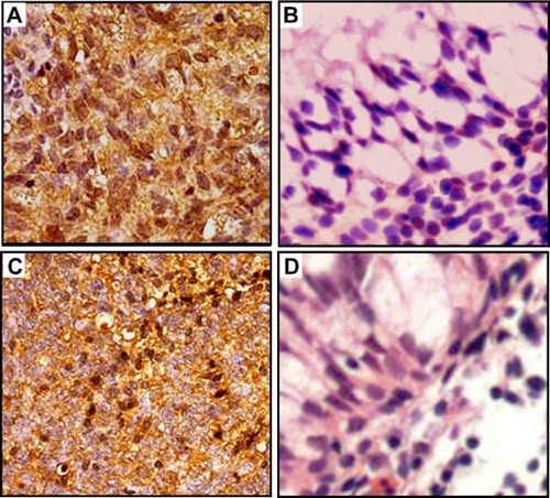
Table 2 Correlations between MMP-1 and PAR-1 expression in nasopharyngeal carcinoma tissues
Association of MMP-1 and/or Par-1 protein expression with the clinicopathological characteristics of human NPC
summarizes the association of MMP-1 and/or PAR-1 protein expression in the 266 NPC specimens, detected by immunohistochemical staining. Expression of MMP-1 was significantly associated with the T-stage (P = 0.02), clinical stage (P = 0.008), and metastatic status (P = 0.01). No significant association between MMP-1 expression and age, gender, N-stage, World Health Organization (WHO) histological type,Citation31 or recurrence was observed (). Regarding PAR-1, its overexpression was significantly associated with advanced clinical stage (P = 0.008), positive recurrence (P = 0.01), and positive metastasis (P = 0.01). Chi-square test showed no significant statistical association of PAR-1 immunostaining with age, gender, T-stage, N-stage, or WHO histological type (all P > 0.05), suggesting that these variables might not affect the expression of PAR-1.
In addition, the biologic significance of the combined expression of MMP-1 and PAR-1 was also evaluated by correlating the expression levels with the clinicopathologic characteristics. As shown in , the coexpression of MMP-1 and PAR-1 in NPC was associated with advanced T-stage (P = 0.01), advanced clinical stage (P = 0.002), positive recurrence (P = 0.01), and metastatic status (P = 0.01), but not with gender, age, N-stage, or WHO histological type (all P > 0.05).
Association of MMP-1 and/or Par-1 protein expression with the prognosis of human NPC
The association of MMP-1 and/or PAR-1 protein expression with the prognosis of human NPC was also evaluated. The 5-year overall survival rate of the cohort of 266 NPC patients was 66.17% (196/266). Kaplan–Meier survival analysis revealed that the NPC patients overexpressing both MMP-1 and PAR-1 proteins exhibited markedly poorer overall survival (P = 0.01) ( and ). Regarding their combined expression, the overall survival in NPC patients with MMP-1 and PAR-1 dual overexpression was significantly shorter than those with dual low expression (P < 0.001) ().
Figure 2 Kaplan–Meier survival plots of MMP-1 (A), PAR-1 (B), and MMP-1/PAR-1 (C) expression. Kaplan–Meier survival analysis revealed that the NPC patients overexpressing MMP-1 and PAR-1 proteins both exhibited markedly poorer overall survival (both P = 0.01). Regarding their combined expression, the overall survival in NPC patients with MMP-1 and PAR-1 dual overexpression was significantly shorter than those with dual low expression (P < 0.001).
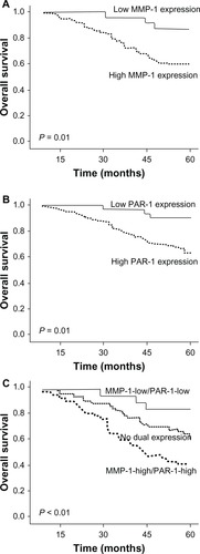
Univariate Cox proportional hazard regression analysis revealed that high MMP-1 expression (hazard ratio [HR] = 5.796, 95% confidence interval [CI]: 0.826–12.013) (P = 0.01), high PAR-1 expression (HR = 5.282, 95% CI: 0.701–11.819) (P = 0.01), and MMP-1-high/PAR-1-high expression (HR = 13.882, 95% CI: 1.986–29.331) (P < 0.001) were significant predictive factors for poor prognosis in NPC patients. Other clinicopathologic parameters, including clinical stage (HR = 7.382, 95% CI: 1.031–16.613) (P = 0.002), recurrence (HR = 8.892, 95% CI: 1.021–18.656) (P = 0.001), and metastasis (HR = 8.788, 95% CI: 1.042–19.052) (P = 0.001), were also found to be prognostic predictors of overall survival in NPC patients (). Furthermore, multivariate Cox proportional hazards regression analysis indicated that combined MMP-1 and PAR-1 overexpression (HR = 9.167, 95% CI: 1.332–23.836) (P = 0.001), clinical stage (HR = 6.193, 95% CI: 1.011–13.392) (P = 0.006), recurrence (HR = 6.928, 95% CI: 0.922–13.556) (P = 0.005), and metastasis (HR = 6.893, 95% CI: 1.023–13.528) (P = 0.005) were independent prognostic factors for overall survival in NPC patients, but the upregulation of MMP-1 and PAR-1 alone was, in each case, not an independent prognostic factor for this disease ().
Table 3 Univariate analysis of different prognostic variables in 266 nasopharyngeal carcinoma patients
Table 4 Multivariate cox regression analysis of different prognostic variables in 266 nasopharyngeal carcinoma patients
Discussion
NPC is an EBV-related cancer with a high metastatic potential compared to other head and neck cancers. There is a significant need for the identification of potential biomarkers to screen patients who are at the great risk of relapse after their primary treatment. In the present study, we analyzed the expression of MMP-1 and/or PAR-1 together with clinicopathologic parameters in NPC patients. We observed the coexpression of MMP-1 and PAR-1 in NPC tissues to be associated with advanced T-stage, advanced clinical stage, positive recurrence and metastatic status, and poor prognosis. These findings suggest that the coexpression of MMP-1 and PAR-1 may be a novel, independent marker for progression and prognosis in NPC patients. To the best of our knowledge, this is the first research on the role of the combined expression of MMP-1 and PAR-1 in a large cohort of NPC patients.
Cellular migration is a complex process that involves the breakdown of ECM, detachment of cells from the basal membrane, migration of cells from the original location, survival of cells during the migration process, intravasation into the target tissue, and finally, interaction of the migrated cells with the target microenvironment.Citation30 Cellular migration is a critical aspect of tumor progression. The degradation of the ECM during the tumor migration and invasion processes requires the action of proteolytic enzymes, such as MMPs, which are critical for remodeling the ECM, thereby affecting cell behavior under physiologic and pathophysiologic circumstances, such as embryogenesis and tumor progression.Citation32 Among the 24 MMP members, the type I collagenase activity of MMP-1 has long been associated with tumor growth, invasion, and metastasis by its dual functions of both modifying the matrix and promoting vessel formation. In addition, recent studies have indicated that MMP-1 may proteolytically activate PAR-1, suggesting that MMP-1 may play a greater role in tumor progression by activating signal transduction pathways and modulating cell behavior.Citation33 PAR-1, as the thrombin receptor, becomes activated when thrombin cleaves a specific residue sequence (R41–S42) within the receptor’s N-terminal extracellular domain.Citation34 PAR-1 expression has been similarly demonstrated to correlate with tumor progression both in vitro and in vivo. In light of the potential significance of MMP-1 and PAR-1 in the tumorigenesis and tumor progression of various cancers, accumulating studiesCitation22–Citation24 have been performed to investigate the role of the MMP-1/PAR-1 axis in cancers. For example, the blockage of the MMP-1/PAR-1 interaction by a monoclonal antibody against PAR-1 has been shown to significantly reduce the migration ability of human mesenchymal stem cells.Citation35 Here, the information for tumor cell invasiveness flows from the stroma, which produces the PAR-1 activator MMP-1, to the tumor cells, which express PAR-1. This result suggests that the level of MMP-1 expression and the specific interaction of MMP-1 with PAR-1 proteins determine the differential migration ability of tumor cells.Citation36,Citation37
Recently, Agarwal et alCitation38 showed a strong correlation between pro-MMP-1 levels in patient fluid samples and malignancy, as well as the ability to induce migration of high PAR1-expressing OVCAR-4 cells, indicating the involvement of MMP-1/PAR-1 signaling in ovarian cancer progression. Peng et alCitation22 reported that the MMP-1/PAR-1 signal transduction axis might be a new therapeutic target for future therapies tailored against esophageal squamous cell carcinoma. Liao et alCitation24 found that both the overexpression of MMP-1 and PAR-1 was significantly associated with poor prognosis in hepatocellular carcinoma. Zhang et alCitation23 demonstrated that the upregulation of MMP-1 and PAR-1 may be correlated with the histological malignancy grade and clinical outcome of glioma patients. Based on these observations, we hypothesized that the MMP-1/PAR-1 axis may play a role in NPC. According to our data in this study, MMP-1 and PAR-1 were both overexpressed in NPC tissues, which is consistent with the previous studies of Ben Nasr et alCitation19 and Zhu et al.Citation30 Interestingly, the expression levels of MMP-1 in the NPC specimens were significantly correlated with those of PAR-1, suggesting a regulatory relationship between MMP-1 and PAR-1. In addition, we found that MMP-1 and/or PAR-1 expression levels were all associated with aggressive tumor progression and poor overall survival of the NPC patients, which is also similar with the previous findings in other cancers.Citation22–Citation24 More importantly, the multivariate analyses here showed that the combined MMP-1 and PAR-1 overexpression was an independent prognostic factor for overall survival (P = 0.001) in NPC patients, but the upregulation of MMP-1 and PAR-1 alone was, in each case, not an independent prognostic factors for this disease, suggesting the importance of the interaction between MMP-1 and PAR-1 in the clinical outcome of NPC patients.
In conclusion, our data provide the convincing evidence, for the first time, that the activation of the MMP-1/PAR-1 axis may be involved in the tumorigenesis and progression of NPC. The upregulation of MMP-1 in combination with PAR-1 overexpression is an unfavorable prognostic marker for NPC, which might offer the possibility of future therapeutic targets.
Disclosure
The authors report no conflicts of interest in this work.
References
- GlastonburyCMSalzmanKLPitfalls in the staging of cancer of nasopharyngeal carcinomaNeuroimaging Clin N Am201323192523199659
- HanBLXuXYZhangCZSystematic review on Epstein-Barr virus (EBV) DNA in diagnosis of nasopharyngeal carcinoma in Asian populationsAsian Pac J Cancer Prev20121362577258122938423
- LeeAWLinJCNgWTCurrent management of nasopharyngeal cancerSemin Radiat Oncol201222323324422687948
- HughesJAlusiGWangYGene therapy and nasopharyngeal carcinomaRhinology201250211512122616071
- HoFCThamIWEarnestALeeKMLuJJPatterns of regional lymph node metastasis of nasopharyngeal carcinoma: a meta-analysis of clinical evidenceBMC Cancer2012129822433671
- PeiDMatrix metalloproteinases target protease-activated receptors on the tumor cell surfaceCancer Cell20057320720815766657
- HeppnerKJMatrisianLMJensenRARodgersWHExpression of most matrix metalloproteinase family members in breast cancer represents a tumor-induced host responseAm J Pathol199614912732828686751
- PoolaIDeWittyRLMarshalleckJJBhatnagarRAbrahamJLeffallLDIdentification of MMP-1 as a putative breast cancer predictive marker by global gene expression analysisNat Med200511548148315864312
- KähäriVMAla-AhoRStromal collagenase in melanoma: a vascular connectionJ Invest Dermatol2009129112545254719826446
- BensonCSBabuSDRadhakrishnaSSelvamuruganNRavi SankarBExpression of matrix metalloproteinases in human breast cancer tissuesDis Markers201334639540523568046
- WangFQFisherJFishmanDAMMP-1-PAR1 axis mediates LPA-induced epithelial ovarian cancer (EOC) invasionGynecol Oncol2011120224725521093894
- LangenskiöldMIvarssonMLHolmdahlLFalkPKåbjörn-GustafssonCAngeneteEIntestinal mucosal MMP-1 – a prognostic factor in colon cancerScand J Gastroenterol201348556356923485198
- ItoTItoMShiozawaJNaitoSKanematsuTSekineIExpression of the MMP-1 in human pancreatic carcinoma: relationship with prognostic factorMod Pathol199912766967410430270
- CaiQWLiJLiXQWangJQHuangYExpression of STAT3, MMP-1 and TIMP-1 in gastric cancer and correlation with pathological featuresMol Med Rep2012561438144222469989
- ShalinskyDRBrekkenJZouHBroad antitumor and antiangiogenic activities of AG3340, a potent and selective MMP inhibitor undergoing advanced oncology clinical trialsAnn N Y Acad Sci199987823627010415735
- BoireACovicLAgarwalAJacquesSSherifiSKuliopulosAPAR1 is a matrix metalloprotease-1 receptor that promotes invasion and tumorigenesis of breast cancer cellsCell2005120330331315707890
- OzdenFSayginCUzunaslanDOnalBDurakHAkiHExpression of MMP-1, MMP-9 and TIMP-2 in prostate carcinoma and their influence on prognosis and survivalJ Cancer Res Clin Oncol201313981373138223708302
- LuJChuaHHChenSYChenJYTsaiCHRegulation of matrix metalloproteinase-1 by Epstein-Barr virus proteinsCancer Res200363125626212517806
- Ben NasrHChahedKRemadiSZakhamaAChouchaneLExpression and clinical significance of latent membrane protein-1, matrix metal-loproteinase-1 and Ets-1 transcription factor in tunisian nasopharyngeal carcinoma patientsArch Med Res200940319620319427971
- HernándezNACorreaEAvilaEPVelaTAPérezVMPAR1 is selectively over expressed in high grade breast cancer patients: a cohort studyJ Transl Med200974719538737
- NaldiniAMorenaEBelottiDCarraroFAllavenaPGiavazziRIdentification of thrombin-like activity in ovarian cancer associated ascites and modulation of multiple cytokine networksThromb Haemost2011106470571121833453
- PengHHZhangXCaoPGMMP-1/PAR-1 signal transduction axis and its prognostic impact in esophageal squamous cell carcinomaBraz J Med Biol Res2012451869222086466
- ZhangYZhanHXuWUpregulation of matrix metalloproteinase-1 and proteinase-activated receptor-1 promotes the progression of human gliomasPathol Res Pract20112071242921087829
- LiaoMTongPZhaoJPrognostic value of matrix metalloproteinase-1/proteinase-activated receptor-1 signaling axis in hepatocellular carcinomaPathol Oncol Res201218239740321909684
- LeonardiSBeckerRCPAR-1 inhibitors: a novel class of antiplatelet agents for the treatment of patients with atherothrombosisHandb Exp Pharmacol201223926022918734
- SerebruanyVLMalininAEisertCOngSAGI-1067, a novel vascular protectant, anti-inflammatory drug and mild antiplatelet agent for treatment of atherosclerosisExpert Rev Cardiovasc Ther20075463564117605642
- SalahZUzielyBJaberMRegulation of human protease-activated receptor 1 (hPar1) gene expression in breast cancer by estrogenFASEB J20122652031204222291441
- ZhangXWangWTrueLDVessellaRLTakayamaTKProtease-activated receptor-1 is upregulated in reactive stroma of primary prostate cancer and bone metastasisProstate200969772773619170048
- ZiglerMKamiyaTBrantleyECVillaresGJBar-EliMPAR-1 and thrombin: the ties that bind the microenvironment to melanoma metastasisCancer Res201171216561656622009534
- ZhuQLuoJWangTRenJHuKWuGThe activation of protease-activated receptor 1 mediates proliferation and invasion of nasopharyngeal carcinoma cellsOncol Rep201228125526122562397
- PanJXuYQiuSZongJGuoQZhangYLinSLuJJA Comparison Between the Chinese 2008 and the 7th Edition AJCC Staging Systems for Nasopharyngeal CarcinomaAm J Clin Oncol2013In press
- BlackburnJSLiuICoonCIBrinckerhoffCEA matrix metalloproteinase-1/protease activated receptor-1 signaling axis promotes melanoma invasion and metastasisOncogene200928484237424819734937
- EckSMBlackburnJSSchmuckerACBurragePSBrinckerhoffCEMatrix metalloproteinase and G protein coupled receptors: co-conspirators in the pathogenesis of autoimmune disease and cancerJ Autoimmun2009333–421422119800199
- Estrada-GutierrezGCappelloREMishraNRomeroRStraussJFWalshSWIncreased expression of matrix metalloproteinase-1 in systemic vessels of preeclamptic women: a critical mediator of vascular dysfunctionAm J Pathol2011178145146021224082
- AustinKMCovicLKuliopulosAMatrix metalloproteases and PAR1 activationBlood2013121343143923086754
- HatziapostolouMPolytarchouCPanutsopulosDCovicLTsichlisPNProteinase-activated receptor-1-triggered activation of tumor progression locus-2 promotes actin cytoskeleton reorganization and cell migrationCancer Res20086861851186118339866
- GoergeTBargASchnaekerEMTumor-derived matrix metalloproteinase-1 targets endothelial proteinase-activated receptor 1 promoting endothelial cell activationCancer Res200666157766777416885380
- AgarwalACovicLSevignyLMTargeting a metalloprotease-PAR1 signaling system with cell-penetrating pepducins inhibits angiogenesis, ascites, and progression of ovarian cancerMol Cancer Ther2008792746275718790755