Abstract
Background
In gastric cancer, poor prognosis is associated with peritoneal dissemination, which often accompanies malignant ascites. We searched for a target molecule in peritoneal metastasis and investigated its clinical utility as a biomarker.
Methods
Biopsy specimens from both primary lesions and peritoneal metastasis, and if possible, malignant ascites, were obtained from 40 patients with gastric cancer. Vascular endothelial growth factor (VEGF) expression was analyzed by immunohistochemical staining and enzyme-linked immunosorbent assay.
Results
VEGF expression was seen in 70% of peritoneal samples. Of the 40 patients, 35 had malignant ascites. These 35 patients were divided into two groups: 15 with ascites found beyond the pelvic cavity (large group) and 20 whose ascites were within the pelvic cavity (small group). The two groups did not significantly differ by serum VEGF levels, but ascites VEGF levels in the large group were significantly higher than in the small group (P < 0.0001). Serum VEGF and ascites VEGF levels were highly correlated in the large group (r = 0.686). A high ascites VEGF level was found to be a risk factor for survival (P = 0.045). We include a report of a patient with chemoresistant refractory gastric cancer and symptomatic ascites who obtained 8 months of palliation from systemic bevacizumab.
Conclusion
Anti-VEGF therapies are promising, and the ascites VEGF level is an important marker in managing patients with gastric cancer and peritoneal metastasis.
Introduction
Peritoneal metastasis is the most life-threatening type of metastasis in gastric cancer.Citation1,Citation2 Recent advances in systemic chemotherapy regimens that combine novel antineoplastic agents have shown encouraging tumor response rates and survival for patients with unresectable or metastatic gastric cancer.Citation3–Citation5 However, the prognosis of patients with peritoneal metastasis includes a median survival time of only 3–6 months.Citation6,Citation7
Several processes are associated with formation of peritoneal metastasis in gastric cancer, including cancer cell attachment, invasion, and proliferation in the peritoneum,Citation8 which are mediated by cytokines, proteases, growth factors, and angiogenic factors.
Vascular endothelial growth factor (VEGF) is a multifunctional cellular factor which can induce neovascularization and increase capillary permeability.Citation9,Citation10 Accordingly, VEGF is implicated in peritoneal metastasis of gastric cancer and subsequent development of malignant ascites, as well as ovarian cancer.Citation11,Citation12 By increasing abdominal pressure due to malignant ascites, severe symptoms, such as abdominal pain, nausea, anorexia, dyspnea, and cachexia decrease quality of life, and even reduce survival.Citation13
Aoyagi et al, using primary tumor specimens, found that VEGF expression correlated with peritoneal metastasis in gastric cancer and appeared to be an essential element in development of peritoneal metastasis.Citation14 However, no reports provide evidence based on peritoneal metastatic specimens themselves. In the present study, we evaluated VEGF expression immunohistochemically, with paired primary gastric cancer and peritoneal metastasis specimens.
Conversely, increased serum VEGF levels are associated with advanced tumor stage and can be used as a prognostic biomarker in a variety of malignancies.Citation15–Citation17 In gastric cancer, serum VEGF levels were also significantly higher in patients with advanced-stage cancer, higher lymph node ratio, and perineural invasion,Citation18 whereas such correlations with ascites VEGF levels were unclear. To understand better the relationship between VEGF levels and malignant ascites, we analyzed and compared VEGF concentrations using an enzyme-linked immunosorbent assay for serum and malignant ascites of individual gastric cancer patients with peritoneal metastasis. VEGF concentration was also considered to assess the effects of VEGF levels on patient survival.
Materials and methods
Patients
Between 2002 and 2010, 64 patients with gastric cancer and peritoneal dissemination were treated at Kanazawa University Hospital. Forty patients underwent both gastroendoscopy and laparoscopy, and biopsy specimens were taken from both primary tumor and peritoneal metastases. Each biopsy specimen was fixed in 10% neutral buffered formalin and embedded in paraffin; if possible, ascetic effusions and serum samples were centrifuged at 1,000× g for 10 minutes and stored at −80°C until analysis.
Immunohistochemical staining
Human epidermal growth factor receptor 2 (HER2), and VEGF were analyzed using immunohistochemical staining with an EnVision™ system (Dako, Carpinteria, CA, USA), which uses dextran polymers conjugated with horseradish peroxidase to avoid any endogenous biotin contamination. Sections were deparaffinized in xylene and rehydrated in a graded ethanol series. Endogenous peroxidase was blocked by immersing the sections in 3% H2O2 in 100% methanol for 20 minutes at room temperature. Sections were then incubated with primary antibody in a humidified chamber at 4°C overnight. As the primary antibody, we used mouse monoclonal antibody CB11 (Invitrogen, Carlsbad, CA, USA) for HER2, diluted at 1:50, mouse monoclonal antibody pY992 (Invitrogen) for epidermal growth factor receptor, diluted at 1:50, and rabbit polyclonal antibody A-20 (Santa Cruz Biotechnology, Santa Cruz, CA, USA) for VEGF, diluted at 1:200. Peroxidase activity was detected with enzyme substrate 3-amino-9-ethyl carbazole. For negative controls, sections were incubated with Tris-buffered saline without primary antibodies. Samples in which ≥10% of tumor cells were slightly counterstained with Mayer’s hematoxylin were defined as positive.
Enzyme-linked immunosorbent assay
A quantitative sandwich enzyme-linked immunosorbent assay with a Quantikine human VEGF immunoassay kit (R&D Systems, Minneapolis, MN, USA) was used in accordance with the manufacturer’s instructions, to measure VEGF in ascites and serum from patients with peritoneal metastasis. The assay quantity limit was 9.0 pg/mL. All experiments were performed in triplicate.
Clinical trial
Patients with refractory peritoneal metastases of gastric cancer and symptomatic ascites resistant to systemic chemotherapy and intraperitoneal docetaxel were treated with single-agent bevacizumab. Bevacizumab was initiated at 5 mg/kg intravenously, every 2 weeks if necessary, for palliation of symptoms. The local committee approved the study protocol (Kanazawa University Hospital, acceptance number 5608) and written informed consent was obtained from the patient.
Statistical analysis
The Mann–Whitney U test was used to compare different groups for continuous and categorical variables. Correlations were analyzed by Pearson and Spearman coefficient analysis. Survival rates were calculated with the Kaplan–Meier method and differences were evaluated by log-rank test. P < 0.05 was considered to be statistically significant. All statistics were carried out using Statistical Package for the Social Sciences version 10 software (SPSS Inc., Chicago, IL, USA).
Results
VEGF expression in gastric cancer
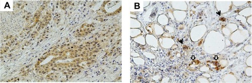
Forty pairs of primary tumor and peritoneal metastases were immunohistochemically examined for HER2, epidermal growth factor receptor, and VEGF expression. Immunoreactivity for HER2 was recognized in cell membranes; positive staining was found in 15% (6/40) of primary tumors and 3% (1/40) of peritoneal metastases. Epidermal growth factor receptor was also observed in cell membranes; positive staining was seen in 18% (7/40) of primary tumors and 3% (1/40) of peritoneal metastases. In contrast, VEGF diffusely stained the cytoplasm of cancer cells (); positive tumor staining was seen in 85% (34/40) of primary tumors and 70% (28/40) of peritoneal metastases.
Figure 1 Representative images of vascular endothelial growth factor immunostaining in gastric cancer tissues. (A) Diffusely stained cytoplasm of cancer cells in primary tumor. (B) Strongly stained cytoplasm of cancer cells (black arrow) and fibroblasts (white arrow) in peritoneal tumor.
Association between VEGF levels and clinicopathological characteristics
Of the 40 patients, 35 had malignant ascites; therefore, 35 pairs of ascites and serum samples were analyzed to quantify VEGF levels. Relationships between VEGF levels and clinicopathological variables are shown in . When patients were grouped as P1–2 and P3 according to the criteria of the Japanese Research Society for Gastric Cancer,Citation19 no significant association between peritoneal metastatic grade and VEGF level was seen in either serum or ascites. The 35 patients were divided into two groups based on whether ascites was found beyond the pelvic cavity or not. The 15 patients with ascites beyond the pelvic cavity were classified as the large group and other 20 patients were classified as the small group. The two groups did not significantly differ for serum VEGF levels, but did significantly vary for ascites VEGF levels (P < 0.0001). The median ascites VEGF level in the small group was 504 (range 82–7,261) pg/mL; for the large group, it was 700 (range 231–7,113) pg/mL. VEGF levels in both serum and ascites showed no association with gender, age, or prior gastrectomy. Ascites VEGF levels and serum VEGF levels correlated in the large group (r = 0.686, P = 0.0034) but not in the small group ().
Figure 2 Correlation of vascular endothelial growth factor levels between serum and ascites. There was a good correlation in the large group.
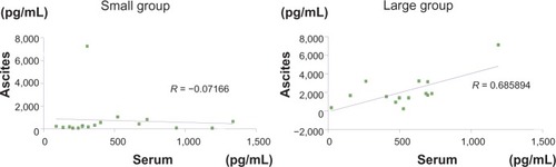
Table 1 Relationship between vascular endothelial growth factor levels and clinicopathological variables
Prognostic factors for overall survival
In the present study, we established cutoff values for serum VEGF (472 pg/mL) and ascites VEGF (660 pg/mL) using median values. The relationship between VEGF levels, volume of ascites, and peritoneal metastatic grade was evaluated using the Kaplan–Meier method. Univariate log-rank analysis showed that high ascites VEGF levels reduced overall survival (P = 0.041, ). Neither ascites volume nor peritoneal metastatic grade showed an association with overall survival. Serum VEGF levels also provided no significant evidence with regard to overall survival. Cox regression analysis showed ascites VEGF levels as a risk factor for survival (hazards ratio 2.21, 95% confidence interval 1.015–4.794, P = 0.045, ).
Figure 3 Kaplan–Meier survival curves for overall survival rate according to vascular endothelial growth factor concentration in ascites.
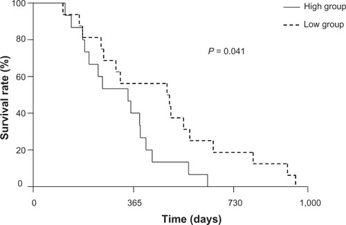
Table 2 Cox regression analysis
Case report
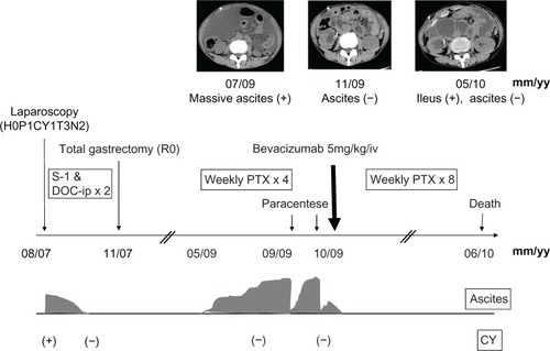
Our patient was a 62-year-old female diagnosed with type 4 gastric cancer, whose clinical course has been partially reported previously by our colleague.Citation20 Exploratory laparoscopy revealed severe peritoneal metastasis with malignant ascites. The patient received two cycles of S-1 plus intraperitoneal docetaxel; disappearance of her peritoneal metastasis was confirmed by second-look laparoscopy. Subsequently, the patient underwent total gastrectomy with D2 lymph node dissection which completed an R0 resection. Eighteen months after surgery, the patient’s cancer recurred and was treated with four cycles of weekly paclitaxel, while the patient suffered from symptomatic ascites requiring frequent paracenteses. Because of lack of response, the patient was administered bevacizumab monotherapy (5 mg/kg) intravenously. Despite only one administration, the patient noted an improvement in abdominal distention and required no paracenteses. After bevacizumab therapy, the patient received eight cycles of weekly paclitaxel. She died of aspiration pneumonia due to ileus ().
Figure 4 Treatment and disease progression for the presented case.
Discussion
Previous reports have implied that VEGF is associated with tumor progression including peritoneal metastasis; however, most of these reports are based in a xenograft model and the status of the primary tumor.Citation14,Citation21–Citation23 In the present study, expression of VEGF was found not only in primary gastric tumors but also in peritoneal metastases, and more frequently than either HER2 or epidermal growth factor receptor. Establishment of peritoneal metastasis needs a multistep process involving detachment of cancer cells from their primary tumor, their attachment to the peritoneal surface, infiltration into the subperitoneal space, and proliferation with angiogenesis.Citation8 VEGF secreted from cancer cells might enhance tumor growth by inducing an angiogenic response in the peritoneal microenvironment. We showed that VEGF is a convincing molecular target for peritoneal metastases. This is the first report to have formed the basis of clinical specimens from peritoneal tumors.
Malignant ascites with peritoneal metastasis seriously affects patients’ quality of life. VEGF mediates formation of malignant ascites by increasing the permeability of blood vessels.Citation6 In this study, levels of circulating VEGF were not correlated with volume of ascites, because circulating VEGF can derive from both a primary tumor and peritoneal metastases, and may depend on total tumor volume. Conversely, VEGF concentration in the ascites statistically correlated with ascites volume. VEGF may be produced by human peritoneal mesothelial cells when stimulated by basic fibroblast growth factor secreted from cancer cells and other human peritoneal mesothelial cells in the microenvironment.Citation24 Thus, human peritoneal mesothelial cells are critical to accumulation of malignant ascites through production of diffusible VEGF. In addition to human peritoneal mesothelial cells, intraperitoneal VEGF may come from various sources, such as subperitoneal capillaries, peritoneal metastatic tumor, fibroblasts,Citation25 and macrophages,Citation26 whereas intraperitoneal VEGF cannot transfer into the systemic circulation due to capillary hyperpermeability. Accordingly, ascites volume correlates with ascites VEGF concentration. This result agrees with a previous report by Rudlowski et al, who showed the same findings in patients with ovarian cancer.Citation27
In the present study, the prognostic value of VEGF levels was also assessed. Although several studies have shown that tumor VEGF is an independent prognostic factor in gastric cancer, measurement of VEGF levels both in serum and ascites is technically simple, does not require a tumor specimen, and is more objective in its evaluation than semi-quantitative immunohistochemistry.Citation28–Citation30 Increased serum VEGF levels have been associated with advanced stage, higher lymph node metastasis, and perineural invasion in gastric cancer.Citation31,Citation32 These data suggest that anti-VEGF therapy might have an effect on gastric cancer. Although Shah et al reported that anti-VEGF therapy using bevacizumab combined with chemotherapy might be a promising therapy for patients with metastatic or unresectable gastric and gastroesophageal junction adenocarcinoma,Citation33 a randomized, double-blind, placebo-controlled, Phase III study (Avastin in Gastric Cancer) failed to establish evidence about the usefulness of bevacizumab in gastric cancer.Citation34 Moreover, Vidal et al found that a high preoperative serum VEGF level was an independent prognostic factor for recurrence and survival after R0 resection of gastric cancer.Citation18 However, we could not find a significant association between serum VEGF level and overall survival. This might be the reason why that all the patients in our study were in stage IV with peritoneal metastasis and did not receive R0 resection. In contrast, the prognostic significance of ascites VEGF level has not been adequately studied. To our knowledge, this is the first study to report that elevated ascites VEGF levels are significantly associated with shorter overall survival in gastric cancer. The principal mechanisms explaining the prognostic significance of VEGF are tumor expansion and massive ascites, which cause severe symptoms, such as bowel obstruction, dyspnea, and cachexia. Additionally, VEGF in malignant ascites may induce immune suppression in cancer by inhibiting dendritic cell maturationCitation35 and increasing tumor-infiltrating regulatory T cells.Citation36 Furthermore, high ascites VEGF levels may be associated with upregulation of multidrug resistance-associated protein, leading to resistance to platinum-based treatment, which is often used in unresectable gastric cancer.Citation37,Citation38 In any case, anti-VEGF therapies should be considered for patients with malignant ascites in gastric cancer.
Our patient experienced successful palliation of symptomatic ascites using intravenous (systemic) bevacizumab. Several studies using mouse models indicate that intraperitoneal (regional) bevacizumab could be useful for peritoneal metastasis.Citation23,Citation39 In clinical case reports, bevacizumab was also administrated regionally for patients with malignant ascites.Citation40,Citation41 Yagi et al reported that bevacizumab had a more pronounced effect on malignant ascites and peritoneal nodules when administered systemically rather than regionally.Citation42 If bevacizumab is administered regionally, most of the antibody will be neutralized in malignant ascites, which contains large amounts of VEGF, resulting in a low blood concentration. These results support the use of systemically administered bevacizumab, with ascites removed before treatment for more efficacy.
In conclusion, VEGF might be correlated with the development of peritoneal metastasis and malignant ascites. The ascites VEGF level appears to be an important prognostic indicator in gastric cancer with peritoneal metastasis. Further prospective studies will be necessary to validate both ascites VEGF as a predictive marker of poor outcome and the efficacy of bevacizumab for chemoresistant malignant ascites.
Disclosure
The authors report no conflicts of interest in this work.
References
- AllumWHPowellDJMcConkeyCCFieldingJWGastric cancer: a 25-year reviewBr J Surg19897665355402758258
- MaruyamaKKaminishiMHayashiKGastric cancer treated in 1991 in Japan: data analysis of nationwide registry. Japanese Gastric Cancer Association Registration CommitteeGastric Cancer200692516616767357
- Van CutsemEMoiseyenkoVMTjulandinSV325 Study GroupPhase III study of docetaxel and cisplatin plus fluorouracil compared with cisplatin and fluorouracil as first-line therapy for advanced gastric cancer: a report of the V325 Study GroupJ Clin Oncol200624314991499717075117
- KoizumiWNaraharaHHataTS-1 plus cisplatin versus S-1 alone for first-line treatment of advanced gastric cancer (SPIRITS trial): a phase III trialLancet Oncol20089321522118282805
- SatoYTakayamaTSagawaTPhase II study of S-1, docetaxel and cisplatin combination chemotherapy in patients with unresectable metastatic gastric cancerCancer Chemother Pharmacol201066472172820041328
- ChuDZLangNPThompsonCOsteenPKWestbrookKCPeritoneal carcinomatosis in nongynecologic malignancies. A prospective study of prognostic factorsCancer19896323643672910444
- SadeghiBArvieuxCGlehenOPeritoneal carcinomatosis from non-gynecologic malignancies: results of the EVOCAPE 1 multicentric prospective studyCancer200088235836310640968
- YonemuraYEndouYYamaguchiTMechanisms of the formation of the peritoneal dissemination in gastric cancerInt J Oncol19968479580221544429
- ConnollyDTHeuvelmanDMNelsonRTumor vascular permeability factor stimulates endothelial cell growth and angiogenesisJ Clin Invest1989845147014782478587
- LeungDWCachianesGKuangWJGoeddelDVFerraraNVascular endothelial growth factor is a secreted angiogenic mitogenScience19892464935130613092479986
- MesianoSFerraraNJaffeRBRole of vascular endothelial growth factor in ovarian cancer. Inhibition of ascites formation by immunoneutralizationAm J Pathol19981534124912569777956
- ByrneATRossLHolashJVascular endothelial growth factor-trap decreases tumor burden, inhibits ascites, and causes dramatic vascular remodeling in an ovarian cancer modelClin Cancer Res20039155721572814654557
- ShererDMEliakimRAbulafiaOThe role of angiogenesis in the accumulation of peritoneal fluid in benign conditions and the development of malignant ascites in the femaleGynecol Obstet Invest200050421722411093042
- AoyagiKKouhujiKYanoSVEGF significance in peritoneal recurrence from gastric cancerGastric Cancer20058315516316086118
- LiLWangLZhangWCorrelation of serum VEGF levels with clinical stage, therapy efficacy, tumor metastasis and patient survival in ovarian cancerAnticancer Res2004243b1973198015274387
- KayaACiledagAGulbayBEThe prognostic significance of vascular endothelial growth factor levels in sera of non-small cell lung cancer patientsRespir Med200498763263615250229
- AlabiAASuppiahAMaddenLAMonsonJRGreenmanJPreoperative serum vascular endothelial growth factor-a is a marker for subsequent recurrence in colorectal cancer patientsDis Colon Rectum200952599399919502868
- VidalOMetgesJPElizaldeIHigh preoperative serum vascular endothelial growth factor levels predict poor clinical outcome after curative resection of gastric cancerBr J Surg200996121443145119918848
- Japanese Research Society for Gastric CancerJapanese Classification of Gastric Carcinoma1st EnglishTokyo, JapanKanehara1995
- KinoshitaJFushidaSMakinoI[The use of bevacizumab in refractory peritoneal dissemination of gastric cancer with malignant ascites – two case reports.]Gan To Kagaku Ryoho2011381223602362 Japanese22202382
- KamiyamaMIchikawaYIshikawaTVEGF receptor antisense therapy inhibits angiogenesis and peritoneal dissemination of human gastric cancer in nude miceCancer Gene Ther20029219720111857038
- BabaMKonnoHMaruoYRelationship of p53 and vascular endothelial growth factor expression of clinicopathological factors in human scirrhous gastric cancerEur Surg Res19983021301379565747
- ImaizumiTAoyagiKMiyagiMShirouzuKSuppressive effect of bevacizumab on peritoneal dissemination from gastric cancer in a peritoneal metastasis modelSurg Today201040985185720740349
- SakoAKitayamaJYamaguchiHVascular endothelial growth factor synthesis by human omental mesothelial cells is augmented by fibroblast growth factor-2: possible role of mesothelial cell on the development of peritoneal metastasisJ Surg Res2003115111312014572781
- PertovaaraLKaipainenAMustonenTVascular endothelial growth factor is induced in response to transforming growth factor-beta in fibroblastic and epithelial cellJ Biol Chem19942699627162748119973
- HermeyJHDimitriadisEKayERdmondHPBouchier-HayesDRegulation of macrophage production of vascular endothelial growth factor (VEGF) by hypoxia and transforming growth factor beta-1Ann Surg Oncol1998532712789607631
- RudlowskiCPickartAKFuhljahnCPrognostic significance of vascular endothelial growth factor expression in ovarian cancer patients: a long-term follow-upInt J Gynecol Cancer200616Suppl 1S183S189
- MaedaKChungYSOgawaYPrognostic value of vascular endothelial growth factor expression in gastric carcinomaCancer19967758588638608475
- SaitoHTsujitaniSKondoAIkeguchiMMaetaMKaibaraNExpression of vascular endothelial growth factor correlates with hematogenous recurrence in gastric carcinomaSurgery1999125219520110026754
- IchikuraTTomimatsuSOhkuraEMochizukiHPrognostic significance of the expression of vascular endothelial growth factor (VEGF) and VEGF-C in gastric carcinomaJ Surg Oncol200178213213711579392
- KarayiannakisAJSyrigosKNPolychrondisACirculating VEGF levels in the serum of gastric cancer patients: correlation with pathological variables, patient survival, and tumor surgeryAnn Surg20022361374212131083
- YoshikawaTTsuburayaAKobayashiOSairenjiMMotohashiHYanomaSPlasma concentration of VEGF and b-FGF in patients with gastric carcinomaCancer Lett20001531–271210779624
- ShahMARamanathanRKIlsonDHMulticenter Phase II study of irinotecan, cisplatin, and bevacizumab in patients with metastatic gastric or gastroesophaeal junction adenocarcinomaJ Clin Oncol200624335201520617114652
- OhtsuAShahMAVan CutsemEBevacizumab in combination with chemotherapy as first-line therapy in advanced gastric cancer: a randomized double-blind, placebo-controlled phase III studyJ Clin Oncol201129303968397621844504
- Della PortaMDanovaMRigolinGMDendritic cells and vascular endothelial growth factor in colorectal cancer: correlations with clinicopathological findingsOncology2005682–327628416015045
- LiBLalaniASHardingTCVascular endothelial growth factor blockade reduces intratumoral regulatory T cells and enhances the efficacy of a GM-CSF-secreting cancer immunotherapyClin Cancer Res200612226808681617121902
- BamiasAKoutsoukouVTerposECorrelation of NK T-like CD3+CD56+ cells and CD4+CD25+(hi) regulatory T cells with VEGF and TNFa in ascites from advanced ovarian cancer: association with platinum resistance and prognosis in patients receiving first-line, platinum-based chemotherapyGynecol Oncol2008108242142718036640
- ZhangXSZhuXFGaoJSMultiple drug resistance phenotype of human endothelial cells induced by vascular endothelial growth factor 165Acta Pharmacol Sin200122873173511749847
- NinomiyaSInomataMTajimaMEffect of bevacizumab monoclonal antibody to vascular endothelial growth factor, on peritoneal matastasis of MKN-45P human gastric cancer in miceJ Surg Res2009154219620219329124
- NumnumTMRocconiRPWhitworthJBarnesMNThe use of bevacizumab to palliate symptomatic ascites in patients with refractory ovarian carcinomaGynecol Oncol2006102342542816797681
- HamiltonCAMaxwellGLChernofskyMRBernsteinSAFarleyJHRoseSIntraperitoneal bevacizumab for the palliation of malignant ascites in refractory ovarian cancerGynecol Oncol2008111353053218561992
- YagiYFushidaSHaradaSBiodistribution of humanized anti-VEGF monoclonal antibody/bevacizumab on peritoneal metastatic models with subcutaneous xenograft of gastric cancer in miceCancer Chemother Pharmacol201066474575320033809