
Abstract
Bioassays that predict clinical outcome are essential to optimize cellular anticancer immunotherapy. We have recently developed a robust and simple skin test to evaluate the capacity of tumor-specific T cells to migrate, recognize their targets and exert effector functions. This bioassay detects T cells with an elevated antineoplastic potential and hence rapidly identifies patients responding to immunotherapy.
Due to the strong immunostimulatory properties of dendritic cells (DCs) and their ability to elicit adaptive immune responses against malignant cells, novel anticancer therapies focus on the efficient generation and activation of this pivotal cell type. Our laboratory has conducted several clinical trials to test the therapeutic profile of DC-based vaccines over the past decade, mainly with autologous DCs generated and educated ex vivo to elicit efficient cellular immunity against neoplastic cells.Citation1-Citation3 Robust T-cell responses against tumor-associated antigens (TAAs) were readily detected in a number of patients upon vaccination, providing a proof-of-principle in support of this immunotherapeutic approach. Despite enormous efforts in research and optimization, however, objective clinical responses could be detected in a minority of patients. Still, such responses were often long-lasting, indicating that enduring protection against neoplastic cells is achievable.Citation1 Interestingly, the fraction of patients who respond to different immunotherapeutic approaches is remarkably constant, pointing to the existence of an “immunologically reactive” subgroup of individuals.Citation4 The identification of such patients early in the course of treatment would greatly improve the clinical efficacy of these novel and costly therapeutic paradigms, but appropriate assays are lacking.
Anticancer immune responses are thought to be primarily mediated by CD8+ T lymphocytes, which are able to trigger the apoptotic demise of neoplastic cells. Thus, current immunomonitoring approaches mainly focus on the assessment of cellular immunity using T cells isolated from the blood at various time points upon vaccination. The list of the biomarkers that are tested in this setting is long and includes the presence of TAA-specific CD4+ and/or CD8+ T cells, the fraction of T cells that secrete interferon γ (IFNγ) upon antigenic stimulation, and the presence of T cells or antibodies against exogenous antigens that are added as control antigens in a number of vaccination protocols, such as keyhole limpet hemocyanin (KLH).Citation5-Citation7 So far, most attempts to predict objective clinical responses using these parameters failed, presumably because individual parameters were not combined in one assay. Moreover, the capacity of T cells to migrate into tissues, which is crucial for efficient anticancer immune responses, is usually not tested.
In order to address these issues, our lab conducted a pilot study in 2005 to investigate the potential value of skin-infiltrating lymphocytes (SKIL) obtained from delayed-type hypersensitivity reactions (DTHs) for predicting clinical responses in metastatic melanoma patients.Citation8 This approach was intended as a very comprehensive analysis of anticancer immunity, simultaneously assessing T-cell migration, effector functions and antigen recognition capability. After encouraging initial results, the SKIL test was included in subsequent vaccination protocols. Recently, we systematically analyzed the general feasibility of the SKIL analysis for the routine immunomonitoring of patients treated with DC-based vaccination in the context of a clinical trial.Citation9
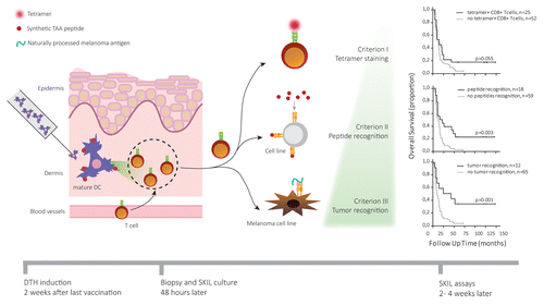
In this study, patients with metastatic melanoma were allocated to receive DCs that have been generated, activated, and pulsed with TAAs plus KLH ex vivo. Patients received 3 intradermal, intravenous or intranodal injections of the vaccine in a biweekly cycle. One to 2 wk after the last injection, mature, autologous DCs pulsed with TAAs were injected intradermally in the back of vaccinated patients to induce DTH reactions. After 48 h, punch biopsies were taken and SKILs emigrating from these tissues were cultured and analyzed for specificity, antigen recognition capability and functionality. In addition, peripheral blood mononuclear cells (PBMCs) collected on the same day than biopsies were analyzed for their ability to proliferate and secrete IFNγ in response to KLH.
We found that neither the KLH-induced proliferation of PBMC-derived CD4+ T cells nor their ability to release IFNγ correlated with the overall survival of patients. This demonstrates that monitoring KLH-elicited responses can indicate the immunological competence of individual patients but does not provide an adequate means to assess antitumor immune responses.
To analyze the potential value of SKILs for predicting clinical response, lymphocytes within skin biopsies were expanded. After 2–4 wk, 80% of SKIL cultures yielded sufficient cell numbers for an extensive assessment of T-cell function and specificity. Thus, the SKIL test appears to be feasible in a large fraction of patients. We developed 3 increasingly stringent criteria to predict clinical responses: (1) the presence of TAA-specific CD8+ T cells in SKIL cultures, (2) the ability of SKIL cultures to specifically recognize cells pulsed with TAA-derived peptides, and (3) the ability of SKIL cultures to specifically recognize cell lines that naturally process and present TAAs. Strikingly, we found that patients whose SKIL cultures contained TAA-specific CD8+ T cells (criterion 1) displayed a survival benefit over patients whose SKIL cultures failed to do so. By adding functional properties to the assessment (criteria 2 and 3), the accuracy of prediction could be further improved ().
Figure 1. Analysis of skin-infiltrating lymphocytes allows for early prediction of clinical responses in cancer patients treated with immunotherapy. Dendritic cells (DCs) generated, activated, and loaded with tumor-associated antigens (TAAs) ex vivo were injected in the dermis of melanoma patients 1 to 2 wk after vaccination. The resulting delayed type hypersensitivity (DTH) reaction promotes the migration of TAA-specific T cells into the skin, which were isolated via punch biopsy 48 h after injection. Isolated skin-infiltrating lymphocytes (SKILs) were expanded and tested for cancer-specificity using tetramer staining (criterion 1). Patients whose SKIL cultures contained antigen-specific CD8+ T cells showed a survival benefit over patients not fulfilling this criterion. Additionally, the specific recognition of TAA-derived peptides by SKILs (criterion II) was measured in terms of cytotoxic activity or secretion of TH1 cytokines, such as interleukin (IL)-2, tumor necrosis factor α (TNFα) and interferon γ (IFNγ), coupled to the release of no TH2 cytokines (i.e., IL-4, IL-5, IL-13). Finally, the ability of SKILs to specifically recognize TAAs naturally processed and presented by a melanoma cell line (criterion III) was assessed. Combining criterion 2 and 3 to criterion 1 further increased the predictive value of our assay.
Remarkably, within SKIL tests we were able to detect distinct T-cell cytokine profiles upon antigenic stimulation. Whereas most of the cultures secreted TH1 cytokines such as IFNγ, tumor necrosis factor α (TNFα) and interleukin (IL)-2, tumor-specific SKILs from two patients were found to secrete IL-5, coinciding with rapid disease progression. This finding highlights the importance of assessing T-cell functionality for the prediction of disease outcome. Interestingly, studies on intracellular pathogens revealed the importance of multifunctional T cells, i.e., individual T cells that are able to exert cytotoxic, pro-inflammatory, and proliferative functions, for disease control.Citation10 Naturally, these T cells would be highly desirable for anticancer therapy as well. Future studies need to clarify which impact multifunctional T cells have on anticancer immunity and what their added value as biomarker for clinical responses is.
In conclusion, we developed a robust and simple assay for the evaluation of the migratory behavior, antigen recognition capability, and effector function of tumor-specific T cells. By identifying highly functional T cells with elevated anticancer potential, this assay allows for the early and reliable prediction of clinical responses in large-scale trials testing the clinical profile of multiple immunotherapeutic interventions.
| Abbreviations: | ||
| DC | = | dendritic cell |
| DTH | = | delayed-type hypersensitivity |
| KLH | = | keyhole limpet hemocyanin |
| SKIL | = | skin infiltrating lymphocyte, TAA, tumor-associated antigen |
Disclosure of Potential Conflicts of Interest
No potential conflicts of interest were disclosed.
Citation: Wimmers F, Aarntzen EH, Schreibelt G, Jacobs JF, JA Punt C, Figdor CG, de Vries IJM. Early predictive value of multifunctional skin-infiltrating lymphocytes in anticancer immunotherapy. OncoImmunology 2013; 2:e27219; 10.4161/onci.27219
References
- Aarntzen EH, Schreibelt G, Bol K, Lesterhuis WJ, Croockewit AJ, de Wilt JH, van Rossum MM, Blokx WA, Jacobs JF, Duiveman-de Boer T, et al. Vaccination with mRNA-electroporated dendritic cells induces robust tumor antigen-specific CD4+ and CD8+ T cells responses in stage III and IV melanoma patients. Clin Cancer Res 2012; 18:5460 - 70; http://dx.doi.org/10.1158/1078-0432.CCR-11-3368; PMID: 22896657
- Jacobs JF, Punt CJ, Lesterhuis WJ, Sutmuller RP, Brouwer HM, Scharenborg NM, Klasen IS, Hilbrands LB, Figdor CG, de Vries IJ, et al. Dendritic cell vaccination in combination with anti-CD25 monoclonal antibody treatment: a phase I/II study in metastatic melanoma patients. Clin Cancer Res 2010; 16:5067 - 78; http://dx.doi.org/10.1158/1078-0432.CCR-10-1757; PMID: 20736326
- Lesterhuis WJ, Schreibelt G, Scharenborg NM, Brouwer HM, Gerritsen MJ, Croockewit S, Coulie PG, Torensma R, Adema GJ, Figdor CG, et al. Wild-type and modified gp100 peptide-pulsed dendritic cell vaccination of advanced melanoma patients can lead to long-term clinical responses independent of the peptide used. Cancer Immunol Immunother 2011; 60:249 - 60; http://dx.doi.org/10.1007/s00262-010-0942-x; PMID: 21069321
- Weide B, Zelba H, Derhovanessian E, Pflugfelder A, Eigentler TK, Di Giacomo AM, Maio M, Aarntzen EH, de Vries IJ, Sucker A, et al. Functional T cells targeting NY-ESO-1 or Melan-A are predictive for survival of patients with distant melanoma metastasis. J Clin Oncol 2012; 30:1835 - 41; http://dx.doi.org/10.1200/JCO.2011.40.2271; PMID: 22529253
- Schumacher K. Keyhole limpet hemocyanin (KLH) conjugate vaccines as novel therapeutic tools in malignant disorders. J Cancer Res Clin Oncol 2001; 127:Suppl 2 R1 - 2; http://dx.doi.org/10.1007/BF01470991; PMID: 11768617
- Coulie PG, van der Bruggen P. T-cell responses of vaccinated cancer patients. Curr Opin Immunol 2003; 15:131 - 7; http://dx.doi.org/10.1016/S0952-7915(03)00009-8; PMID: 12633661
- Janetzki S, Panageas KS, Ben-Porat L, Boyer J, Britten CM, Clay TM, Kalos M, Maecker HT, Romero P, Yuan J, et al, Elispot Proficiency Panel of the CVC Immune Assay Working Group. Results and harmonization guidelines from two large-scale international Elispot proficiency panels conducted by the Cancer Vaccine Consortium (CVC/SVI). Cancer Immunol Immunother 2008; 57:303 - 15; http://dx.doi.org/10.1007/s00262-007-0380-6; PMID: 17721781
- de Vries IJ, Bernsen MR, Lesterhuis WJ, Scharenborg NM, Strijk SP, Gerritsen MJ, Ruiter DJ, Figdor CG, Punt CJ, Adema GJ. Immunomonitoring tumor-specific T cells in delayed-type hypersensitivity skin biopsies after dendritic cell vaccination correlates with clinical outcome. J Clin Oncol 2005; 23:5779 - 87; http://dx.doi.org/10.1200/JCO.2005.06.478; PMID: 16110035
- Aarntzen EH, Bol K, Schreibelt G, Jacobs JF, Lesterhuis WJ, Van Rossum MM, Adema GJ, Figdor CG, Punt CJ, De Vries IJ. Skin-test infiltrating lymphocytes early predict clinical outcome of dendritic cell-based vaccination in metastatic melanoma. Cancer Res 2012; 72:6102 - 10; http://dx.doi.org/10.1158/0008-5472.CAN-12-2479; PMID: 23010076
- Betts MR, Nason MC, West SM, De Rosa SC, Migueles SA, Abraham J, Lederman MM, Benito JM, Goepfert PA, Connors M, et al. HIV nonprogressors preferentially maintain highly functional HIV-specific CD8+ T cells. Blood 2006; 107:4781 - 9; http://dx.doi.org/10.1182/blood-2005-12-4818; PMID: 16467198