
Abstract
Multiple experimental and human trials have shown that microcirculatory alterations are frequent in sepsis. In this review, we discuss the various mechanisms that are potentially involved in their development and the implications of these alterations. Endothelial dysfunction, impaired inter-cell communication, altered glycocalyx, adhesion and rolling of white blood cells and platelets, and altered red blood cell deformability are the main mechanisms involved in the development of these alterations. Microcirculatory alterations increase the diffusion distance for oxygen and, due to the heterogeneity of microcirculatory perfusion in sepsis, may promote development of areas of tissue hypoxia in close vicinity to well-oxygenated zones. The severity of microvascular alterations is associated with organ dysfunction and mortality. At this stage, therapies to specifically target the microcirculation are still being investigated.
Introduction
Septic shock is characterized by profound hemodynamic alterations associated with organ dysfunction. These hemodynamic alterations include some degree of hypovolemia and a decrease in vascular tone and myocardial depression. Even when systemic hemodynamic variables seems to have been corrected and are within therapeutic goals, signs of impaired tissue perfusion may persist. Recently, alterations in microcirculatory blood flow have been identified in severe sepsisCitation1 and the severity of these alterations is associated with a poor outcome.Citation2,Citation3 The impact of therapeutic interventions on microcirculatory function is beginning to be reported.Citation4-Citation6 In this manuscript, we discuss the pathophysiology of the alterations in microvascular perfusion in sepsis, and their implications for organ function and for therapy.
Microcirculatory Alterations in Sepsis: Where is the Evidence?
Multiple experimental studies have found that sepsis induces marked alterations in the microcirculation. Compared with normal conditions in which there is a dense network of capillaries, most of which are perfused, sepsis is associated with a decrease in capillary density in association with an increase in heterogeneity of perfusion because of the presence of intermittently or not perfused capillaries in close proximity to well perfused capillaries.Citation7-Citation10 Importantly, this is a dynamic process, as capillaries in which there is no flow at a given time may be perfused a few minutes later. These alterations have been reported after administration of endotoxin or live bacteria and during bacterial peritonitis,Citation8,Citation9,Citation11 and have been observed in rodentsCitation12,Citation13 as well as in large animals.Citation9,Citation11 In addition, all studied organs have been affected, including the skin,Citation14 muscle,Citation12,Citation13 eye,Citation15 tongue,Citation9 gut,Citation9,Citation10 liver,Citation7 heart,Citation16 and even the brain.Citation11 Hence, these changes seem to be ubiquitous and to have common pathophysiologic mechanisms.
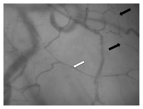
In humans, demonstration of these alterations has been longer in coming, mostly because of technical limitations that prevented exposure of the human microcirculation. The development of new imaging techniques has enabled direct visualization of the human microcirculation with small handheld microscopes.Citation17,Citation18 We demonstrated that microcirculatory perfusion is altered in patients with severe sepsis and septic shock.Citation1 An example of altered human microcirculation in sepsis is shown in . These alterations in microvascular perfusion are very similar to those occurring in experimental conditions, and are characterized by a decrease in vascular density together with an increased number of capillaries with stopped or intermittent flow. Since this initial study, more than 30 studies have shown similar results.
Figure 1. Sublingual microcirculation in sepsis. Photograph of the sublingual microcirculation in a patient with septic shock using a sidestream dark field (SDF) imaging device. The white arrow shows a perfused capillary, the black arrows identify a stopped flow capillary.
What are the Consequences of These Alterations?
The decreased capillary density results in an increased diffusion distance for oxygen.Citation16 More importantly, microvascular blood flow is heterogeneous, with perfused capillaries in close vicinity to non-perfused capillaries, leading to alterations in oxygen extraction and hypoxic zones even when total blood flow to the organ is preserved.Citation19 Heterogeneity of microvascular perfusion is a crucial aspect. Heterogeneous perfusion leads to more severe alterations in tissue oxygenation than does homogenously decreased perfusion.Citation20,Citation21 Heterogeneity of perfusion is associated with heterogeneity in oxygenationCitation22 but also with altered oxygen extraction capabilities.Citation10,Citation20,Citation21,Citation23,Citation24 During episodes of hypoperfusion, the heterogeneity of microvascular perfusion further increases in sepsis instead of being minimized as in normal conditions.Citation24
These alterations play an important role in the development of organ dysfunction and are not just an indication of the severity of sepsis. Microvascular alterations can lead to cellular injuryCitation25 and reversal of these alterations is associated with improvement in lactateCitation5 and NADHCitation26 levels, suggesting that microvascular alterations directly impair tissue oxygenation. In addition, several trials have demonstrated an association between the severity of microvascular dysfunction and the development of organ dysfunctionCitation27-Citation29 and mortality.Citation1,Citation2,Citation19,Citation28,Citation30-Citation33
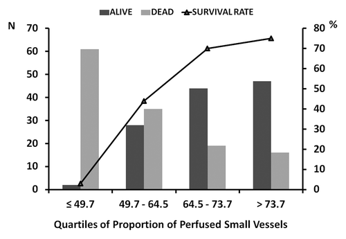
In a large series of 252 patients with septic shock, microvascular perfusion was an independent factor associated with survival.Citation3 Of note this is not an on-and-off but rather a progressive phenomenon. Dividing the population into quartiles of proportions of perfused capillaries, mortality rates markedly increased with alterations in the microcirculation ().Citation3 Looking at which variables differed between survivors and non-survivors, the proportion of perfused capillaries was the strongest predictor of outcome. Vascular density, and especially vascular density of perfused capillaries, have been associated with outcome,Citation1,Citation3,Citation19 as well as the heterogeneity index,Citation3,Citation19 but not the velocity in perfused capillaries.Citation19 More importantly, evolution over time of these alterations also differs in patients with good or poor outcomes,Citation2,Citation33 rapidly improving in survivors but remaining disturbed in non-survivors, whether these patients die from acute circulatory failure or from organ failure after resolution of shock.Citation2 In children with septic shock, the microcirculation improved from day 1 to day 2 in survivors but remained altered in non-survivors.Citation33 Interestingly, the severity of microvascular alterations was an independent factor associated with outcome in the early and later phases of sepsis, but the cut-off value separating survivors from non-survivors was lower in the early phase.Citation3
Figure 2. Relationship between sublingual microcirculation and ICU mortality in patients with severe sepsis. In this series of 252 patients with severe sepsis, the sublingual microcirculation was assessed either with an orthogonal polarization spectral (OPS) or a sidestream dark field (SDF) imaging device. The patients were grouped into quartiles of proportion of perfused capillaries. From reference Citation3 with permission.
Although one may consider that the microcirculation is just adapting to direct cellular alterations, several factors suggest that microcirculatory alterations are the primary event leading to cellular dysfunction. First, microcirculatory alterations are co-localized with low PO2, production of hypoxia-inducible factorCitation16 or redox potentialCitation26 in experimental conditions. Second, oxygen saturation at the capillary end of well-perfused capillaries is low, not elevated, suggesting that the tissues are using the delivered oxygen.Citation23 Third, the tissue to arterial PCO2 gradient, the PCO2 gap, is increased in sepsis.Citation34-Citation38 In addition, there is an inverse relationship between sublingual microvascular perfusion and the PCO2 gap.Citation35 A similar inverse relationship was found between ileal mucosal perfusion and ileal to arterial PCO2 gap.Citation38 If flow were just matching metabolism, CO2 production would be low because the primary alteration is the decrease in metabolism, and PCO2 gap would be normal, even at low flows. Fourth, perfusion abnormalities precede alterations in organ function.Citation39 Fifth, improvement in the sublingual microcirculation in response to initial resuscitation procedures was associated with an improvement of organ function 24 h later.Citation29 Finally, the decrease in lactate levels is proportional to the improvement of the microcirculation during dobutamine administration.Citation5
Admittedly, microcirculatory alterations are not the sole mechanism contributing to organ dysfunction in sepsis. Cellular metabolic alterations and in particular mitochondrial dysfunction may also contribute. Discussion of these factors is beyond the scope of this review. Importantly, there is an interplay between hypoxia and inflammation and mitochondrial dysfunction.Citation40 Limiting perfusion abnormalities is associated with reduced expression of inflammatory molecules, caspases, and mitochondrial abnormalities.Citation41,Citation42
What Mechanisms Could Be Involved in the Development of These Alterations?
Endothelial dysfunction is one of the key mechanisms underlying these alterations. We have seen earlier that endothelial reactivity to vasoconstrictive and vasodilating substances is decreased in sepsis, and both constriction and dilation are important for the regulation of microvascular blood flow. This aspect of endothelial involvement is illustrated by the alteration in post-ischemic hyperemic response, which is blunted in patients with sepsis.Citation27,Citation32,Citation43-Citation47 Furthermore, alterations in the descending (oxygen consumption) or ascending slope are associated with development of organ failure.Citation27,Citation32 In addition, and perhaps more importantly, communication between endothelial cells may be altered. In normal conditions, matching of tissue perfusion with metabolism is obtained by backward communication through perivascular nerves but also by transmission of information between endothelial cells. Indeed, the stimulation of endothelial cells in a given area results in a change in membrane potential which is transmitted to contiguous cells, resulting in transmission of the information to upstream arterioles up to a distance of 1000 µm.Citation48 During endotoxic conditions, the communication rate between microvessels 500 microns apart is markedly impaired, but this phenomenon is transient and fully reversible after recovery from endotoxin exposure.Citation49 The interaction between the endothelial surface and circulating cells is also impaired in sepsis. In particular, the glycocalyx is altered in sepsis. The glycocalyx is a thin layer of glucosaminoglycans that covers the endothelial surface and in which various substances, such as superoxide dismutase and antithrombin, are embedded. The glycocalyx facilitates the flow of red blood cells and limits adhesion of white blood cells and platelets to the endothelium. The size of the glycocalyx is markedly decreased in sepsisCitation50-Citation52 and byproducts of its degradation are found in the blood.Citation50,Citation53 In addition, the glycocalyx is made more permeable to labeled substances.Citation51 Alteration in the glycocalyx layer promotes leukocyte rolling and adhesion to the endothelium.Citation51,Citation54 Administration of hyaluronidase, which partially destroys the glycocalyx layer, mimics sepsis-induced alterations in microcirculatory perfusion.Citation55
Activation of coagulation may also play a key role in the pathogenesis of microcirculatory alterationsCitation8,Citation56 even though microthrombi formation is rarely documented in experimental sepsis.Citation7 In mice challenged with endotoxin, fibrin deposition occurred in a significant proportion of capillaries; the addition of antithrombin decreased the number of non-perfused capillaries, whereas this number was increased after addition of FeCl3, a factor locally activating coagulation.Citation8
Finally, circulating cells also play a key role in these alterations. Leukocyte and platelet rolling and adhesion to the endothelial surface is increased in sepsis,Citation7,Citation8 which may impair the circulation of other cells. Red blood cells also may play a role, with alterations in red blood cell deformability,Citation57 impaired release of nitric oxide (NO), and/or adhesion of red blood cells to the endothelium.Citation58 Altogether, these data suggest that multiple mechanisms are involved in the development of microvascular dysfunction, and that it is unlikely that an intervention focused on a single pathway would be effective. As an example, chemotherapy-induced neutropenia and thrombopenia does not prevent microcirculatory alterations from occurring,Citation59 which nicely illustrates that this mechanism in isolation is not enough to generate microvascular alterations.
In septic patients who have severe microvascular alterations, we demonstrated that topical administration of a large dose of acetylcholine, an endothelium-dependent vasodilating agent, restored the microcirculation to a state similar to that of healthy volunteers and non-septic ICU patients.Citation1 This observation has profound implications. First, sepsis-associated microcirculatory alterations are functional and can be totally reversed. Complete obstruction of microvessels by clots is thus unlikely. Second, the endothelium, although dysfunctional, is still able to respond to a supraphysiological stimulation. These observations suggest that therapeutic interventions could be used to try to reverse these alterations.
The mechanisms involved in microvascular alterations differ from those involved in the development of systemic hemodynamic alterations. Accordingly, it is expected that microcirculatory derangements may be present even when systemic hemodynamics are within acceptable limits.
It should be noted that many of the mechanisms involved in microcirculatory dysfunction are probably mandatory for the control of infection. Activation of inflammation and coagulation are important for compartmentalization of infection; rolling and adhesion of white blood cells and increased permeability are needed to allow these cells to enter the tissue and kill bacteria; intravascular neutrophil extracellular traps (NETs) bind circulating bacteria and are useful for bacterial clearance even though they may impair circulation of blood cells.Citation60 Hence, totally inhibiting the factors responsible for the activations of these processes does not seem to be a rational approach, rather it should be modulated, to maintain a limited, beneficial level.
Therapeutic Interventions Targeted at the Microcirculation
Given the heterogeneous nature of the microvascular alterations, it is more important to recruit the microcirculation than to increase total flow to the organ. Ideally, the intervention should affect one or several of the mechanisms involved in the development of these microvascular alterations. Nevertheless, most interventions that are currently used for their impact on systemic hemodynamics may also influence the microcirculation to some degree.
Fluids and vasoactive agents are key components of hemodynamic resuscitation, given with the aim of improving tissue perfusion. However, improved cellular oxygen supply implies an improvement in microvascular perfusion. Two recent trials have demonstrated that fluids can improve microvascular perfusion, increasing the proportion of perfused capillaries and decreasing perfusion heterogeneity.Citation4,Citation61 Importantly, in both trials the microcirculatory effects were relatively independent of the systemic effects. The microcirculatory effects of fluids seem to be mostly present in the early phase of sepsis (within 24 h of diagnosis) whereas later (after 48 h) fluid administration failed to improve the microcirculation even when cardiac output increased.Citation4 Whether different types of fluid are associated with different microvascular responses is still debated. In some experimental conditions, colloids may increase microcirculatory perfusion more than crystalloids,Citation62 but this difference has not been confirmed in septic patients.Citation4 The mechanisms by which fluids may improve the microcirculation are not well understood, but may be related to a decrease in viscosity, a decrease in white blood cell adhesion and rolling, or, indirectly, to a decrease in endogenous vasoconstrictive substances. Whether the effects of fluids, when observed, will persist or be transient, and also whether this effect can be “saturated”, i.e., only the initial effects would be beneficial and further administration of fluids would have minimal effect, requires further study. This “saturable” effect is suggested by the observations of Pottecher et al.Citation61 who reported that the first bolus of fluids improved microvascular perfusion but the second had no effect even though cardiac output increased further.
The effects of red blood cell transfusions are variable and may depend on the severity of underlying microcirculatory alterations. In patients with sepsis, transfusions failed to improve the microcirculation in the entire population; however, transfusions did improve microvascular perfusion in patients with the most severely altered microcirculation at baseline and even worsened the microcirculation in patients with microcirculation closer to normal values.Citation63
Beta-adrenergic agents have been shown to improve microvascular perfusion, increasing not only convective but also diffusive transport.Citation5,Citation64 These effects were dissociated from the systemic effects of these agents.Citation5 Similar effects were observed with milrinoneCitation65 but the effects of levosimendan may be even more pronounced.Citation6,Citation65
Vasopressor agents also have variable effects. Correction of severe hypotension does not impair and may even improve microvascular perfusion,Citation66,Citation67 probably through the restoration of organ perfusion by restoring a minimal perfusion pressure. However, increasing blood pressure further (mean arterial pressure from 65 to 75 and 85 mmHg) may not improve microvascular perfusion. Of note, these data were obtained in small cohorts of patients and there was large inter-individual variability.Citation68-Citation70 Interestingly, the increase in arterial pressure impaired the sublingual microcirculation in patients with close to normal microcirculation at baseline, whereas it was beneficial in the most severe cases.Citation69
Vasodilating substances may have a role in manipulation of the microcirculation because local constriction–dilation is involved in the regulation of flow and capillary recruitment and decreased vascular density and stopped-flow capillaries may be the result of excessive vasoconstriction. We demonstrated that topical administration of a large dose of acetylcholine (10−2 M directly on the sublingual area) reversed microvascular alterations in patients with severe sepsis.Citation1 In a small series of patients, Spronk et al.Citation71 reported in a research letter that nitroglycerin administration rapidly improved the microcirculation. These results have been challenged. In an experimental trial in endotoxic sheep, nitroglycerin administration at a fixed rate of 0.2 µg/kg.min did not improve gut mucosal microcirculation or gut mucosal PCO2.Citation72 A randomized trial that included 70 patients with septic shock showed no effect of nitroglycerin on the microcirculation.Citation73 Does this second trial close the issue? Probably not, because important differences exist between the studies. In particular, Spronk et al.Citation71 assessed the microcirculation 2 min after administration of a bolus dose of 0.5 mg nitroglycerin whereas Boerma et al.Citation73 evaluated the microcirculation 30 min after initiation of a continuous infusion of 4 mg/h (0.07 mg/min). Dosing may be crucial, as illustrated in cardiogenic shock.Citation74 In the first trial,Citation71 the bolus dose of nitroglycerin was associated with marked hypotension and fluid boluses were administered rapidly. Importantly, the microcirculation was minimally altered at baseline in the trial by Boerma et al.,Citation73 because the proportion of perfused capillaries was already normal (98%), leaving no room for further improvement. Other vasodilating agents have been used, especially in experimental models. Salgado et al.Citation75 recently evaluated the effects of angiotensin converting enzyme inhibition in an ovine model of septic shock. The sublingual microcirculation was slightly less severely altered in treated animals compared with controls but these effects were not accompanied by an improvement in organ function. Administration of other vasodilatory agents, such as magnesium sulfate, also failed to improve the microcirculation.Citation76 Accordingly, at this stage, the use of vasodilating agents cannot be recommended. One of the reasons for this relative failure is the lack of selectivity of these agents, which dilate both perfused and non-perfused vessels, thus leading to luxury perfusion of some areas and diverting flow from areas that require it most.
The variability in the response to fluids, red blood cell transfusions, inotropic, and vasopressor agents suggests that systematic use of these agents cannot be recommended and that a patient centered approach with evaluation of the impact of each intervention on the microcirculation should be preferred. Vasodilating agents cannot be recommended at this stage.
Modulation of endothelial NO synthase (eNOS) appears attractive. eNOS is actively involved in the control of blood flow at the microcirculatory level, its stimulation leading to an increase in perfusion in the concerned vessels. In sepsis, eNOS may be dysfunctional, which results not only in impaired perfusion and endothelial reactivity but also in overproduction of reactive oxygen species, including peroxynitrite.Citation77 Modulation of eNOS, enabling NOS to locally produce NO could thus be beneficial for tissue perfusion but also for cellular function. Tetrahydrobiopterin (BH4) is an important cofactor of endothelial NOS and the ratio of BH4 to dihydrobiopterin determines production of NO rather than superoxide and peroxynitrite production.Citation78 In human healthy volunteers challenged by low doses of endotoxin, BH4 administration restored the forearm blood flow response to acetylcholine. This property of BH4 to restore endothelial function has been observed in various models, including acute hyperglycemiaCitation79 and ischemia–reperfusion injury.Citation80 In a rodent model of septic shock, BH4 improved microvascular perfusion,Citation81 and this effect was not observed in endothelial NOS knockout mice, demonstrating the involvement of eNOS in this effect. In a sheep model of septic shock induced by fecal peritonitis, BH4 administered 4 and 12 h after the onset of sepsis blunted the decrease in proportion of perfused capillaries and in functional capillary density, and limited the increase in heterogeneity in capillary perfusion.Citation82 There was also indirect evidence of blunted increased microvascular permeability in this model. More importantly, BH4 administration was associated with an improvement in organ function and increased survival duration. Accordingly, BH4 seems to be a promising agent to manipulate the microcirculation.
Vitamin C is another agent active on eNOS, in part by increasing BH4 levels.Citation83 Interestingly, administration of vitamin C improved the microcirculation in various experiments in rats with peritonitis.Citation81,Citation84,Citation85 It was not only beneficial when administered just after,Citation84 but also when administered 6 hCitation81 and even 24 hCitation85 after the insult.
Various anticoagulant agents have been shown to improve the microcirculation, including activated protein CCitation50,Citation86-Citation88 antithrombin,Citation89 and low molecular weight heparin.Citation90,Citation91 The anticoagulant effect seems not to be crucial for the microcirculatory effects of these agents: a modified antithrombin, deprived of its ligation site for the endothelium but with preserved anticoagulant activity, failed to improve the microcirculation in endotoxic animals.Citation89 In line with these findings, hirudin, a pure thrombin inhibitor, did not improve the microcirculation of septic animals.Citation92 Some experimental data suggest that decreased white blood cell and platelet rolling and adhesion,Citation86,Citation87 preservation of glycocalyx size,Citation50 and improvement in endothelial reactivityCitation93 are the most likely mechanisms involved in the improvement in microvascular perfusion induced by these agents.
Conclusions
Multiple experimental and clinical trials have shown that microcirculatory alterations occur in sepsis and that they may play a role in the development of organ dysfunction. Various mechanisms can be involved in the development of these alterations, including endothelial dysfunction and failure of communication between endothelial cells, glycocalyx alterations, and altered interactions between the endothelium and circulating cells. Although observation of microcirculatory alterations has helped us to better understand the pathophysiology of sepsis and multiple organ failure, monitoring of the microcirculation is not yet ready for routine clinical practice because microcirculatory endpoints for resuscitation and the impact of many therapeutic interventions have not yet been defined.
Disclosure of Potential Conflicts of Interest
No potential conflicts of interest were disclosed.
References
- De Backer D, Creteur J, Preiser JC, Dubois MJ, Vincent JL. Microvascular blood flow is altered in patients with sepsis. Am J Respir Crit Care Med 2002; 166:98 - 104; http://dx.doi.org/10.1164/rccm.200109-016OC; PMID: 12091178
- Sakr Y, Dubois MJ, De Backer D, Creteur J, Vincent JL. Persistent microcirculatory alterations are associated with organ failure and death in patients with septic shock. Crit Care Med 2004; 32:1825 - 31; http://dx.doi.org/10.1097/01.CCM.0000138558.16257.3F; PMID: 15343008
- De Backer D, Donadello K, Sakr Y, Ospina-Tascon G, Salgado D, Scolletta S, Vincent JL. Microcirculatory alterations in patients with severe sepsis: impact of time of assessment and relationship with outcome. Crit Care Med 2013; 41:791 - 9; http://dx.doi.org/10.1097/CCM.0b013e3182742e8b; PMID: 23318492
- Ospina-Tascon G, Neves AP, Occhipinti G, Donadello K, Büchele G, Simion D, Chierego ML, Silva TO, Fonseca A, Vincent JL, et al. Effects of fluids on microvascular perfusion in patients with severe sepsis. Intensive Care Med 2010; 36:949 - 55; http://dx.doi.org/10.1007/s00134-010-1843-3; PMID: 20221744
- De Backer D, Creteur J, Dubois MJ, Sakr Y, Koch M, Verdant C, Vincent JL. The effects of dobutamine on microcirculatory alterations in patients with septic shock are independent of its systemic effects. Crit Care Med 2006; 34:403 - 8; http://dx.doi.org/10.1097/01.CCM.0000198107.61493.5A; PMID: 16424721
- Morelli A, Donati A, Ertmer C, Rehberg S, Lange M, Orecchioni A, Cecchini V, Landoni G, Pelaia P, Pietropaoli P, et al. Levosimendan for resuscitating the microcirculation in patients with septic shock: a randomized controlled study. Crit Care 2010; 14:R232; http://dx.doi.org/10.1186/cc9387; PMID: 21182783
- Croner RS, Hoerer E, Kulu Y, Hackert T, Gebhard MM, Herfarth C, Klar E. Hepatic platelet and leukocyte adherence during endotoxemia. Crit Care 2006; 10:R15; http://dx.doi.org/10.1186/cc3968; PMID: 16420661
- Secor D, Li F, Ellis CG, Sharpe MD, Gross PL, Wilson JX, Tyml K. Impaired microvascular perfusion in sepsis requires activated coagulation and P-selectin-mediated platelet adhesion in capillaries. Intensive Care Med 2010; 36:1928 - 34; http://dx.doi.org/10.1007/s00134-010-1969-3; PMID: 20689935
- Verdant CL, De Backer D, Bruhn A, Clausi CM, Su F, Wang Z, Rodriguez H, Pries AR, Vincent JL. Evaluation of sublingual and gut mucosal microcirculation in sepsis: a quantitative analysis. Crit Care Med 2009; 37:2875 - 81; http://dx.doi.org/10.1097/CCM.0b013e3181b029c1; PMID: 19770750
- Farquhar I, Martin CM, Lam C, Potter R, Ellis CG, Sibbald WJ. Decreased capillary density in vivo in bowel mucosa of rats with normotensive sepsis. J Surg Res 1996; 61:190 - 6; http://dx.doi.org/10.1006/jsre.1996.0103; PMID: 8769965
- Taccone FS, Su F, Pierrakos C, He X, James S, Dewitte O, Vincent JL, De Backer D. Cerebral microcirculation is impaired during sepsis: an experimental study. Crit Care 2010; 14:R140; http://dx.doi.org/10.1186/cc9205; PMID: 20667108
- Hollenberg SM, Broussard M, Osman J, Parrillo JE. Increased microvascular reactivity and improved mortality in septic mice lacking inducible nitric oxide synthase. Circ Res 2000; 86:774 - 8; http://dx.doi.org/10.1161/01.RES.86.7.774; PMID: 10764411
- McKinnon RL, Lidington D, Tyml K. Ascorbate inhibits reduced arteriolar conducted vasoconstriction in septic mouse cremaster muscle. Microcirculation 2007; 14:697 - 707; http://dx.doi.org/10.1080/10739680701410389; PMID: 17885995
- Wester T, Häggblad E, Awan ZA, Barratt-Due A, Kvernebo M, Halvorsen PS, Mollnes TE, Kvernebo K. Assessments of skin and tongue microcirculation reveals major changes in porcine sepsis. Clin Physiol Funct Imaging 2011; 31:151 - 8; http://dx.doi.org/10.1186/cc9271; PMID: 21087397
- Pranskunas A, Pilvinis V, Dambrauskas Z, Rasimaviciute R, Planciuniene R, Dobozinskas P, Veikutis V, Vaitkaitis D, Boerma EC. Early course of microcirculatory perfusion in eye and digestive tract during hypodynamic sepsis. Crit Care 2012; 16:R83; http://dx.doi.org/10.1186/cc11341; PMID: 22587828
- Bateman RM, Tokunaga C, Kareco T, Dorscheid DR, Walley KR. Myocardial hypoxia-inducible HIF-1alpha, VEGF, and GLUT1 gene expression is associated with microvascular and ICAM-1 heterogeneity during endotoxemia. Am J Physiol Heart Circ Physiol 2007; 293:H448 - 56; http://dx.doi.org/10.1152/ajpheart.00035.2007; PMID: 17369472
- Groner W, Winkelman JW, Harris AG, Ince C, Bouma GJ, Messmer K, Nadeau RG. Orthogonal polarization spectral imaging: a new method for study of the microcirculation. Nat Med 1999; 5:1209 - 12; http://dx.doi.org/10.1038/13529; PMID: 10502828
- Goedhart PT, Khalilzada M, Bezemer R, Merza J, Ince C. Sidestream Dark Field (SDF) imaging: a novel stroboscopic LED ring-based imaging modality for clinical assessment of the microcirculation. Opt Express 2007; 15:15101 - 14; http://dx.doi.org/10.1364/OE.15.015101; PMID: 19550794
- Edul VS, Enrico C, Laviolle B, Vazquez AR, Ince C, Dubin A. Quantitative assessment of the microcirculation in healthy volunteers and in patients with septic shock. Crit Care Med 2012; 40:1443 - 8; http://dx.doi.org/10.1097/CCM.0b013e31823dae59; PMID: 22430243
- Walley KR. Heterogeneity of oxygen delivery impairs oxygen extraction by peripheral tissues: theory. J Appl Physiol 1996; 81:885 - 94; PMID: 8872660
- Goldman D, Bateman RM, Ellis CG. Effect of decreased O2 supply on skeletal muscle oxygenation and O2 consumption during sepsis: role of heterogeneous capillary spacing and blood flow. Am J Physiol Heart Circ Physiol 2006; 290:H2277 - 85; http://dx.doi.org/10.1152/ajpheart.00547.2005; PMID: 16399873
- Legrand M, Bezemer R, Kandil A, Demirci C, Payen D, Ince C. The role of renal hypoperfusion in development of renal microcirculatory dysfunction in endotoxemic rats. Intensive Care Med 2011; 37:1534 - 42; http://dx.doi.org/10.1007/s00134-011-2267-4; PMID: 21695476
- Ellis CG, Bateman RM, Sharpe MD, Sibbald WJ, Gill R. Effect of a maldistribution of microvascular blood flow on capillary O(2) extraction in sepsis. Am J Physiol Heart Circ Physiol 2002; 282:H156 - 64; PMID: 11748059
- Humer MF, Phang PT, Friesen BP, Allard MF, Goddard CM, Walley KR. Heterogeneity of gut capillary transit times and impaired gut oxygen extraction in endotoxemic pigs. J Appl Physiol 1996; 81:895 - 904; PMID: 8872661
- Eipel C, Bordel R, Nickels RM, Menger MD, Vollmar B. Impact of leukocytes and platelets in mediating hepatocyte apoptosis in a rat model of systemic endotoxemia. Am J Physiol Gastrointest Liver Physiol 2004; 286:G769 - 76; http://dx.doi.org/10.1152/ajpgi.00275.2003; PMID: 14715524
- Kao R, Xenocostas A, Rui T, Yu P, Huang W, Rose J, Martin CM. Erythropoietin improves skeletal muscle microcirculation and tissue bioenergetics in a mouse sepsis model. Crit Care 2007; 11:R58; http://dx.doi.org/10.1186/cc5920; PMID: 17509156
- Doerschug KC, Delsing AS, Schmidt GA, Haynes WG. Impairments in microvascular reactivity are related to organ failure in human sepsis. Am J Physiol Heart Circ Physiol 2007; 293:H1065 - 71; http://dx.doi.org/10.1152/ajpheart.01237.2006; PMID: 17483235
- Shapiro NI, Arnold R, Sherwin R, O’Connor J, Najarro G, Singh S, Lundy D, Nelson T, Trzeciak SW, Jones AE, Emergency Medicine Shock Research Network (EMShockNet). The association of near-infrared spectroscopy-derived tissue oxygenation measurements with sepsis syndromes, organ dysfunction and mortality in emergency department patients with sepsis. Crit Care 2011; 15:R223; http://dx.doi.org/10.1186/cc10463; PMID: 21939529
- Trzeciak S, McCoy JV, Phillip Dellinger R, Arnold RC, Rizzuto M, Abate NL, Shapiro NI, Parrillo JE, Hollenberg SM, Microcirculatory Alterations in Resuscitation and Shock (MARS) investigators. Early increases in microcirculatory perfusion during protocol-directed resuscitation are associated with reduced multi-organ failure at 24 h in patients with sepsis. Intensive Care Med 2008; 34:2210 - 7; http://dx.doi.org/10.1007/s00134-008-1193-6; PMID: 18594793
- De Backer D, Creteur J, Dubois MJ, Sakr Y, Vincent JL. Microvascular alterations in patients with acute severe heart failure and cardiogenic shock. Am Heart J 2004; 147:91 - 9; http://dx.doi.org/10.1016/j.ahj.2003.07.006; PMID: 14691425
- den Uil CA, Lagrand WK, van der Ent M, Jewbali LS, Cheng JM, Spronk PE, Simoons ML. Impaired microcirculation predicts poor outcome of patients with acute myocardial infarction complicated by cardiogenic shock. Eur Heart J 2010; 31:3032 - 9; http://dx.doi.org/10.1093/eurheartj/ehq324; PMID: 20829210
- Creteur J, Carollo T, Soldati G, Buchele G, De Backer D, Vincent JL. The prognostic value of muscle StO2 in septic patients. Intensive Care Med 2007; 33:1549 - 56; http://dx.doi.org/10.1007/s00134-007-0739-3; PMID: 17572876
- Top AP, Ince C, de Meij N, van Dijk M, Tibboel D. Persistent low microcirculatory vessel density in nonsurvivors of sepsis in pediatric intensive care. Crit Care Med 2011; 39:8 - 13; http://dx.doi.org/10.1097/CCM.0b013e3181fb7994; PMID: 21076287
- Marik PE. Sublingual capnography: a clinical validation study. Chest 2001; 120:923 - 7; http://dx.doi.org/10.1378/chest.120.3.923; PMID: 11555530
- Creteur J, De Backer D, Sakr Y, Koch M, Vincent JL. Sublingual capnometry tracks microcirculatory changes in septic patients. Intensive Care Med 2006; 32:516 - 23; http://dx.doi.org/10.1007/s00134-006-0070-4; PMID: 16485092
- Vallée F, Mateo J, Dubreuil G, Poussant T, Tachon G, Ouanounou I, Payen D. Cutaneous ear lobe Pco₂ at 37°C to evaluate microperfusion in patients with septic shock. Chest 2010; 138:1062 - 70; http://dx.doi.org/10.1378/chest.09-2690; PMID: 20472858
- Levy B, Gawalkiewicz P, Vallet B, Briancon S, Nace L, Bollaert PE. Gastric capnometry with air-automated tonometry predicts outcome in critically ill patients. Crit Care Med 2003; 31:474 - 80; http://dx.doi.org/10.1097/01.CCM.0000050445.48656.28; PMID: 12576954
- Dubin A, Edul VS, Pozo MO, Murias G, Canullán CM, Martins EF, Ferrara G, Canales HS, Laporte M, Estenssoro E, et al. Persistent villi hypoperfusion explains intramucosal acidosis in sheep endotoxemia. Crit Care Med 2008; 36:535 - 42; http://dx.doi.org/10.1097/01.CCM.0000300083.74726.43; PMID: 18216603
- Rosengarten B, Hecht M, Auch D, Ghofrani HA, Schermuly RT, Grimminger F, Kaps M. Microcirculatory dysfunction in the brain precedes changes in evoked potentials in endotoxin-induced sepsis syndrome in rats. Cerebrovasc Dis 2007; 23:140 - 7; http://dx.doi.org/10.1159/000097051; PMID: 17124395
- Eltzschig HK, Carmeliet P. Hypoxia and inflammation. N Engl J Med 2011; 364:656 - 65; http://dx.doi.org/10.1056/NEJMra0910283; PMID: 21323543
- Corrêa TD, Vuda M, Blaser AR, Takala J, Djafarzadeh S, Dünser MW, Silva E, Lensch M, Wilkens L, Jakob SM. Effect of treatment delay on disease severity and need for resuscitation in porcine fecal peritonitis. Crit Care Med 2012; 40:2841 - 9; http://dx.doi.org/10.1097/CCM.0b013e31825b916b; PMID: 22890256
- Rivers EP, Kruse JA, Jacobsen G, Shah K, Loomba M, Otero R, Childs EW. The influence of early hemodynamic optimization on biomarker patterns of severe sepsis and septic shock. Crit Care Med 2007; 35:2016 - 24; http://dx.doi.org/10.1097/01.CCM.0000281637.08984.6E; PMID: 17855815
- Pareznik R, Knezevic R, Voga G, Podbregar M. Changes in muscle tissue oxygenation during stagnant ischemia in septic patients. Intensive Care Med 2006; 32:87 - 92; http://dx.doi.org/10.1007/s00134-005-2841-8; PMID: 16261341
- De Blasi RA, Palmisani S, Alampi D, Mercieri M, Romano R, Collini S, Pinto G. Microvascular dysfunction and skeletal muscle oxygenation assessed by phase-modulation near-infrared spectroscopy in patients with septic shock. Intensive Care Med 2005; 31:1661 - 8; http://dx.doi.org/10.1007/s00134-005-2822-y; PMID: 16205889
- Girardis M, Rinaldi L, Busani S, Flore I, Mauro S, Pasetto A. Muscle perfusion and oxygen consumption by near-infrared spectroscopy in septic-shock and non-septic-shock patients. Intensive Care Med 2003; 29:1173 - 6; http://dx.doi.org/10.1007/s00134-003-1805-0; PMID: 12774160
- Skarda DE, Mulier KE, Myers DE, Taylor JH, Beilman GJ. Dynamic near-infrared spectroscopy measurements in patients with severe sepsis. Shock 2007; 27:348 - 53; http://dx.doi.org/10.1097/01.shk.0000239779.25775.e4; PMID: 17414414
- Neviere R, Mathieu D, Chagnon JL, Lebleu N, Millien JP, Wattel F. Skeletal muscle microvascular blood flow and oxygen transport in patients with severe sepsis. Am J Respir Crit Care Med 1996; 153:191 - 5; http://dx.doi.org/10.1164/ajrccm.153.1.8542115; PMID: 8542115
- Beach JM, McGahren ED, Duling BR. Capillaries and arterioles are electrically coupled in hamster cheek pouch. Am J Physiol 1998; 275:H1489 - 96; PMID: 9746501
- Tyml K, Wang X, Lidington D, Ouellette Y. Lipopolysaccharide reduces intercellular coupling in vitro and arteriolar conducted response in vivo. Am J Physiol Heart Circ Physiol 2001; 281:H1397 - 406; PMID: 11514312
- Marechal X, Favory R, Joulin O, Montaigne D, Hassoun S, Decoster B, Zerimech F, Neviere R. Endothelial glycocalyx damage during endotoxemia coincides with microcirculatory dysfunction and vascular oxidative stress. Shock 2008; 29:572 - 6; PMID: 18414231
- Henry CB, Duling BR. TNF-alpha increases entry of macromolecules into luminal endothelial cell glycocalyx. Am J Physiol Heart Circ Physiol 2000; 279:H2815 - 23; PMID: 11087236
- Nieuwdorp M, Meuwese MC, Mooij HL, van Lieshout MH, Hayden A, Levi M, Meijers JC, Ince C, Kastelein JJ, Vink H, et al. Tumor necrosis factor-alpha inhibition protects against endotoxin-induced endothelial glycocalyx perturbation. Atherosclerosis 2009; 202:296 - 303; http://dx.doi.org/10.1016/j.atherosclerosis.2008.03.024; PMID: 18550063
- Hofmann-Kiefer KF, Kemming GI, Chappell D, Flondor M, Kisch-Wedel H, Hauser A, Pallivathukal S, Conzen P, Rehm M. Serum heparan sulfate levels are elevated in endotoxemia. Eur J Med Res 2009; 14:526 - 31; PMID: 20149986
- Constantinescu AA, Vink H, Spaan JA. Endothelial cell glycocalyx modulates immobilization of leukocytes at the endothelial surface. Arterioscler Thromb Vasc Biol 2003; 23:1541 - 7; http://dx.doi.org/10.1161/01.ATV.0000085630.24353.3D; PMID: 12855481
- Cabrales P, Vázquez BY, Tsai AG, Intaglietta M. Microvascular and capillary perfusion following glycocalyx degradation. J Appl Physiol 2007; 102:2251 - 9; http://dx.doi.org/10.1152/japplphysiol.01155.2006; PMID: 17347383
- De Backer D, Donadello K, Favory R. Link between coagulation abnormalities and microcirculatory dysfunction in critically ill patients. Curr Opin Anaesthesiol 2009; 22:150 - 4; http://dx.doi.org/10.1097/ACO.0b013e328328d1a1; PMID: 19390244
- Piagnerelli M, Boudjeltia KZ, Vanhaeverbeek M, Vincent JL. Red blood cell rheology in sepsis. Intensive Care Med 2003; 29:1052 - 61; http://dx.doi.org/10.1007/s00134-003-1783-2; PMID: 12802488
- Eichelbrönner O, Sielenkämper A, Cepinskas G, Sibbald WJ, Chin-Yee IH. Endotoxin promotes adhesion of human erythrocytes to human vascular endothelial cells under conditions of flow. Crit Care Med 2000; 28:1865 - 70; http://dx.doi.org/10.1097/00003246-200006000-00030; PMID: 10890634
- Karvunidis T, Chvojka J, Lysak D, Sykora R, Krouzecky A, Radej J, Novak I, Matejovic M. Septic shock and chemotherapy-induced cytopenia: effects on microcirculation. Intensive Care Med 2012; 38:1336 - 44; http://dx.doi.org/10.1007/s00134-012-2582-4; PMID: 22584795
- Fuchs TA, Abed U, Goosmann C, Hurwitz R, Schulze I, Wahn V, Weinrauch Y, Brinkmann V, Zychlinsky A. Novel cell death program leads to neutrophil extracellular traps. J Cell Biol 2007; 176:231 - 41; http://dx.doi.org/10.1083/jcb.200606027; PMID: 17210947
- Pottecher J, Deruddre S, Teboul JL, Georger JF, Laplace C, Benhamou D, Vicaut E, Duranteau J. Both passive leg raising and intravascular volume expansion improve sublingual microcirculatory perfusion in severe sepsis and septic shock patients. Intensive Care Med 2010; 36:1867 - 74; http://dx.doi.org/10.1007/s00134-010-1966-6; PMID: 20725823
- Hoffmann JN, Vollmar B, Laschke MW, Inthorn D, Schildberg FW, Menger MD. Hydroxyethyl starch (130 kD), but not crystalloid volume support, improves microcirculation during normotensive endotoxemia. Anesthesiology 2002; 97:460 - 70; http://dx.doi.org/10.1097/00000542-200208000-00025; PMID: 12151938
- Sakr Y, Chierego M, Piagnerelli M, Verdant C, Dubois MJ, Koch M, Creteur J, Gullo A, Vincent JL, De Backer D. Microvascular response to red blood cell transfusion in patients with severe sepsis. Crit Care Med 2007; 35:1639 - 44; http://dx.doi.org/10.1097/01.CCM.0000269936.73788.32; PMID: 17522571
- Secchi A, Wellmann R, Martin E, Schmidt H. Dobutamine maintains intestinal villus blood flow during normotensive endotoxemia: an intravital microscopic study in the rat. J Crit Care 1997; 12:137 - 41; http://dx.doi.org/10.1016/S0883-9441(97)90043-5; PMID: 9328853
- Schwarte LA, Picker O, Bornstein SR, Fournell A, Scheeren TW. Levosimendan is superior to milrinone and dobutamine in selectively increasing microvascular gastric mucosal oxygenation in dogs. Crit Care Med 2005; 33:135 - 42, discussion 246-7; http://dx.doi.org/10.1097/01.CCM.0000150653.89451.6F; PMID: 15644660
- Nakajima Y, Baudry N, Duranteau J, Vicaut E. Effects of vasopressin, norepinephrine, and L-arginine on intestinal microcirculation in endotoxemia. Crit Care Med 2006; 34:1752 - 7; http://dx.doi.org/10.1097/01.CCM.0000218812.73741.6C; PMID: 16625119
- Georger JF, Hamzaoui O, Chaari A, Maizel J, Richard C, Teboul JL. Restoring arterial pressure with norepinephrine improves muscle tissue oxygenation assessed by near-infrared spectroscopy in severely hypotensive septic patients. Intensive Care Med 2010; 36:1882 - 9; http://dx.doi.org/10.1007/s00134-010-2013-3; PMID: 20689910
- Deruddre S, Cheisson G, Mazoit JX, Vicaut E, Benhamou D, Duranteau J. Renal arterial resistance in septic shock: effects of increasing mean arterial pressure with norepinephrine on the renal resistive index assessed with Doppler ultrasonography. Intensive Care Med 2007; 33:1557 - 62; http://dx.doi.org/10.1007/s00134-007-0665-4; PMID: 17486316
- Dubin A, Pozo MO, Casabella CA, Pálizas F Jr., Murias G, Moseinco MC, Kanoore Edul VS, Pálizas F, Estenssoro E, Ince C. Increasing arterial blood pressure with norepinephrine does not improve microcirculatory blood flow: a prospective study. Crit Care 2009; 13:R92; http://dx.doi.org/10.1186/cc7922; PMID: 19534818
- Thooft A, Favory R, Salgado DR, Taccone FS, Donadello K, De Backer D, Creteur J, Vincent JL. Effects of changes in arterial pressure on organ perfusion during septic shock. Crit Care 2011; 15:R222; http://dx.doi.org/10.1186/cc10462; PMID: 21936903
- Spronk PE, Ince C, Gardien MJ, Mathura KR, Oudemans-van Straaten HM, Zandstra DF. Nitroglycerin in septic shock after intravascular volume resuscitation. Lancet 2002; 360:1395 - 6; http://dx.doi.org/10.1016/S0140-6736(02)11393-6; PMID: 12423989
- Edul VS, Ferrara G, Pozo MO, Murias G, Martins E, Canullén C, Canales HS, Estenssoro E, Ince C, Dubin A. Failure of nitroglycerin (glyceryl trinitrate) to improve villi hypoperfusion in endotoxaemic shock in sheep. Crit Care Resusc 2011; 13:252 - 61; PMID: 22129287
- Boerma EC, Koopmans M, Konijn A, Kaiferova K, Bakker AJ, van Roon EN, Buter H, Bruins N, Egbers PH, Gerritsen RT, et al. Effects of nitroglycerin on sublingual microcirculatory blood flow in patients with severe sepsis/septic shock after a strict resuscitation protocol: a double-blind randomized placebo controlled trial. Crit Care Med 2010; 38:93 - 100; http://dx.doi.org/10.1097/CCM.0b013e3181b02fc1; PMID: 19730258
- den Uil CA, Caliskan K, Lagrand WK, van der Ent M, Jewbali LS, van Kuijk JP, Spronk PE, Simoons ML. Dose-dependent benefit of nitroglycerin on microcirculation of patients with severe heart failure. Intensive Care Med 2009; 35:1893 - 9; http://dx.doi.org/10.1007/s00134-009-1591-4; PMID: 19639300
- Salgado DR, He X, Su F, de Sousa DB, Penaccini L, Maciel LK, Taccone F, Rocco JR, Silva E, De Backer D, et al. Sublingual microcirculatory effects of enalaprilat in an ovine model of septic shock. Shock 2011; 35:542 - 9; http://dx.doi.org/10.1097/SHK.0b013e3182115e6a; PMID: 21283060
- Pranskunas A, Vellinga NA, Pilvinis V, Koopmans M, Boerma EC. Microcirculatory changes during open label magnesium sulphate infusion in patients with severe sepsis and septic shock. BMC Anesthesiol 2011; 11:12; http://dx.doi.org/10.1186/1471-2253-11-12; PMID: 21672227
- Schmidt TS, Alp NJ. Mechanisms for the role of tetrahydrobiopterin in endothelial function and vascular disease. Clin Sci (Lond) 2007; 113:47 - 63; PMID: 17555404
- Crabtree MJ, Smith CL, Lam G, Goligorsky MS, Gross SS. Ratio of 5,6,7,8-tetrahydrobiopterin to 7,8-dihydrobiopterin in endothelial cells determines glucose-elicited changes in NO vs. superoxide production by eNOS. Am J Physiol Heart Circ Physiol 2008; 294:H1530 - 40; http://dx.doi.org/10.1152/ajpheart.00823.2007; PMID: 18192221
- Ihlemann N, Rask-Madsen C, Perner A, Dominguez H, Hermann T, Køber L, Torp-Pedersen C. Tetrahydrobiopterin restores endothelial dysfunction induced by an oral glucose challenge in healthy subjects. Am J Physiol Heart Circ Physiol 2003; 285:H875 - 82; PMID: 12730050
- Maglione M, Hermann M, Hengster P, Schneeberger S, Mark W, Obrist P, Werner-Felmayer G, Werner ER, Margreiter R, Brandacher G. Tetrahydrobiopterin attenuates microvascular reperfusion injury following murine pancreas transplantation. Am J Transplant 2006; 6:1551 - 9; http://dx.doi.org/10.1111/j.1600-6143.2006.01345.x; PMID: 16827855
- Tyml K, Li F, Wilson JX. Septic impairment of capillary blood flow requires nicotinamide adenine dinucleotide phosphate oxidase but not nitric oxide synthase and is rapidly reversed by ascorbate through an endothelial nitric oxide synthase-dependent mechanism. Crit Care Med 2008; 36:2355 - 62; http://dx.doi.org/10.1097/CCM.0b013e31818024f6; PMID: 18596627
- He X, Su F, Velissaris D, Salgado DR, de Souza Barros D, Lorent S, Taccone FS, Vincent JL, De Backer D. Administration of tetrahydrobiopterin improves the microcirculation and outcome in an ovine model of septic shock. Crit Care Med 2012; 40:2833 - 40; http://dx.doi.org/10.1097/CCM.0b013e31825b88ba; PMID: 22846780
- Wilson JX. Mechanism of action of vitamin C in sepsis: ascorbate modulates redox signaling in endothelium. Biofactors 2009; 35:5 - 13; http://dx.doi.org/10.1002/biof.7; PMID: 19319840
- Armour J, Tyml K, Lidington D, Wilson JX. Ascorbate prevents microvascular dysfunction in the skeletal muscle of the septic rat. J Appl Physiol 2001; 90:795 - 803; PMID: 11181585
- Tyml K, Li F, Wilson JX. Delayed ascorbate bolus protects against maldistribution of microvascular blood flow in septic rat skeletal muscle. Crit Care Med 2005; 33:1823 - 8; http://dx.doi.org/10.1097/01.CCM.0000172548.34622.DE; PMID: 16096461
- Gierer P, Hoffmann JN, Mahr F, Menger MD, Mittlmeier T, Gradl G, Vollmar B. Activated protein C reduces tissue hypoxia, inflammation, and apoptosis in traumatized skeletal muscle during endotoxemia. Crit Care Med 2007; 35:1966 - 71; http://dx.doi.org/10.1097/01.CCM.0000275270.14835.2A; PMID: 17568327
- Gupta A, Berg DT, Gerlitz B, Sharma GR, Syed S, Richardson MA, Sandusky G, Heuer JG, Galbreath EJ, Grinnell BW. Role of protein C in renal dysfunction after polymicrobial sepsis. J Am Soc Nephrol 2007; 18:860 - 7; http://dx.doi.org/10.1681/ASN.2006101167; PMID: 17301189
- De Backer D, Verdant C, Chierego M, Koch M, Gullo A, Vincent JL. Effects of drotrecogin alfa activated on microcirculatory alterations in patients with severe sepsis. Crit Care Med 2006; 34:1918 - 24; http://dx.doi.org/10.1097/01.CCM.0000220498.48773.3C; PMID: 16715034
- Hoffmann JN, Vollmar B, Römisch J, Inthorn D, Schildberg FW, Menger MD. Antithrombin effects on endotoxin-induced microcirculatory disorders are mediated mainly by its interaction with microvascular endothelium. Crit Care Med 2002; 30:218 - 25; http://dx.doi.org/10.1097/00003246-200201000-00031; PMID: 11902265
- Iba T, Takayama T. Enoxaparin attenuates endothelial damage with less bleeding compared with unfractionated heparin in endotoxemic rats. Shock 2009; 32:530 - 4; http://dx.doi.org/10.1097/SHK.0b013e3181a2e279; PMID: 19295478
- Iba T, Okamoto K, Ohike T, Tajirika T, Aihara K, Watanabe S, Kayhanian H. Enoxaparin and fondaparinux attenuates endothelial damage in endotoxemic rats. J Trauma Acute Care Surg 2012; 72:177 - 82; PMID: 22310127
- Hoffmann JN, Vollmar B, Inthorn D, Schildberg FW, Menger MD. The thrombin antagonist hirudin fails to inhibit endotoxin-induced leukocyte/endothelial cell interaction and microvascular perfusion failure. Shock 2000; 14:528 - 34; http://dx.doi.org/10.1097/00024382-200014050-00006; PMID: 11092685
- Donati A, Romanelli M, Botticelli L, Valentini A, Gabbanelli V, Nataloni S, Principi T, Pelaia P, Bezemer R, Ince C. Recombinant activated protein C treatment improves tissue perfusion and oxygenation in septic patients measured by near-infrared spectroscopy. Crit Care 2009; 13:Suppl 5 S12; http://dx.doi.org/10.1186/cc8010; PMID: 19951384