Figures & data
Table 1. Potency and affinity characterization of mAb X3
Figure 1. Potency determination of mAb X3 on human and cynomolgus IL-1α. The MRC-5 cell line produces IL-8 in response to stimulation with both human and cynomolgus IL-1α and IL-1β. In this potency assay, MRC-5 cells were stimulated with 3 pM of either human (•) or cynomolgus (▪) IL-1α in the presence of increasing concentrations of the IL-1α-specific mAb X3.
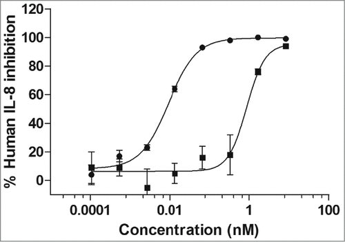
Table 2. Characterization of affinity-matured versions of SK48-E26
Figure 2. Design of human IL-1α/β dual variable domain immunoglobulins (DVD-Igs). (A) Representation of a DVD-Ig with an anti-IL-1α variable domain on the “outer” position linked to an anti-IL-1β variable domain in the “inner” position. (B) Representation of a DVD-Ig with an anti-IL-1β variable domain in the “outside” position linked to an anti-IL-1α variable domain in the “inner” position. Several DVD-Igs were constructed, expressed in HEK 293 cells, and purified from cell supernatants using Protein A affinity chromatography. The architecture of ABT-981 is based on design B.
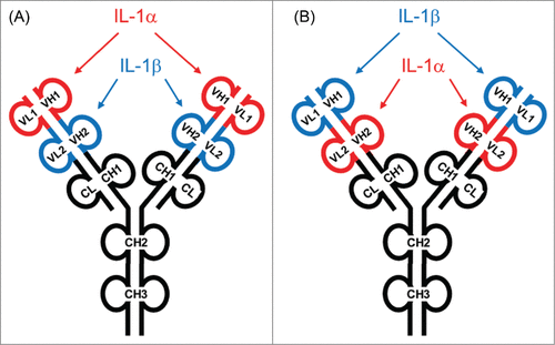
Table 3. Binding affinity and neutralization potency of IL-1α/β DVD-Igs to human and cynomolgus IL-1α and IL-1β
Table 4. Apparent stoichiometry for consecutive binding of IL‑1α and IL‑β to ABT-981
Figure 3. Consecutive binding of IL-1α and IL-1β to ABT-981. To generate this representative sensogram, ABT-981 was immobilized on a Biacore sensor chip via goat anti-human IgG and 500 nM human IL-1β was injected followed by injection of 500 nM human IL-1α. No dissociation time of the IL-1β was allowed prior to the injection of the IL-1α. The sensogram indicates that human IL-1β binds to ABT-981, and IL-1α variable domains are still accessible for binding to IL-1α. The reciprocal of this study (binding of IL-1α followed by IL-1β) showed identical results (data not shown). The Y-axis of the plot is response units and the X-axis is time in seconds.
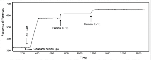
Table 5. Specificity of ABT-981 to human IL-1α and IL-1β
Table 6. ED50 and ED80 for ABT‑981 to neutralize human or cynomolgus IL‑1α or IL‑1β
Figure 4. In vivo neutralization of human and cynomolgus IL‑1α (left panel) and IL‑1β (right panel) with ABT‑981. A pharmacodynamic mouse model was established to demonstrate in vivo activity of ABT-981. Upon injection with 30 ng of either human (•) or cynomolgus (5) IL‑1α and IL‑1β, mouse IL-6 was measured in serum. The loss of IL-6 in serum was correlated to the dose of ABT-981 given to each animal and a percent inhibition by ABT-981 was calculated.
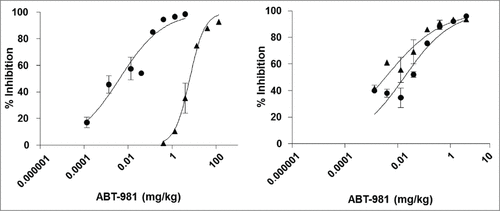
Table 7. Summary of main pharmacokinetic parameters of ABT-981 following a single intravenous or subcutaneous dose in BALB/c mouse, Sprague‑Dawley rat and cynomolgus monkey
Figure 5. (A) Mean (±SD) serum concentrations of ABT-981 following a 5 mg/kg intravenous () or subcutaneous () dose in male BALB/c mice. (B) Mean (±SD) serum concentrations of ABT-981 following a 4 mg/kg intravenous (![]()
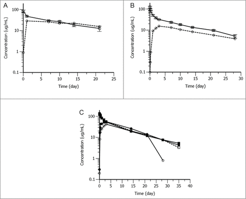
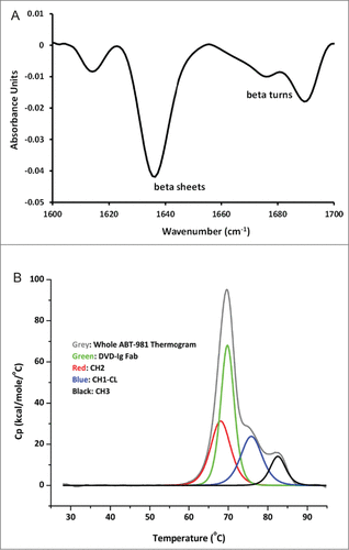
Figure 6. Biophysical characterization of ABT-981 in solution. (A) Fourier transform infrared spectroscopy chromatogram for secondary structure characterization indicating ABT-981 is comprised primarily of beta sheets supplemented by beta turns. (B) Differential scanning calorimetry thermogram following thermal stability analysis. The unfolding transitions for the individual domains of ABT-981 are assigned according to the legend.