
Abstract
The early detection of invading viruses by the host depends on their identification by pathogen sensors. These include Toll-like receptors (TLRs) as well as cytoplasmic RNA helicases such as retinoic acid inducible protein I (RIG-I) and melanoma differentiation associated gene 5 (MDA-5). These pathogen sensors recognize specific molecular patterns found in viruses and trigger inflammatory and antiviral responses that result in the eradication of invading pathogens. In this study we investigated the specific recognition of Human rhinovirus 6 (HRV6) the common cold pathogen by the innate immune response in lung epithelial cells. Our experiments established that in the first stages on infection the TLRs play a crucial role in HRV recognition and that different constituents of HRV6 are recognized by different TLRs, while upon viral replication and generation of dsRNA the type I IFN inflammatory response is mediated by MDA-5. The HRV6 capsid is recognized via TLR2, whereas upon HRV6 ssRNA internalization the virus genome is recognized by TLR7 and TLR8. Upon generation of dsRNA the type I IFN response is mediated by MDA-5. The combined recognition by different TLRs and MDA5 and their upregulation concurs with the huge inflammatory response seen in the common cold caused by human rhinoviruses.
Figures and Tables
Figure 1 TLR/helicase expression in human airway epithelial cells. Primary human bronchial cells were either not stimulated (0 h) or stimulated at different time points with HRV6 (1 × 10³ PFU/ml) (A) HRV6 ssRNA (B) or UV HRV6 (C). The cells were fixed and permeabilized, followed by antibody staining against the particular receptor molecule and incubation with the appropriate secondary antibody conjugated to FITC. Fluorescence was detected using a FACSC alibur (BectonDickinson). The data presented are the mean of three independent experiments.
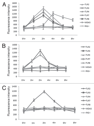
Figure 2 HRV6 activation of human airway epithelial cells. Human bronchial cells were either not stimulated (grey bar charts) or stimulated with HRV6 virions (1 × 10³ PFU/ml) (black bar charts), UV HRV6 (white bar charts) or HRV6 ssRNA (stripped bar charts) for different hours. The supernatants were harvested and assayed for cytokine secretion using the Cytometric Bead Array (CBA) system (Becton Dickinson). Fluorescence was detected using a FACSC alibur (BectonDickinson). IL6 secretion is depicted in graph (A) while IFNβ in graph (B). The data presented is the mean of three independent experiments. Asterisks denote statistical significance (p < 0.001).
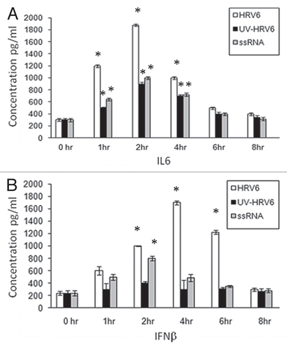
Figure 3 Inhibition of HRV6 activation of human airway cells by silencing TLRs MDA-5 and RIG-I. Following RNA interference (receptor expression levels were reduced by 80% by RNA interference A) human airway cells were either not stimulated (grey bar charts), incubated with HRV6 (black bar charts), incubated with UV-inactivated HRV6 (white bar charts) and HRV6 ssRNA (stripped bar charts). The supernatants were harvested and assayed for IL6 cytokine secretion in 2 h (B) and IFNβ secretion in 4 h (C) using the Cytometric Bead Array (CBA) system (Becton Dickinson). Fluorescence was detected using a FACSC alibur (BectonDickinson). The data represents the mean of three independent experiments. Asterisks denote statistical significance (p < 0.001).
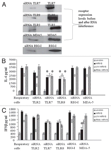
Figure 4 Receptor dependent activation in response to HRV6. HEK293 cells transfected with either TLR2, TLR3, TLR4, TLR7, TLR8, MDA-5 and RIG-I and a luciferase reporter gene were incubated with either no stimulus (grey bar charts), HRV6 virions (1 × 10³ PFU/ml) (black bar charts), UV-inactivated HRV6 (white bar charts), ssRNA HRV6 (stripped black charts), specific receptor activating ligands (black dot charts). After stimulation, the cells were lysed and analyzed for luciferase activity. The data shown represent a mean of three independent experiments. Asterisks denote statistical significance (p < 0.001).
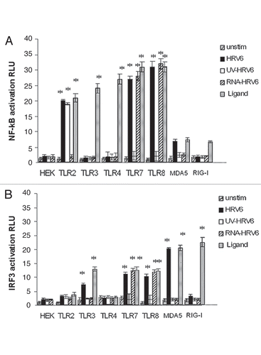
Figure 5 HRV6 recognition by TLR2. HEK293 cells transfected with TLR2 and a luciferase reporter gene were pre-incubated with TLR2-specific mAb (TLR2.1) prior to incubation with HRV6 (white bar charts) and UV HRV6 (black bar charts) (A). The cells were also incubated with an isotype control IgG2a from BD Biosciences (B). To determine NFκB activation the cells were lysed and analyzed for luciferase activity. The data shown represent a mean of three independent experiments. Asterisks denote statistical significance (p < 0.001).
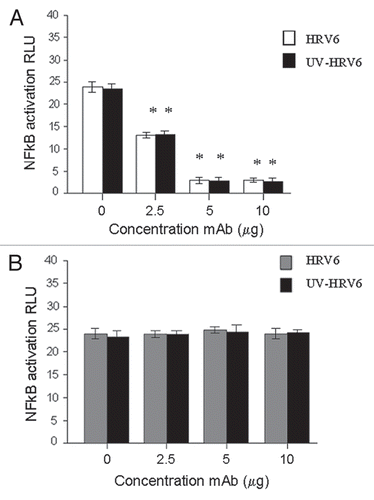
Figure 6 HRV6 ssRNA induced IL-6 secretion. HEK293 cells transfected with TLR7, TLR8 were either stimulated with HRV6 ssRNA (25 µg/ml) for 1 hour (stripped bar charts) or treated with 50 nM NH4Cl and then stimulated with HRV6 ssRNA (black bar charts). The supernatants were harvested and assayed for cytokine secretion using the Cytometric Bead Array (CBA) system (Becton Dickinson). Fluorescence was detected using a FACSCalibur (BectonDickinson). The data presented is the mean of three independent experiments. Asterisks denote statistical significance (p < 0.001).
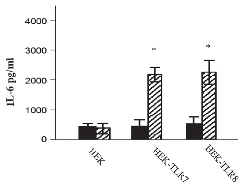
Table 1 Energy transfer efficiency values between donor-acceptor pairs