
Abstract
Background: Despite advances in traumatic wound care and management, infections remain a leading cause of mortality, morbidity, and economic disruption in millions of wound patients around the world. Animal models have become standard tools for studying a wide array of external traumatic wound infections and testing new antimicrobial strategies.
Methods: This review covers experimental infections in animal models of surgical wounds, skin abrasions, burns, lacerations, excisional wounds, and open fractures.
Results: Animal models of external traumatic wound infections reported by different investigators vary in animal species used, microorganism strains, the number of microorganisms applied, the size of the wounds, and, for burn infections, the length of time the heated object or liquid is in contact with the skin.
Conclusions: As antibiotic resistance continues to increase, more new antimicrobial approaches are urgently needed. These should be tested using standard protocols for infections in external traumatic wounds in animal models.
Acknowledgments
Research in the Hamblin laboratory is supported by the NIH (grant RO1AI050875) and the US Air Force MFEL program (contract FA9550-04-1-0079). T.D. was partially supported by a Bullock-Wellman Fellowship Award and an Airlift Research Foundation Extremity Trauma Research Grant (grant #109421).
Figures and Tables
Figure 1 Surgical site infections (SSIs) are classified as superficial incisional SSI, deep incisional SSI and organ/space SSIs.Citation26
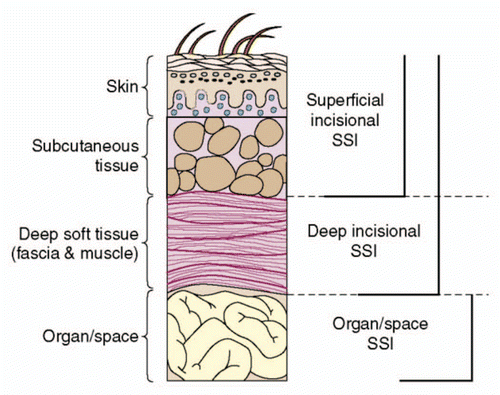
Figure 2 (A) Wound morphology at day 4 post-infection of a representative mouse wound. (B) A Gram-stained section of a mouse skin abrasion specimen showing the biofilms formed by Gram-positive MRSA near the skin surface. Dark blue area: biofilms of MRSA. The mouse skin abrasion specimen was harvested at day 3 post-infection.
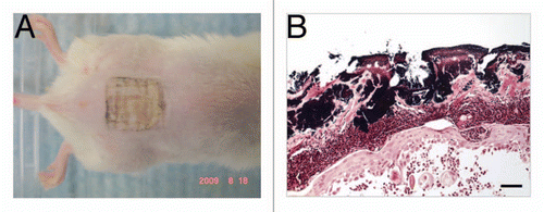
Figure 3 Full-thickness burn was created by applying two pre-heated brass blocks (92–95°C) to the opposing sides of an elevated skin fold on the back of a shaved mouse. Square: burned area. Bacteria were applied to surface of burn as a suspension in PBS.
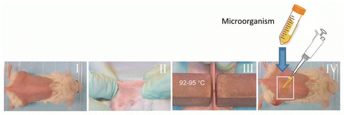
Figure 4 Schematic depiction of laceration wound.
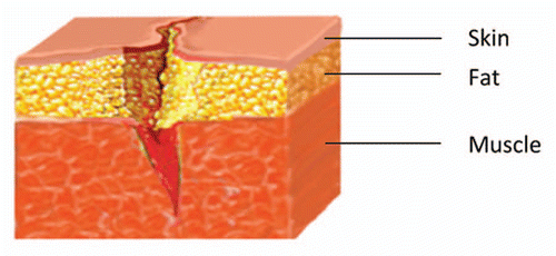
Figure 5 A complex wound involving injury to muscle, fascia, periosteum and bone.Citation126
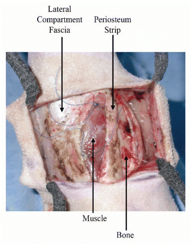
Table 1 Representative animal models of surgical site infections (SSIs)
Table 2 Representative animal models of skin abrasion wound infection
Table 3 Representative animal models of burn wound and infections
Table 4 Representative animal models of laceration wound infections
Table 5 Representative animal models of excisional wound infections
Table 6 Representative animal models of open fracture infections