Figures & data
Figure 1. The structure of the milk fat globule membrane (MFGM) (A). molecular structure of MFGM phospholipids (B).
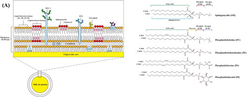
Table 1. Healthful properties of MFGM lipids, proteins, and carbohydrates.
Table 2. MFGM Brands and components on the market.
Figure 2. Isolation and purification methods of MFGM.
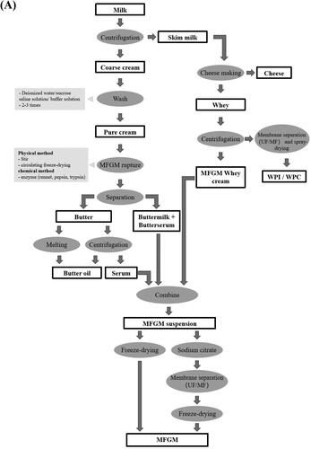
Figure 3. Liposomal structures for encapsulation of bioactive substances (A). TEM analysis of strains grown in AOAC medium (B). (a) L. rhamnosus GG wild-type (LGG WT); (b) welE mutant CMPG5351 (Lebeer et al. Citation2009). TEM analysis of LGG grown after 3 h of incubation (C). (a) in MRS broth; (b) MRS broth with 5 g/L MFGM-10; (c) 0.5% bile in MRS broth; (d) 0.5% bile in MRS broth with 5 g/L of MFGM-10 (Zhang et al. Citation2020). TEM analysis of the effect of MFGM phospholipids in the cell morphology (D). (b) and (d) have MFGM phospholipids adhered to L. delbrueckii and L. plantarum obviously (the arrows indicate accumulation of MFGM phospholipids on the cell wall) (Ortega-Anaya, Marciniak, and Jiménez-Flores Citation2021).
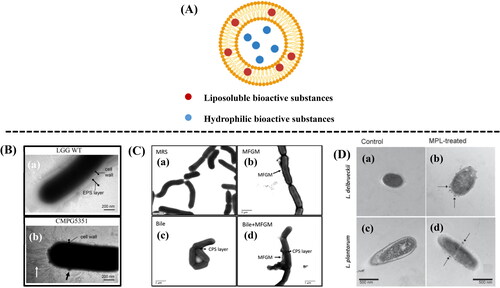
Table 3. Use of MFGM for encapsulation of bioactive substances.
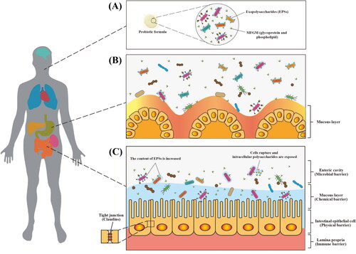
Figure 4. Schematic diagram of proposed gastrointestinal digestion of MFGM-encapsulated probiotics. Encapsulation of probiotics (e.g., LGG-welE, LGG-spa CBA, L. reuteri, L. delbrueckii, and L. plantarum) by MFGM (e.g., glycoprotein and phospholipid) (A). proposed protection mechanism of probiotics against gastric acid (B). proposed gastrointestinal digestion of MFGM-encapsulated probiotics (C).