Figures & data
Figure 1 Whole plant of M. maderaspatana. (Color figure available online.)
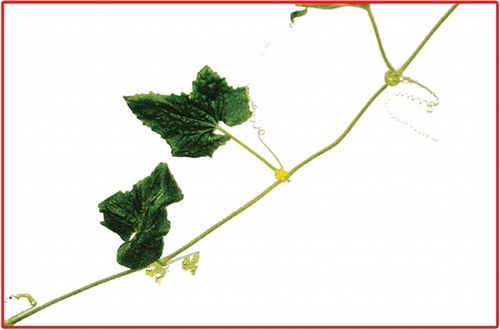
Figure 2 Transverse section of the basal region of petiole (a), schematic diagram of basal region of petiole (b), and isolated elements of the petiole (c). In Fig. 2a: Col: collenchyma; Cu: cuticle; Epi: epidermis; Par: parenchyma; Par.cort: parenchymatous cortex; Phl: phloem; Scl: sclerenchyma; Scl1: sclerenchymatous pericycle; Scl2: cortical parenchyma tendency of thickening; Tri: trichome; Xv: xylem vessel; Xyl: xylem. In Fig. 2c: a–b: xylem vessels; c–e: fibres; f: trichome; g: parenchyma.
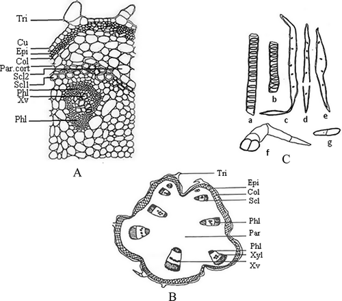
Figure 3 Transverse section of midrib region of leaf (a), schematic diagram of tendril (b), and trichomes and xylem vessel in isolated elements of leaf (c). In Fig. 3a: A1: midrib and part of lamina; A2: lamina; A3: margin of lamina; Col: collenchyma; Epi: epidermis; L.Epi: lower epidermis; Pal: palisade cells; Par: parenchyma; Phl: phloem; Pi: pith; S.P: spongy parenchyma; Scl: sclerenchyma; St: stomata; Tri: trichome; U.Epi: upper epidermis; V: vessel; Xyl: xylem. In Fig. 3c: a–c: trichomes of basal region of midrib; d–e: trichomes of middle region of midrib; f–h: trichomes of apical region of midrib; i–j: trichomes and xylem vessel in isolated elements.
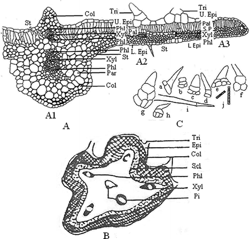
Table 1 Numerical values of the leaf of M. maderaspatana
Figure 4 Transverse section of mature stem (a) and isolated elements of mature stem (b). In Fig. 3b: Camb: cambium; C.c: cork cambium; Col: collenchyma; M.r: medullary rays; Par: parenchyma; Par.scl: parenchymatous sclerenchyma; Sec.cort: secondary cortex; Xv: xylem vessel; Xyl: xylem. In Fig. 3b: a–d: xylem vessels; e–h: fibres; i–j: tracheids; k: xylem parenchyma; l: trichome.
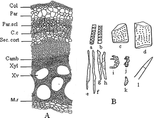
Figure 5 Transverse section of the mature root (a) and isolated elements of the mature root (b). In Fig. 4a: Ck: cork; Phl: phloem; M.r: medullary rays; Sec.cort: secondary cortex; S.g: starch grain; Xv: xylem vessel; Xyl: xylem. In Fig. 4b: a: part of cortex; b: xylem parenchyma; c: cork fragments; d–h: fibres; i–j: stone cells; k–m: tracheids; n–o: xylem vessels; p: starch grain.
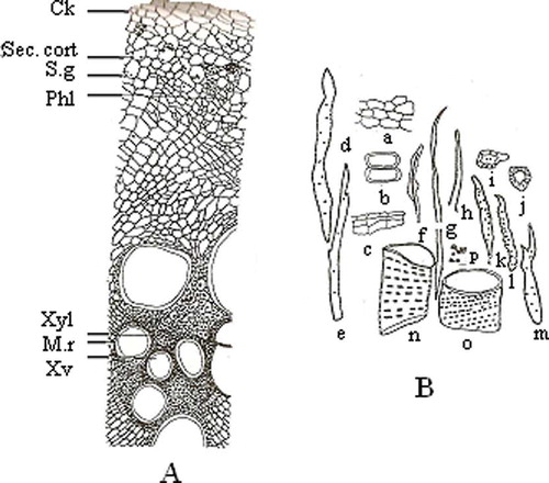
Table 2 Physicochemical standards
Table 3 Fluorescence characteristics
Table 4 Preliminary phytochemical screening
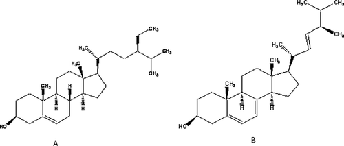
Figure 6 Structures of (a) β–sitosterol and (b) ergosterol.