Figures & data
FIGURE 1 SEM. Surface of the Prunus fruits; A, D, G, J—“Bluefre;” B, E, H, K—“Sweet Common Prune;” C, F, I, L—“President.” Visible microwrinkles (A, C) and undulations (B) centered around the stomata. D–F—visible wax platelets (E; arrow heads) and stomata (D–F; arrows) surrounded by wrinkles. G–L—visible crystalline wax in the form of vertical plates (arrows; G, H, J, K) and microgranules (I–L), and numerous microcracks in the wax layer (H, K), asterisks—wax microgranules in high density.
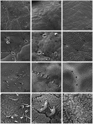
FIGURE 2 SEM. Surface of the Prunus fruits with microcracks and stomata; A–C—“Bluefre;” D–F—“Sweet Common Prune;” G–I—“President.” F, L—LM. Note stomata with wax microgranules visible on the cell surface and in the interior of stomata (A, D, G), and stomata with microcracks (arrows; B, C, E, H, I). F—A microcrack (arrows) in the skin of “Sweet Common Prune.”
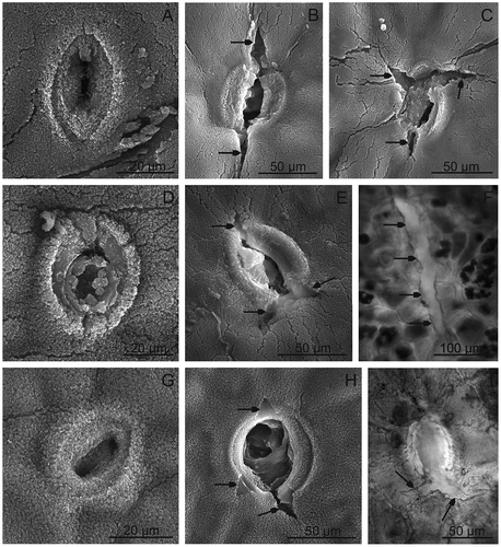
TABLE 2 Fruit skin characteristics and antioxidant contents of Prunus fruits
FIGURE 3 The cuticle on the “President” fruit surface. A—visible a brightly shining layer of the cuticle observed under LM. B–D—The cuticle observed under TEM. B, C—Note two layers of the cuticle: brighter—cuticle proper and darker—cuticular layer and the border of the occurrence of the epicuticular wax layer (arrow heads). D—Visible cuticle penetrating between the cells of the epidermis (asterisk). c: cuticle, cp: cuticle proper, cl: cuticular layer, cw: cell wall, e: epidermis, n: nucleus, v: vacuole.
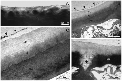
FIGURE 4 LM. Fragments of sections through the fruit surface layer; A—‘”Bluefre,” B—“Sweet Common Prune,” C—“President.” Visible chloroplasts in many hypodermal and parenchymal cells (arrows). e: epidermis, h: hypodermis, p: parenchyma.
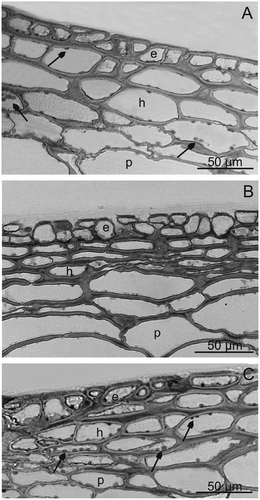
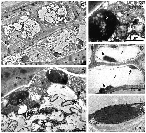
FIGURE 5 TEM. Skin and parenchymal cells of “President” fruits; A—epidermis and hypodermis cells with nuclei, plastids with starch grains and numerous vacuoles with fibrous and granular deposits. B, C—fragments of epidermis cells with plastids containing starch grains, mitochondria, and vacuoles with membranes forming compartments of various shapes (asterisks). D, E—chloroplasts (arrows) with parenchymal cells. c: cuticle, cw: cell wall, n: nucleus, p: plastid with starch grains, m: mitochondrion, v: vacuole.