Figures & data
Figure 1. Solubility of AMP in distilled water or buffer solution at various pH values.
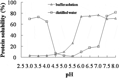
Figure 2. SDS-PAGE of soluble protein fractions of AMP at various pH values. M: Molecular weight marker; PM: Paramyosin; T: Tropomyosin; MHC: Myosin heavy chains; A: Actin, MLC: Myosin light chains.
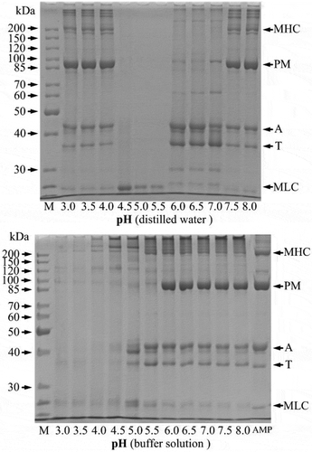
Figure 3. Effect of pH on the breaking force (a), deformation (b) of AMP gels. Error bars represent the standard deviation of triplicate determinations, and different letters on the top of each column indicated significant difference (p < 0.05).
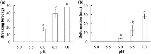
Figure 4. Water holding capacity of AMP gels at various pH values. Error bars represent the standard deviation of triplicate determinations, and different letters on the top of each column indicated significant difference (p < 0.05).
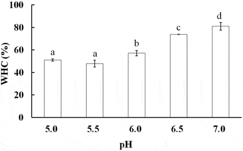
Figure 5. Scanning electron micrographs of AMP gels at various pH values (5000 × magnifications).
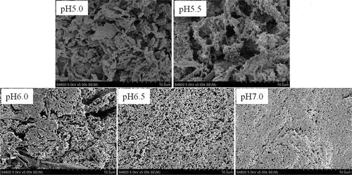
Figure 6. Differential scanning calorimetry thermograms of AMP gels at various pH values.
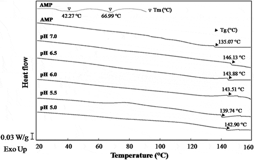
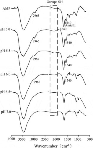
Figure 7. Fourier transform infrared spectroscopy of AMP gels at various pH values.