Figures & data
Table 1. Estimated secondary structures of yogurt antioxidant peptides.
Figure 1. Antioxidant capacity of yogurt antioxidant peptides under the following treatments: (A) pH6, pH7, and pH8; (B) heat treat with 38°C, 50 oC, and 65 oC; (C) urea on the concentration of 1mol/L, 3mol/L,5mol/L,7mol/L, and 8mol/L; (D) trifluoroethanol on the concentration of 10%, 30%, and 50% (v/v). Different label letters indicate significant differences among samples at a significant level of 0.05, and the same label alphabet means insignificant.
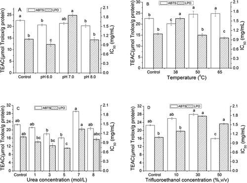
Figure 2. Fluorescence spectrum of yogurt antioxidant peptides under the following treatments: (A) pH6, pH7, and pH8; (B) heat treat with 38°C, 50 oC, and 65 oC; (C) urea on the concentration of 1mol/L, 3mol/L,5mol/L,7mol/L, and 8mol/L; (D) trifluoroethanol on the concentration of 10%, 30%, and 50% (v/v). Different label letters indicate significant differences among samples at a significant level of 0.05, and the same label alphabet means insignificant.
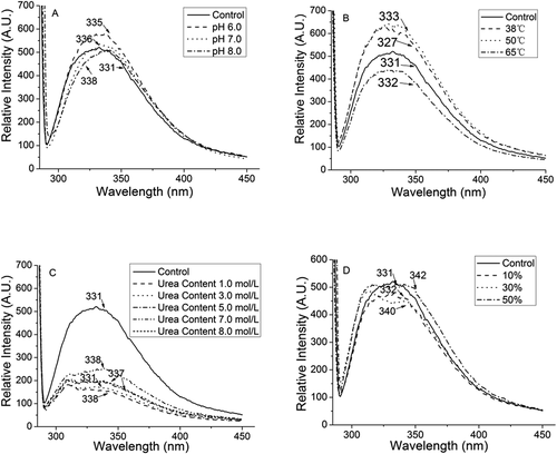
Figure 3. FTIR spectrum of yogurt antioxidant peptides under the following treatments: (A) pH6, pH7, and pH8; (B) heat treat with 38°C, 50 oC, and 65 oC; (C) urea on the concentration of 1mol/L, 3mol/L,5mol/L,7mol/L, and 8mol/L; (D) trifluoroethanol on the concentration of 10%, 30%, and 50% (v/v). Different label letters indicate significant differences among samples at a significant level of 0.05, and the same label alphabet means insignificant.
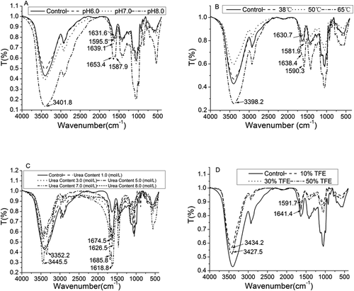
Figure 4. Spectrum of amide I (1700–1600 cm−1) in of yogurt antioxidant peptides: (A) Control, (B) pH 6.0 yogurt peptide, (C) pH 7.0 yogurt peptide, (D) pH 8.0 yogurt peptide, (E) 38°C treated yogurt peptide, (F) 50°C treated yogurt peptide, (G)65°C treated yogurt peptide, (H) 1.0 mol/L urea treated yogurt peptide, (I) 3.0 mol/L urea treated yogurt peptide, (J) 5.0 mol/L urea treated yogurt peptide, (K) 7.0 mol/L urea treated yogurt peptide, (L) 8.0 mol/L urea treated yogurt peptide, (M) 10% (v/v) trifluoroethanol treated yogurt peptide, (N) 30% (v/v) trifluoroethanol treated yogurt peptide, and (O) 50% (v/v) trifluoroethanol treated yogurt peptide. Different label letters indicate significant differences among samples at a significant level of 0.05, and the same label alphabet means insignificant.
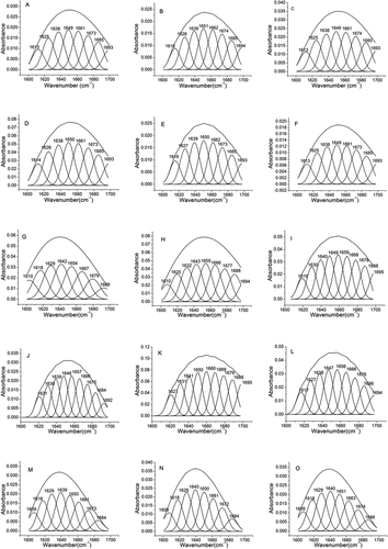
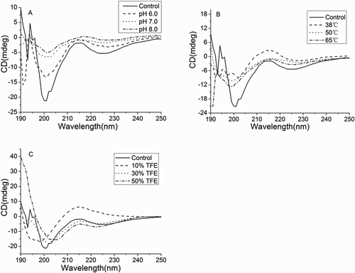
Figure 5. Circular dichromatic spectrogram of yogurt antioxidant peptides under the following treatments: (A) pH6, pH7, and pH8; (B) heat treat with 38°C, 50 oC, and 65 oC; (C) urea on the concentration of 1mol/L, 3mol/L,5mol/L,7mol/L, and 8mol/L; (D) trifluoroethanol on the concentration of 10%, 30%, and 50% (v/v). Different label letters indicate significant differences among samples at a significant level of 0.05, and the same label alphabet means insignificant.