Figures & data
Figure 1. Inhibition halos of the growth of L. monocytogenes in BHI agar during the quantification of bacteriocin activity by the critical dilution method. (A) Antilisterial activity of the antimicrobial peptide (AM) produced by S. infantarius, 21 to 212 were the dilutions examined. (B) Antilisterial activity of nisin, 21 to 28 were the dilutions examined.
Figura 1. Halos de inhibición del crecimiento de L. monocytogenes en agar BHI durante la cuantificación de la actividad de bacteriocinas por el método de dilución crítica. (A) Actividad antilisterial del péptido antimicrobiano (AM) producido por S. infantarius, 20 a 212 fueron las diluciones examinadas. (B) Actividad antilisterial de nisina, 20 a 28 fueron las diluciones examinadas.
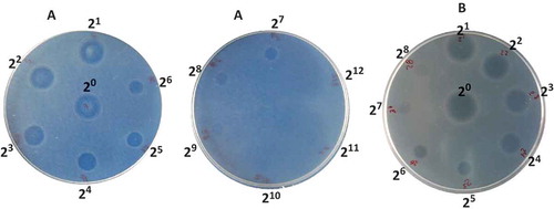
Figure 2. Growth evolution of L. monocytogenes in Oxford agar plates without films (white circles) and with: C-AM film (grey squares); C-AM-control film (white squares); C-Nis film; (grey triangles); C-Nis-control film (white triangles). Plates incubated at 35°C and 32% RH.
Figura 2. Evolución del crecimiento de L. monocytogenes en placas de agar Oxford sin película (círculos blancos) y con: películas C-AM (cuadros grises); película C-AM-control (cuadros blancos); película C-Nis (triángulos grises) y película C-Nis-control (triángulos blancos). Placas incubadas a 35°C y 32% HR.
Figure 3. Growth evolution of L. monocytogenes in Oxford agar plates without films (white circles) and with: C-AM film (grey squares); C-AM-control film (white squares); C-Nis film; (grey triangles); C-Nis-control film (white triangles). Plates incubated at 4°C and 98% RH.
Figura 3. Evolución del crecimiento de L. monocytogenes en placas de agar Oxford sin película (círculos blancos) y con: películas C-AM (cuadros grises); película C-AM-control (cuadros blancos); película C-Nis (triángulos grises) y película C-Nis-control (triángulos blancos). Placas incubadas a 4°C y 98% HR.
Table 1. Physical properties of caseinate films with antimicrobial agents from S. infantarius isolated from pozol (AM) and nisin (Nis).
Tabla 1. Propiedades físicas de películas de caseinato con agentes antimicrobianos de S. infantarius aislado del Pozol (AM) y nisina (Nis).
Figure 4. Typical true stress (σT) vs. Hencky strain (ЄH) curves obtained in tensile test carried out on some representative caseinate films at 25°C. Film containing antimicrobial agent from S. infantarius (C-AM); control film of C-AM (C-AM-control); film with nisin (C-Nis) and control film of C-Nis (C-Nis-control).
Figura 4. Curvas típicas de esfuerzo verdadero (σT) vs. deformación de Hencky (εH) obtenidas en pruebas de tensión realizadas a 25°C en algunas películas representativas de caseinato. Películas con agente antimicrobiano de S. infantarius (C-AM); película control de C-AM (C-AM-control); película con nisina (C-Nis) y película control de C-Nis (C-Nis-control).