Figures & data
Figure 1. Integument properties and candidate gene expression of Galleria mellonella morphs. Appearance (a), cuticle thickness (n = 170, *** = p < 0.001), and patterns of melanin deposition (c, d) of melanic and non-melanic morphs. Enhanced expression (mRNA levels) of genes encoding immune factors (gloverin, gallerimycin, apolipophorin III, IMPI, DDC, PPO), stress management (HSP90), detoxification, and cell proliferation of naive melanic (M) larvae compared to non-melanic (NM) larvae (e).
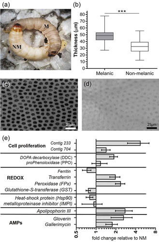
Figure 2. Biochemical profiles of Galleria mellonella morphs. Quantities and compositions of epicuticular hydrocarbons (a) and fatty acids (b) from un-stimulated (i.e., naive) melanic and non-melanic wax moth larvae. Unpaired t-tests were used to assess differences between each insect morph (* = p < 0.05).
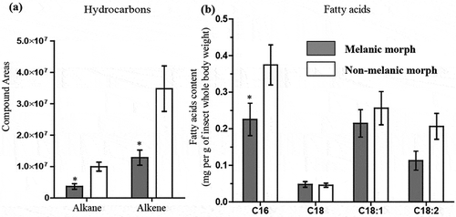
Figure 3. Development of mycosis (Metarhizium brunneum) in melanic (M) and non-melanic (NM) morphs of Galleria mellonella larvae after topical inoculation. The survival of each insect morph was recorded over 12 days following exposure to two doses of M. brunneum conidia. LС30 values demonstrate similar mortality levels of M and NM larvae in the presence of 107 conidia (p < 0.01, **) and 105 conidia (p < 0.001, ***), respectively (n = 60). The numbers of conidia per 0.25 mm2 cuticle were counted on M and NM morphs during infection with M. brunneum (107) (b), and the percentage of germinating conidia (c) that successfully adhered (*** = p < 0.001; n = 22). Penetration of the integument by germinating conidia led to the formation of melanotic lesions (white arrows) on NM larvae – representing de novo synthesis of melanin (d). Conversely, the pattern of melanin deposition on M larvae remained unchanged (e), indicating the melanic-defenses are preformed (images were taken 24 h.p.i. with 107 conidia).
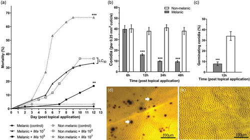
Figure 4. Gene expression (mRNA levels) of Galleria mellonella integumental tissues exposed to Metarhizium brunneum conidia (LС30) relative to uninfected (control) insects. Insect metalloproteinase inhibitor (IMPI), heat-shock protein (HSP 90), DOPA decarboxylase (DDC), prophenoloxidase (PPO), antifungal peptide gallerimycin, antibacterial peptide gloverin, β-glucan binding protein apolipophorin III, growth factors contig 233 (growth-blocking peptide) and contig 704 (pleiotrophin-like protein). (* = p < 0.05; melanic versus non-melanic at the respective time point).
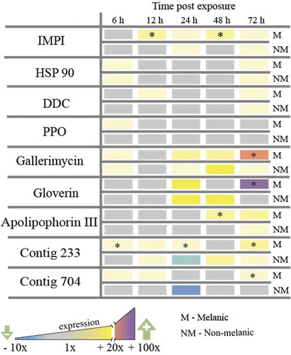
Figure 5. Detoxification-associated gene expression in Galleria mellonella morphs exposed to Metarhizium brunneum. Antioxidant enzymes, peroxidase (FPx) (a), glutathione-S-transferase (GST) (b) and transferrin, (c) in the integuments of M and NM larvae at 72 h post fungal infection. The asterisk [*] indicates a significant difference (p < 0.05) between NM (107) and M (105). The hashtag [#] indicates a significant difference (p < 0.01) between NM (107) and NM (105).
![Figure 5. Detoxification-associated gene expression in Galleria mellonella morphs exposed to Metarhizium brunneum. Antioxidant enzymes, peroxidase (FPx) (a), glutathione-S-transferase (GST) (b) and transferrin, (c) in the integuments of M and NM larvae at 72 h post fungal infection. The asterisk [*] indicates a significant difference (p < 0.05) between NM (107) and M (105). The hashtag [#] indicates a significant difference (p < 0.01) between NM (107) and NM (105).](/cms/asset/1aa4bddc-c3c4-422f-99ba-49593fd76cd6/kvir_a_1693230_f0005_b.gif)
Figure 6. Gene expression (mRNA levels) of Metarhizium brunneum during the infection cycle on melanic and non-melanic morphs of Galleria mellonella. Scheme representing the broad stages of host colonization (a). Subtilisin-like proteases (Pr1a, Pr1b, Pr2), heat-shock proteins (HSP30, HSP70), adhesin-like proteins (MAD1 and MAD2), conidiation – associated gene (cag 8) and nitrogen regulator response (nrr) expression on the insect integuments from 6 to 72-h post-exposure.
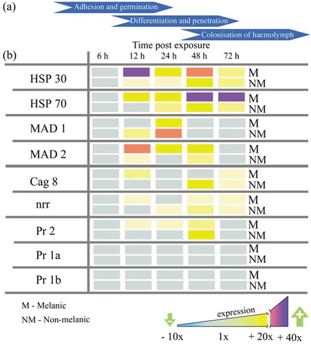
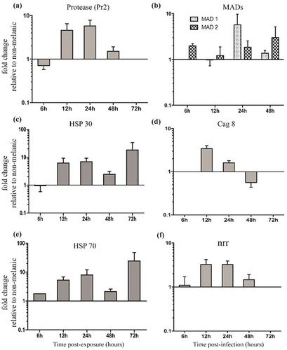
Figure 7. Relative expression patterns of Metarhizium brunneum genes on the integuments of melanic larvae compared directly to non-melanic larvae. Fungal protease Pr2 (a), adhesin-like proteins (MAD1 and MAD2) (b), heat-shock proteins 30/70 (HSP30/70, c/e), conidiation-associated gene (cag 8, d) and nitrogen regulator response (nrr, f).