
Figures & data
Figure 1. The sequence analysis of aad riboswitch RNAs
(a) The conserved 5′ conserved segment of class 1 integrons. The strong promoter (Pc) [Citation9] transcribes the inserted gene cassette (i.e., aad resistance gene). While the divergent promoter Pint transcribes the integrase (int) gene. Single gene cassettes are inserted by int-mediated recombination between int recognition sites. The int expression is linked to the SOS response that can only be induced by non-aminoglycoside antibiotics in E. coli. (b) The leader aad riboswitch RNAs from different pathogens. The RNA is located between the divergent int and aad genes at the conserved 5′ end of the integron. (c) The blast search results of the aac/aad riboswitch sequence from P. fluorescens. A total of the 141 of the top 150 BLAST sequences were annotated to identify the neighboring genes. The majority of neighboring resistance genes encoded AAD (n = 97) and AAC (n = 41). One non-aminoglycoside resistance gene dihydrofolate reductase (dhfR, 1) and a nonfunctional multidrug exporter (qacH, 2) were also noted.
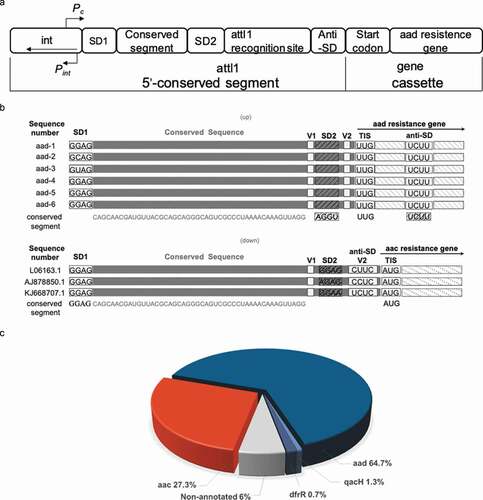
Figure 2. Reporter gene expression controlled by aad riboswitches
(a) Agar diffusion assay of E.coli transformed with aad-4 reporter construct plasmid with aminoglycoside antibiotics in the presence of IPTG. (b) Agar diffusion assay of the reporter construct plasmid with aminoglycoside antibiotics for the aad-4 A18G point mutation in the presence of IPTG. (c) IPTG control agar diffusion assay of the aad-4 reporter construct plasmid with aminoglycoside antibiotics in the absence of IPTG. (d) Control RNA agar diffusion assay of the NarJ reporter construct plasmid with aminoglycoside antibiotics in the presence of IPTG. (e) The highest induction fold change for the aad riboswitch constructs (aad 1–6) in the presence of amikacin. The aac-siso* refers to the highest induction fold of the P. fluorescens riboswitch RNA under sisomicin [Citation35]. (f) The β-gal activity (Miller units) of the reporter gene with aad-4 riboswitch sequence upon titration of aminoglycoside antibiotics. (g) Comparison of the fold change in β-gal activity of the wild-type aad-4 sequence with the inactive A18G point mutation on titration of amikacin to 5 µM. NB for all β-gal activity measurements, error bars are standard deviations of at least three independent experiments.
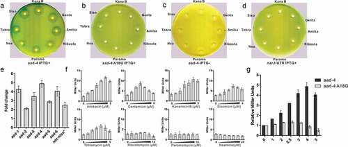
Figure 3. The binding affinity of aad-4 riboswitch RNA with aminoglycoside antibiotics
(a) The 4–6 deoxystreptamine aminoglycoside Amikacin. (b) Binding curve generated by MST for binding of amikacin to the aad-4 riboswitch. Error bars are standard deviations of at least three independent experiments. (c) Binding affinity of aad-4 riboswitch RNA with different aminoglycoside antibiotics measured by MST. Aminoglycoside dependent induction of gene expression for aad-4 (). Error bars are the standard deviations of at least three independent experiments.
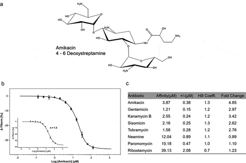
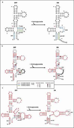
Figure 4. The structure transition model of aac riboswitch published and consensus model of aac/aad riboswitch in the presence or absence of aminoglycosides
(a) The secondary-structure of the aac riboswitch from P. fluorescens in the paper published by Jia et al [Citation35]. The red arrow indicates the position of the inactive A18G point mutation. (b) The covariance model of aac riboswitch in the paper published by Wang et al (36). Red, black and gray nucleotides indicate the conservation of at least 97%, 90%, and 75%, respectively. The circles with red, gray, white color represent the positions in which nucleotide identity is less conserved as 97%, 75% and 50%, respectively. Base pairs shaded in red exhibit natural covariation, while those in blue exhibit compatible mutations. Purines and pyrimidines are identified by R and Y. The red arrow indicates the position of the inactive A18G point mutation. (c) The covariance model of the aad riboswitch RNA. Red, black and gray nucleotides indicate the conservation of at least 97%, 90%, and 75%, respectively. The circles with red, gray and white color represent the positions in which nucleotide identity is less conserved as 97%, 75% and 50%, respectively. Base pairs shaded in red exhibit natural covariation, while those in blue exhibit compatible mutations. Purines and pyrimidines are identified by R and Y. The red arrow indicates the position of the inactive A18G point mutation. The covariant structures were drawn using R2R and Adobe Illustrator.
Supplemental material