17,839
Views
45
CrossRef citations to date
0
Altmetric
Review Article

Pathogenicity and virulence of Clostridium perfringens

, , , , &
Pages 723-753 | Received 01 Sep 2020, Accepted 29 Jan 2021, Published online: 12 Apr 2021

Figures & data

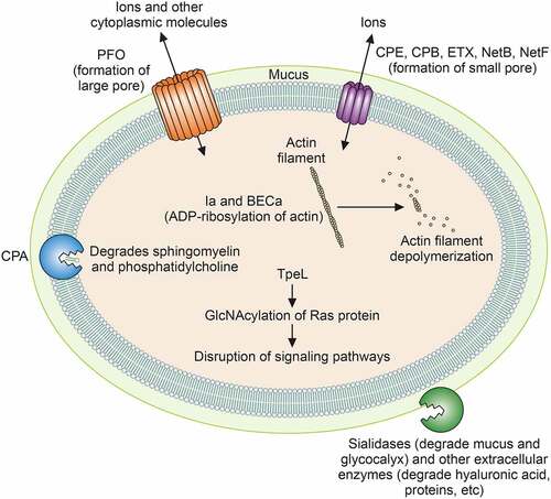
Table 1. Characteristics of toxins and extracellular degradative enzymes produced by C. perfringens

Table 2. Current C. perfringens toxinotyping scheme

Table 3. C. perfringens toxinotype: disease associations

Figure 1. Actions of C. perfringens toxins and degradative enzymes. The cellular sites of action and mechanisms of action of major toxins and sialidases are depicted. See text for details

Figure 1. Actions of C. perfringens toxins and degradative enzymes. The cellular sites of action and mechanisms of action of major toxins and sialidases are depicted. See text for details

Figure 2. Diagrams of the major known plasmid families of C. perfringens. Orange depicts the replication (rep) region, green depicts the conjugative transfer regions in pCW3-like plasmids and pCP13-like plasmids and yellow depicts the regions carrying variable genes encoding toxins, antimicrobial resistance (AMR) factors or bacteriocins. See text for further details

Figure 2. Diagrams of the major known plasmid families of C. perfringens. Orange depicts the replication (rep) region, green depicts the conjugative transfer regions in pCW3-like plasmids and pCP13-like plasmids and yellow depicts the regions carrying variable genes encoding toxins, antimicrobial resistance (AMR) factors or bacteriocins. See text for further details

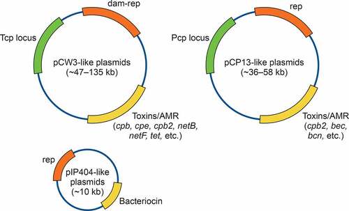
Table 4. Size and diversity C. perfringens plasmids encoding key-toxins1.

Figure 3. Current model for cross-talk between the VirS/VirR two-component regulatory system and the Agr-like quorum sensing system in C. perfringens. The AgrD peptide is processed by AgrB (and perhaps other unidentified factors) to form a cyclic autoinducing signaling peptide (AIP). AIP then binds to VirS, which in turn phosphorylates (p) VirR. The phosphorylated VirR protein then binds to VirR boxes upstream of some toxins genes (e.g. the pfoA gene) and upstream of the vrr gene encoding VR-RNA. VR-RNA then leads to increased transcription of genes encoding toxins such as CPA. This reults in increased production of those toxins. Based upon [Citation162,Citation308]

Figure 3. Current model for cross-talk between the VirS/VirR two-component regulatory system and the Agr-like quorum sensing system in C. perfringens. The AgrD peptide is processed by AgrB (and perhaps other unidentified factors) to form a cyclic autoinducing signaling peptide (AIP). AIP then binds to VirS, which in turn phosphorylates (p) VirR. The phosphorylated VirR protein then binds to VirR boxes upstream of some toxins genes (e.g. the pfoA gene) and upstream of the vrr gene encoding VR-RNA. VR-RNA then leads to increased transcription of genes encoding toxins such as CPA. This reults in increased production of those toxins. Based upon [Citation162,Citation308]

Figure 4. Gas gangrene by C. perfringens type A in an experimentally infected mouse. This animal was challenged with 109 washed vegetative cells of C. perfringens strain 13 and euthanized 4 h later. There is severe skeletal muscle necrosis (asterisks) and myriad intralesional bacilli (arrows and insert). Notice that there is minimal inflammatory infiltrate. Hematoxylin and eosin. Scale bar = 50 μm

Figure 4. Gas gangrene by C. perfringens type A in an experimentally infected mouse. This animal was challenged with 109 washed vegetative cells of C. perfringens strain 13 and euthanized 4 h later. There is severe skeletal muscle necrosis (asterisks) and myriad intralesional bacilli (arrows and insert). Notice that there is minimal inflammatory infiltrate. Hematoxylin and eosin. Scale bar = 50 μm

Figure 5. Naturally-acquired necrotic enteritis caused by C. perfringens type C in a neonatal piglet. A. Diffuse necrosis of mucosa (**), which is covered by a pseudomembrane (*) that is composed mostly by fibrin, cell debris and inflammatory cells. These effects are a consequence of CPB (see text). The intestinal lumen is indicated (L). B. This higher magnification of image A shows thrombosis of mucosal vessels (↗) and myriad neutrophils admixed with fibrin and cell debris forming a pseudomembrane (*) on the surface of the necrotic mucosa (**). Scale bar=50 μm. Hematoxylin and eosin

Figure 5. Naturally-acquired necrotic enteritis caused by C. perfringens type C in a neonatal piglet. A. Diffuse necrosis of mucosa (**), which is covered by a pseudomembrane (*) that is composed mostly by fibrin, cell debris and inflammatory cells. These effects are a consequence of CPB (see text). The intestinal lumen is indicated (L). B. This higher magnification of image A shows thrombosis of mucosal vessels (↗) and myriad neutrophils admixed with fibrin and cell debris forming a pseudomembrane (*) on the surface of the necrotic mucosa (**). Scale bar=50 μm. Hematoxylin and eosin

Figure 6. Perivascular proteinaceous edema (PVE) in the cerebellar white matter of a sheep experimentally infected with C. perfringens type D. This lesion is a consequence of the action of epsilon toxin on the vascular endothelial cells, which increases vascular permeability allowing albumin and water to leave the vascular lumen. An arteriole (solid arrow) and two venules (hollow arrows) are indicated. Scale bar = 50 μm. Hematoxylin and eosin

Figure 6. Perivascular proteinaceous edema (PVE) in the cerebellar white matter of a sheep experimentally infected with C. perfringens type D. This lesion is a consequence of the action of epsilon toxin on the vascular endothelial cells, which increases vascular permeability allowing albumin and water to leave the vascular lumen. An arteriole (solid arrow) and two venules (hollow arrows) are indicated. Scale bar = 50 μm. Hematoxylin and eosin

Figure 7. Small intestine of a chicken with naturally acquired necrotic enteritis produced by C. perfringens type G. Notice the necrotic and denudated villi (v), lined by large number of bacilli (arrow heads). A wide band of heterophils (arrows) separates the superficial necrotic villi from the more normal looking deep part of the tissue (bottom of the photograph). Scale bar = 100 μm. Hematoxylin and eosin

Figure 7. Small intestine of a chicken with naturally acquired necrotic enteritis produced by C. perfringens type G. Notice the necrotic and denudated villi (v), lined by large number of bacilli (arrow heads). A wide band of heterophils (arrows) separates the superficial necrotic villi from the more normal looking deep part of the tissue (bottom of the photograph). Scale bar = 100 μm. Hematoxylin and eosin

Figure 8. Effect of C. perfringens type F enterotoxin (CPE) on the small intestinal loops of a rabbit. A. CPE (50 micrograms) was injected into the lumen of a ligated small intestinal loop of an anesthetized rabbit. The animal was kept under anesthesia during 6 h after which it was euthanized and the intestinal loop was collected and processed for histology. There is almost complete loss of the mucosa (asterisk); no villi are observed. Scale bar = 100 μm. B. Normal control shown for comparison. This ligated small intestinal loop of the same rabbit was inoculated with buffer instead of CPE. Notice the intact mucosa with long villi (v). Scale bar = 70 μm. Hematoxylin and eosin

Figure 8. Effect of C. perfringens type F enterotoxin (CPE) on the small intestinal loops of a rabbit. A. CPE (50 micrograms) was injected into the lumen of a ligated small intestinal loop of an anesthetized rabbit. The animal was kept under anesthesia during 6 h after which it was euthanized and the intestinal loop was collected and processed for histology. There is almost complete loss of the mucosa (asterisk); no villi are observed. Scale bar = 100 μm. B. Normal control shown for comparison. This ligated small intestinal loop of the same rabbit was inoculated with buffer instead of CPE. Notice the intact mucosa with long villi (v). Scale bar = 70 μm. Hematoxylin and eosin