Figures & data
Figure 1. Overexpression of YPT6 rescues the hyphal invasive growth defect in an arl1 deletion mutant. (A-D) Overexpression of YPT6 specifically rescues invasive growth in arl1/arl1 cells. Indicated strains were grown on agar-containing YEPD with 50% FCS (A, C, D) or on Spider media (B) and images were taken after 5–6 days. Similar results were observed in 2 independent experiments. (C) Rescue of invasive growth in arl1/arl1 cells depends on Ypt6 activity. Indicated strains were grown on agar-containing YEPD with FCS as in A. (D) Overexpression of ARL1 does not rescue invasive growth in ypt6/ypt6 cells. Indicated strains were grown on agar-containing YEPD with FCS as in A. (E) Overexpression of YPT6 partially rescues arl1/arl1 hyphal length defect. Cells from the indicated strains were incubated with FCS for 90 min and the graph shows the average hyphal length (mean of 200–400 cells each strain, from 3 experiments); error bars indicate SD. Student t test was used to calculate the p values: arl1 + ARL1 vs arl1: 0.0028 and arl1 + YPT6 vs arl1: 0.0042. (F) YPT6 and ARL1 transcripts in the overexpression mutants. mRNA and cDNA were prepared from the indicated strains and the transcripts were quantified by RT-PCR; actin (ACT1) was used for normalization. (G) Overexpression of YPT6 does not rescue cell wall defects in arl1/arl1 cells. Serial dilutions of the indicated strains were spotted on YEPD media (Ctrl) containing 400 μg/ml Congo red (CR) or 1 mg/ml hygromycin B (HygB). Images were taken after 2 days.
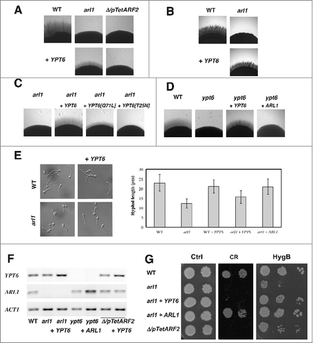
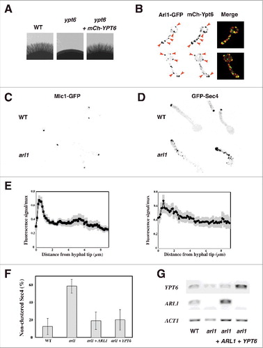
Figure 2. Overexpression of YPT6 rescues the altered Sec4 distribution in the arl1 deletion mutant. (A-B) Arl1 and Ypt6 partially co-localize in hyphae. The mCh-Ypt6 fusion is functional (A) Cells from the indicated strains were incubated on Spider media and images were taken after 5 days. Similar results were observed in 2 independent experiments. Maximum projections of 21 deconvolved z-sections of representative cells expressing Arl1-GFP together with mCh-Ypt6 after 90 min FCS-induced hyphal growth; GFP and mCherry signals were acquired simultaneously. (B) (C-E) Sec4, but not Mlc1, distribution is altered in arl1/arl1 cells. WT and arl1/arl1 cells, expressing Mlc1-GFP (C) or GFP-Sec4 (D), were incubated with FCS for 45 and 90 min, respectively. Representative images are shown. Cell fluorescence concentration profiles were analyzed with the HyphalPolarity program,Citation47 to quantify the GFP-Sec4 signal concentration along the major axis of hyphal cells, from sum projection images generated with Image J (E). (F) Overexpression of YPT6 restores Sec4 clustering in arl1/arl1 cells. The graph shows averages of 3 independent experiments (n = 10–30 cells each) for each indicated strain, with p values: arl1 vs WT: 0.0004, arl1 + ARL1 vs arl1: 0.0028 and arl1 + YPT6 vs arl1: 0.0042; no statistically significant difference was observed between the values for WT, arl1 + ARL1 and arl1 + YPT6. (G) YPT6 and ARL1 transcripts in strains expressing GFP-Sec4. Transcripts were determined as in .