
Figures & data
Table 1. Mouse injection groups and quantities.
Table 2. Liposome sizes, zeta potentials, polydispersity indexes (PDI), and fusion protein encapsulation efficiencies in the various liposomal formulations (means ± SDs, n = 3).
Figure 1. Cytokine concentrations in immunized mice three weeks after the last injection. Mice were immunized three times at two-week intervals. Their spleens were removed and the splenocytes were cultured and stimulated in vitro with the HspX, PPE44, EsxV fusion protein (FP) (5 μg/ml). IFN-γ (A), IL-17 (B), IL-12 (C) and IL-4 (D) concentrations in splenocyte supernatants were measured by sandwich ELISAs after 72 h of in vitro incubation. DDA: Dimethyldioctadecylammonium; TDB: Trehalose-6,6'-dibehenate; CHO: Cholesterol; FP: Fusion protein; BCG: Bacillus Calmette-Guerin.
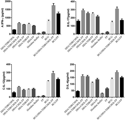
Figure 2. IgG1 (A), IgG2a (B), and IgG2b (C) titers in sera of immunized BALB/c mice. Mice were immunized subcutaneously three times at two-week intervals. Blood samples were collected from the mice three weeks after the last Injection. The IgG1, IgG2a, and IgG2b titers were determined by ELISAs. The assays were performed in triplicate at 200, 2000 and 20,000 -fold dilutions for each serum sample. DDA: Dimethyldioctadecylammonium; TDB: Trehalose-6,6'-dibehenate; CHOL: Cholesterol; FP: Fusion protein; BCG: Bacillus Calmette-Guerin.
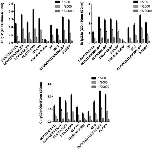
Table 3. IgG2a/IgG1 Ratioes of diluted serum from immunized mice (OD: 450 nm–630 nm).